Comparative Study
. 2007 May 30;27(22):5967-75.
doi: 10.1523/JNEUROSCI.0110-07.2007.

Epigenetic modulation of seizure-induced neurogenesis and cognitive decline

Affiliations
Comparative Study

Epigenetic modulation of seizure-induced neurogenesis and cognitive decline

Sebastian Jessberger et al. J Neurosci. .

Abstract

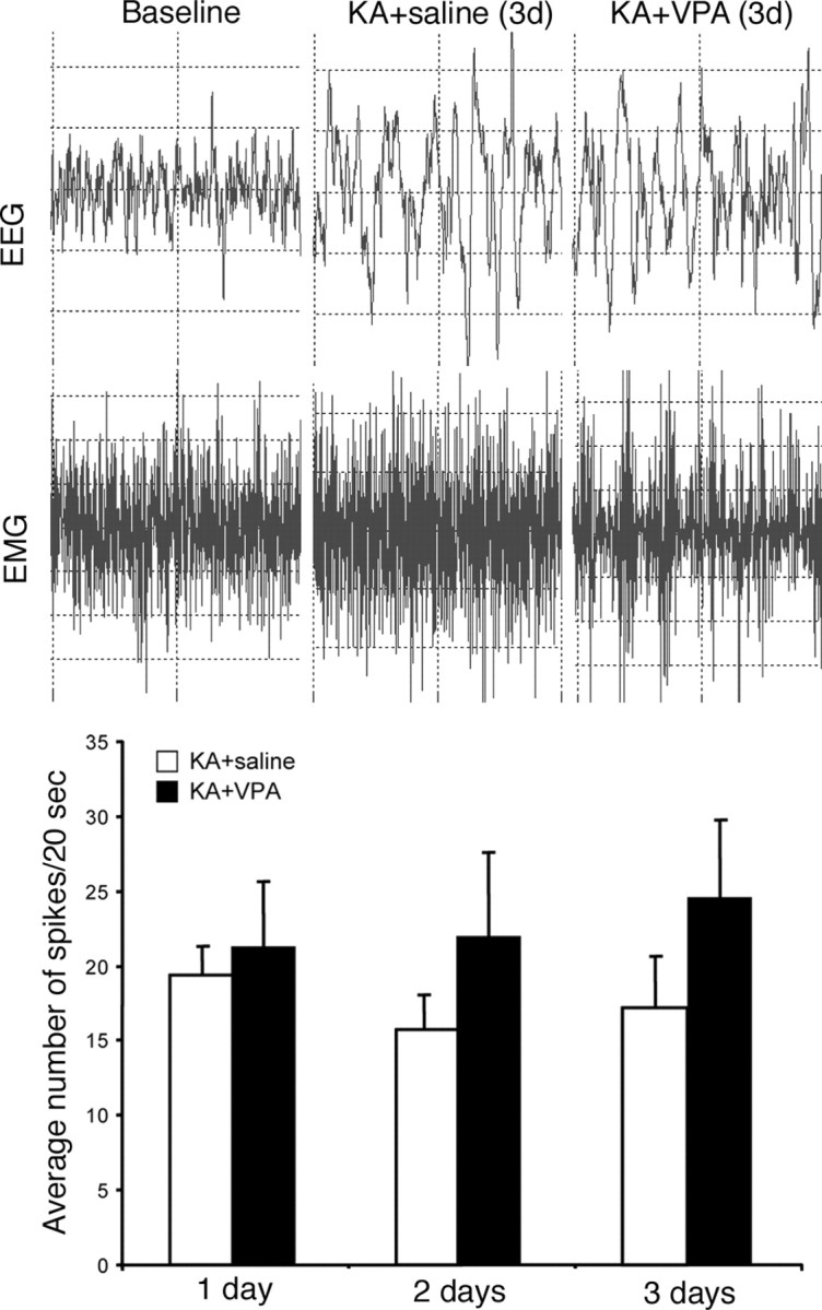
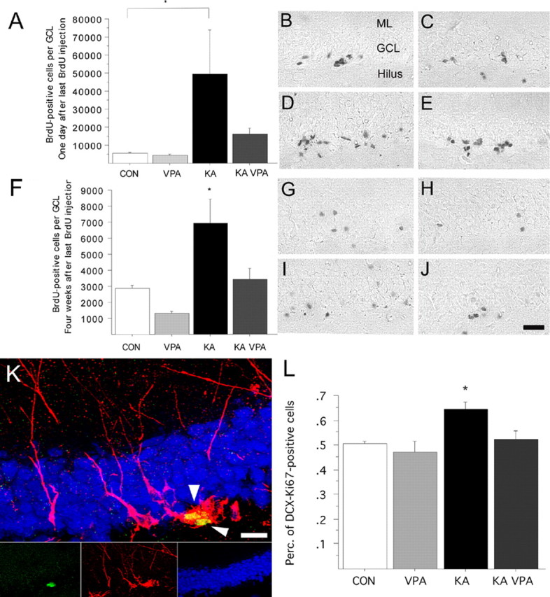
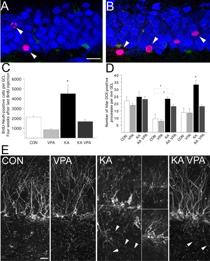
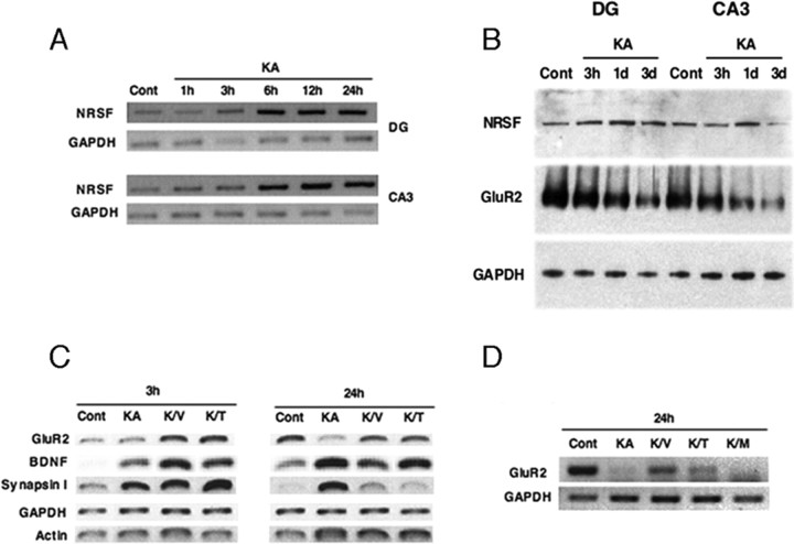
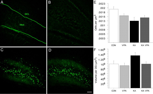
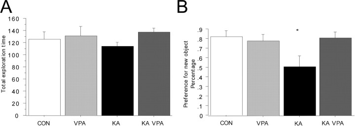
The conceptual understanding of hippocampal function has been challenged recently by the finding that new granule cells are born throughout life in the mammalian dentate gyrus (DG). The number of newborn neurons is dynamically regulated by a variety of factors. Kainic acid-induced seizures, a rodent model of human temporal lobe epilepsy, strongly induce the proliferation of DG neurogenic progenitor cells and are also associated with long-term cognitive impairment. We show here that the antiepileptic drug valproic acid (VPA) potently blocked seizure-induced neurogenesis, an effect that appeared to be mainly mediated by inhibiting histone deacetylases (HDAC) and normalizing HDAC-dependent gene expression within the epileptic dentate area. Strikingly, the inhibition of aberrant neurogenesis protected the animals from seizure-induced cognitive impairment in a hippocampus-dependent learning task. We propose that seizure-generated granule cells have the potential to interfere with hippocampal function and contribute to cognitive impairment caused by epileptic activity within the hippocampal circuitry. Furthermore, our data indicate that the effectiveness of VPA as an antiepileptic drug may be partially explained by the HDAC-dependent inhibition of aberrant neurogenesis induced by seizure activity within the adult hippocampus.


Figures

Figure 1.
Figure 1.
Representative examples of EEG monitoring at baseline and spike-wave activity characteristic of seizures in KA-treated animals receiving saline or VPA injections after 3 d. Quantification of the average number of spikes per 20 s time period between VPA- and saline-treated seizure animals at 1, 2, and 3 d, showing no significant difference between the two groups.
Figure 2.
Figure 2.
VPA treatment blocks seizure-induced neurogenesis and prevents aberrant DCX-expressing progenitor cell activation. A–E, Number of BrdU-positive cells 1 d after the last BrdU injection, indicative of proliferative activity after SE (A). Representative BrdU staining of saline (B), VPA-treated (C), KA-injected (D), and KA-injected plus VPA-treated (E) animals killed 1 d after last BrdU injection. F–J, Number of BrdU-positive cells 4 weeks after the last BrdU injection, indicative of stable cell genesis after SE (F). Representative BrdU staining of saline (G), VPA-treated (H), KA-injected (I), and KA-injected plus VPA-treated (J) animals killed 4 weeks after last BrdU injection. K, Example of two DCX (red)/Ki-67 (green) colabeled cells (arrowheads) in the subgranular zone (blue, NeuN) 8 d after SE. L, The percentage of DCX/Ki-67 colabeled cells is increased 8 d after SE compared with controls. VPA treatment prevents aberrant DCX-positive progenitor cell activation. Scale bars: (in J) B–J, 100 μm; K, 20 μm. *p < 0.01.
Figure 3.
Figure 3.
VPA treatment abates the number of seizure-generated neurons and decreases the formation of hilar basal dendrites. A–C, Compared with control animals (A), the number of new neurons (arrowheads) 4 weeks after the last BrdU injection is dramatically increased after KA injection (B; red, BrdU; blue, NeuN; green, S100β). VPA treatment significantly reduced the number of new neurons after SE (C). D, E, VPA treatment inhibited the formation of basal dendrites reaching into the polymorphic cell layer (D). Representative examples of DCX-immunoreactive cells in saline (CON), VPA-treated (VPA), KA-injected (KA), and KA-injected plus VPA-treated (KA VPA) animals killed 8 d after SE (E). Arrowheads point toward hilar basal dendrites. Scale bars: A, E, 20 μm. *p < 0.01.
Figure 4.
Figure 4.
VPA inhibits seizure-mediated neurogenesis by normalizing changes in gene expression. A–D, RT-PCR and Western blot analysis of DG and CA3 from saline-injected (Cont) and KA-injected (KA) rats from 1 h to 3 d (A, B). RT-PCR of DG and CA3 from control (Cont) and seizure (KA) rats from 1–24 h (A). Representative Western blots of DG and CA3 from control (Cont) and seizure (KA) animals from 3 h to 3 d (B). RT-PCR of DG from saline-injected (Cont) and KA-injected animals receiving either VPA (K/V) or TSA (K/T) (1 h before treatment) and RNA harvested 3 and 24 h after onset of seizures (C). RT-PCR of DG from control (Cont) and KA-treated rats with VPA (K/V), TSA (K/T), or valpromide (K/M) given 5 h after seizure onset, and RNA was harvested 24 h later (D). GAPDH and/or β-actin (data not shown) were used as normalization controls. Data are representative of at least three independent experiments.
Figure 5.
Figure 5.
Seizure-induced cell death in the dentate area is not affected by VPA treatment. A–D, Fluoro-Jade B staining of saline (A), VPA-treated (B), KA-injected (C), and KA-injected plus VPA-treated (D) animals killed 5 weeks after SE shows degenerating neuronal cells in the hilus in the KA-injected animals. E, F, Cell density and DG volume in control animals and seizure animals 5 weeks after SE. Scale bar, 100 μm.
Figure 6.
Figure 6.
Seizure-associated cognitive impairment is prevented by VPA treatment. A, B, Although the total exploration time was comparable between all groups (A), KA-challenged animals did not show a preference for the new object 3 h after the presentation of the initial objects (B). VPA treatment prevented this phenotype, and KA-challenged animals that received VPA injections showed the same level of preference for the new objects as controls did. *p < 0.01.

References

    1. Ballas N, Battaglioli E, Atouf F, Andres ME, Chenoweth J, Anderson ME, Burger C, Moniwa M, Davie JR, Bowers WJ, Federoff HJ, Rose DW, Rosenfeld MG, Brehm P, Mandel G. Regulation of neuronal traits by a novel transcriptional complex. Neuron. 2001;31:353–365. - PubMed
    1. Ben-Ari Y, Cossart R. Kainate, a double agent that generates seizures: two decades of progress. Trends Neurosci. 2000;23:580–587. - PubMed
    1. Bengzon J, Kokaia Z, Elmér E, Nanobashvili A, Kokaia M, Lindvall O. Apoptosis and proliferation of dentate gyrus neurons after single and intermittent limbic seizures. Proc Natl Acad Sci USA. 1997;94:10432–10437. - PMC - PubMed
    1. Bengzon J, Mohapel P, Ekdahl CT, Lindvall O. Neuronal apoptosis after brief and prolonged seizures. Prog Brain Res. 2002;135:111–119. - PubMed
    1. Beuckmann CT, Sinton CM, Williams SC, Richardson JA, Hammer RE, Sakurai T, Yanagisawa M. Expression of a poly-glutamine-ataxin-3 transgene in orexin neurons induces narcolepsy-cataplexy in the rat. J Neurosci. 2004;24:4469–4477. - PMC - PubMed

Publication types

MeSH terms