. 2007 Oct 1;110(7):2600-9.
doi: 10.1182/blood-2006-01-028647. Epub 2007 May 30.

NPM/ALK binds and phosphorylates the RNA/DNA-binding protein PSF in anaplastic large-cell lymphoma

Affiliations

NPM/ALK binds and phosphorylates the RNA/DNA-binding protein PSF in anaplastic large-cell lymphoma

Annamaria Galietta et al. Blood. .

Abstract

The oncogenic fusion tyrosine kinase nucleophosmin/anaplastic lymphoma kinase (NPM/ALK) induces cellular transformation in anaplastic large-cell lymphomas (ALCLs) carrying the t(2;5) chromosomal translocation. Protein-protein interactions involving NPM/ALK are important for the activation of downstream signaling pathways. This study was aimed at identifying novel NPM/ALK-binding proteins that might contribute to its oncogenic transformation. Using a proteomic approach, several RNA/DNA-binding proteins were found to coimmunoprecipitate with NPM/ALK, including the multifunctional polypyrimidine tract binding proteinassociated splicing factor (PSF). The interaction between NPM/ALK and PSF was dependent on an active ALK kinase domain and PSF was found to be tyrosine-phosphorylated in NPM/ALK-expressing cell lines and in primary ALK(+) ALCL samples. Furthermore, PSF was shown to be a direct substrate of purified ALK kinase domain in vitro, and PSF Tyr293 was identified as the site of phosphorylation. Y293F PSF was not phosphorylated by NPM/ALK and was not delocalized in NPM/ALK(+) cells. The expression of ALK fusion proteins induced delocalization of PSF from the nucleus to the cytoplasm and forced overexpression of PSF-inhibited proliferation and induced apoptosis in cells expressing NPM/ALK. PSF phosphorylation also increased its binding to RNA and decreased the PSF-mediated suppression of GAGE6 expression. These results identify PSF as a novel NPM/ALK-binding protein and substrate, and suggest that PSF function may be perturbed in NPM/ALK-transformed cells.


Figures

Figure 1
Figure 1
Identification of proteins coimmunoprecipitating with NPM/ALK by mass spectrometry. (A) Silver-stained SDS-PAGE gel of anti-ALK1 immunoprecipitates from NPM/ALK K562 cells (lane 1), lysis buffer alone (lane 2), and NPM/ALK+ SUDHL-1 cells (lane 3). Bands were excised from the gel, trypsin-digested, and analyzed by MALDI-TOF. Identified proteins are indicated by arrows. (B) MS/MS sequencing of peptides identifying PSF (GenBank accession no. P23246) and NPM/ALK (GenBank accession no. AAA58698) proteins.
Figure 2
Figure 2
PSF coimmunoprecipitates with ALK fusion proteins expressed in cell lines and ALCL tumors. Reciprocal immunoprecipitation (IP) and Western blotting (WB) experiments were performed using anti-ALK1 and anti-PSF antibodies on lysates derived from (A) NPM/ALK+ SUDHL-1 cells and NPM/ALK K562 cells; (B) BaF3-NPM/ALK (BaF3-N/A), BaF3-Parental (BaF3-PAR), and BaF3 cells expressing kinase-dead NPM/ALK (BaF3-KD); (C) lymph node tissue taken from 2 patients with NPM/ALK+ ALCL and NPM/ALK spleen tissue; and (D) BaF3 cells expressing GCN4/ALK (BaF3-GCN4/ALK). As an internal control, SUDHL-1 and K562 cells were subjected to IP with an anti-IgG antibody (A).
Figure 3
Figure 3
PSF is tyrosine-phosphorylated in NPM/ALK-expressing cells and is a substrate of ALK tyrosine kinase activity. (A) Anti-ALK1 and anti-PSF immunoprecipitates (IPs) from SUDHL-1, K562, BaF3-Par, BaF3-KD, and BaF3-NA cells were Western blotted with an antiphosphotyrosine (anti-pTyr) antibody. (B) Anti-ALK1 immunoprecipitates from lymph nodes derived from patient 1 with NPM/ALK+ ALCL and NPM/ALK spleen tissue were Western blotted using the anti-pTyr antibody. (C) A radioactive in vitro kinase assay was performed with purified recombinant 6xHisALK kinase domain (rALK) and purified full-length 6xHis-PSF. Protein purity was checked by coomassie staining (Ci). Kinase assay was performed in the presence of rALK kinase domain alone (0.4 μg), PSF alone (0.25 μg), or rALK and PSF together. Samples were analyzed by SDS-PAGE and visualized by autoradiography (Cii). Bands corresponding to PSF and ALK are indicated by arrows. (D) Time courses of the phosphorylation of PSF peptides (400 μM) by GST-ALK. The peptide sequences are as follows: Tyr251, RGGRQHHPPYHQQHHQGP; Tyr293, PGEKTYTQRCRLFVGNLPADIT; Tyr470, EKLAQKNPMYQKERETPTR; Tyr488-490, TFEYEYSQRWKSLDEMEKQQR; Tyr527, EMEDAYHEHQANLLRQDLMRRQ; Tyr597-602, REESYSRMGYMDPRERDMR; Tyr624, MNMGDPYGSGGQKFPPLGGG; and Tyr698, GRGREEYEGPNKKPRF (target tyrosine in bold). (E) Immunoprecipitation with anti-HA (lanes 2,3) and anti-ALK1 (lanes 4,5) monoclonal antibodies of lysate derived from 293T cells transiently transfected with wild-type or mutant Y293F HA-PSF together with NPM/ALK. Immunoprecipitates were resolved by SDS-PAGE, and Western blotting with anti-pTyr and anti-HA antibodies was performed. Lane 1 shows lysate from nontransfected 293T cells.
Figure 4
Figure 4
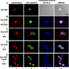
PSF is delocalized to the cytoplasm in cells expressing ALK fusion proteins. Indirect immunofluorescence was performed on 293T cells transiently transfected with HA-PSF alone, or together with kinase-dead NPM/ALK (KD-N/A), NPM/ALK (N/A), GCN4/ALK (G/A), or with mutant HA-PSF-Y293F together with NPM/ALK (N/A). Fluorescence was detected by confocal microscopy. Nuclei were detected using propidium iodide (PI) shown in red (column 1). HA-PSF was detected by FITC staining shown in green (column 2). NPM/ALK was detected by Cy5 staining shown in blue (column 3). A transmission image in place of Cy5 staining is shown for cells transfected with HA-PSF alone. Images were acquired using a Nikon 60×/1.4 NA oil-immersion objective lens, a medium solution of 95% glycerol in PBS, and a Biorad Laser Sharp 2000 camera; image processing was done with Adobe Photoshop CS2 version 9.0.
Figure 5
Figure 5
PSF content in cytoplasmic and nuclear extracts derived from human cell lines. Nuclear and cytoplasmic extracts were prepared from the NPM/ALK cell line, Jurkat, and the NPM/ALK+ cell lines, SUDHL-1 and JB6. (A) Total cell lysates (lanes 1-3), cytoplasmic extracts (lanes 4-6), and nuclear extracts (lanes 7-9) were resolved by SDS-PAGE, and PSF was detected by Western blotting (top panel). The purity of the extracts were controlled by Western blotting with anti-β-actin for the cytoplasm (middle panel) and anti-histone H1 for the nucleus (bottom panel). (B) Nuclear and cytoplasmic extracts from SUDHL-1 cells were probed with antiphosphotyrosine antibody.
Figure 6
Figure 6
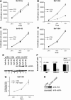
PSF overexpression reduces the growth rate and clonogenic potential of NPM/ALK-expressing cells. BaF3-Par (A), BaF3-NA (B), BaF3-KD (C), and BaF3-BA (D) cells were transiently transfected with pCR3.1 encoding HA-PSF (PSF) or with empty vector (CON). Cell proliferation was measured by 3H-thymidine incorporation at 24, 48, and 72 hours after transfection. Results are shown as the mean (± SD) and are representative of 3 independent experiments. (E) Expression of HA-PSF was verified by Western blotting using anti-HA antibody at 72 hours, and protein loading was controlled by antiactin Western blotting. (F) The NPM/ALK+ cell lines, JB6 and SUDHL-1, and the NPM/ALK cell line, Jurkat, were transduced with the MigR1-HA-PSF construct (▩), or the MigR1 retrovirus alone (■). Cells were selected for GFP positivity, and clonogenic potential was measured by colony formation in methylcellulose. Results are expressed as the mean (± SEM) of 3 independent clonogenic assays. Significance was determined by comparing MigR1 alone versus MigR1-HA-PSF in a 2-tailed paired samples t test. (G) BaF3-NA cells were transiently transfected with pCR3.1 encoding HA-Y293T PSF or with empty vector (CON). Error bars are standard deviation. (H) Transfection efficiency was controlled by anti-HA immunoblotting at 48 hours in control (CON) and HA-PSF Y293F samples loaded on the same gel. Total protein loading was controlled by antiactin immunoblotting.
Figure 7
Figure 7
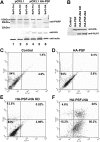
PSF overexpression induces apoptosis in NPM/ALK-expressing cell lines. (A) BaF3-Par, BaF3-KD, and Baf3-NA cells were electroporated with pCR3.1 plasmid alone (lanes 1-3) and with pCR3.1 HA-PSF (lanes 4-6). After 30 hours of culture, the cell extracts from 5 × 10⁶ cells were lysed and subjected to denaturing SDS-PAGE and Western blotting with an anti-PARP antibody. PSF expression and protein loading were controlled by anti-HA and antiactin Western blotting. (B-F) 293T cells were transiently transfected with pCR3.1 empty vector (CON) or HA-PSF alone or together with kinase-dead or wild-type NPM/ALK. The efficiency of transfection was determined by anti-HA and anti-ALK1 Western blotting at 72 hours after transfection (B). Apoptosis was assessed at the same time point by Annexin V analysis in control cells (C), cells transfected with HA-PSF alone (D), or together with kinase-dead NPM/ALK (E) (HA-PSF + N/A-KD), or wild-type NPM/ALK (F) (HA-PSF + NA). C-F, live cell percentages are shown in the bottom left panels; apoptotic cell percentages in the bottom right panels, and dead cell percentages in the top right panels.

Similar articles

Cited by

References

    1. Stein H, Foss HD, Durkop H, et al. CD30(+) anaplastic large cell lymphoma: a review of its histopathologic, genetic, and clinical features. Blood. 2000;96:3681–3695. - PubMed
    1. Morris SW, Xue L, Ma Z, Kinney MC. Alk+ CD30+ lymphomas: a distinct molecular genetic subtype of non-Hodgkin's lymphoma. Br J Haematol. 2001;113:275–295. - PubMed
    1. Morris SW, Naeve C, Mathew P, et al. ALK, the chromosome 2 gene locus altered by the t(2;5) in non-Hodgkin's lymphoma, encodes a novel neural receptor tyrosine kinase that is highly related to leukocyte tyrosine kinase (LTK). Oncogene. 1997;14:2175–2188. - PubMed
    1. Iwahara T, Fujimoto J, Wen D, et al. Molecular characterization of ALK, a receptor tyrosine kinase expressed specifically in the nervous system. Oncogene. 1997;14:439–449. - PubMed
    1. Pulford K, Morris SW, Turturro F. Anaplastic lymphoma kinase proteins in growth control and cancer. J Cell Physiol. 2004;199:330–358. - PubMed

Publication types

MeSH terms