. 2007 Aug;18(8):3193-203.
doi: 10.1091/mbc.e06-12-1147. Epub 2007 May 30.

The clathrin adaptor complex AP-1 binds HIV-1 and MLV Gag and facilitates their budding

Affiliations

The clathrin adaptor complex AP-1 binds HIV-1 and MLV Gag and facilitates their budding

Grégory Camus et al. Mol Biol Cell. 2007 Aug.

Abstract

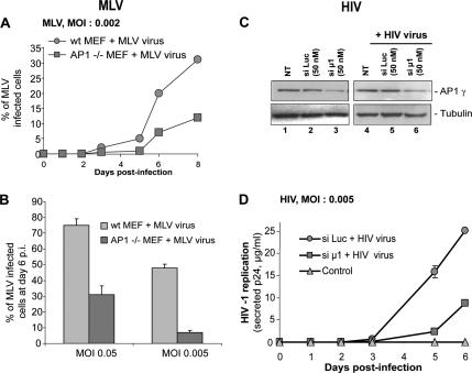
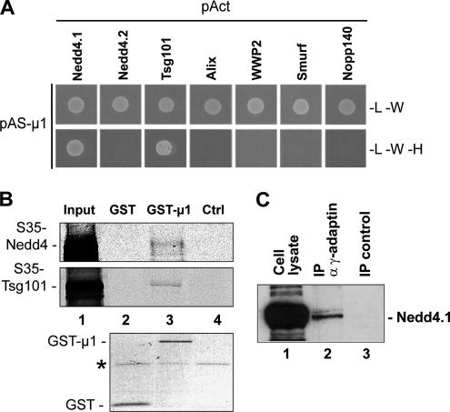
Retroviral assembly is driven by Gag, and nascent viral particles escape cells by recruiting the machinery that forms intralumenal vesicles of multivesicular bodies. In this study, we show that the clathrin adaptor complex AP-1 is involved in retroviral release. The absence of AP-1mu obtained by genetic knock-out or by RNA interference reduces budding of murine leukemia virus (MLV) and HIV-1, leading to a delay of viral propagation in cell culture. In contrast, overexpression of AP-1mu enhances release of HIV-1 Gag. We show that the AP-1 complex facilitates retroviral budding through a direct interaction between the matrix and AP-1mu. Less MLV Gag is found associated with late endosomes in cells lacking AP-1, and our results suggest that AP-1 and AP-3 could function on the same pathway that leads to Gag release. In addition, we find that AP-1 interacts with Tsg101 and Nedd4.1, two cellular proteins known to be involved in HIV-1 and MLV budding. We propose that AP-1 promotes Gag release by transporting it to intracellular sites of active budding, and/or by facilitating its interactions with other cellular partners.


Figures

Figure 1.
Figure 1.
Two-hybrid interactions of AP-1μ with MLV and HIV-1 Gag. (A) Schematic representation of retroviral Gag proteins. Matrix (MA), capsid (CA), and nucleocapsid (NC) domains are shown respectively in gray, light gray, and dark gray, respectively. (B) MLV and HIV-1 Gag interact with AP-1μ in two-hybrid assays. HIV-1 and MLV Gag were fused to the Gal4 DNA-binding domain and tested against μ1, μ2, μ3 fused to the Gal4 activation domain. Nopp140 was used as a negative control. Strains producing the hybrid proteins were analyzed for histidine auxotrophy. -L-W, mating control; -L-W-H, growth only if the tested proteins interact. (C) MLV MA-p12 mediates the interaction of Gag with AP-1μ. The indicated portions of MLV Gag were fused to the Gal4 DNA-binding domain and tested as in B.
Figure 2.
Figure 2.
MLV and HIV-1 Gag interact with the AP-1 complex in vitro and in vivo. (A) In vitro binding of AP-1μ to HIV-1 Gag. His-μ1–purified from bacteria was incubated with equal amounts of immobilized GST, GST-Gag, GST-MA, or GST-CA. Top panel, bound material was analyzed by Western blot using anti-His antibodies. Bottom panel corresponds to the ponceau red staining of the membrane; 20% of input was loaded in lane 1. (B) Coimmunoprecipitation of HIV-1 Gag with the AP-1 complex. 293T cells were transfected with HIV-1 HXB2R (HIV) or HXB2RΔEnv (HIVΔEnv) proviral DNAs. The endogenous AP-1 complex was immunoprecipitated with anti-AP-1γ or with anti-α-tubulin antibodies as a control. Bound material was analyzed by Western blot with anti-AP-1γ (top panel) and anti-CAp24 antibodies (bottom panel). Left panel, 10% of input. (C) In vitro binding of AP-1μ to MLV Gag. 35S-labeled MLV Gag was translated in vitro and incubated with GST or GST-μ1 immobilized on beads. Top panel, the autoradiogram of bound material; the bottom panel corresponds to the Coomassie blue staining of purified GST and GST-μ1. Asterisk shows a nonspecific protein that copurified; 5% of input was loaded in lane 1. (D) Interaction of the endogenous AP-1 complex with MLV MA-p12. GST-MA-p12 and GST were expressed in 293T cells and purified on glutathione-Sepharose beads, and bound proteins were analyzed by Western blotting using anti-AP-1γ antibodies.
Figure 3.
Figure 3.
MLV and HIV-1 replication requires AP-1. (A and B) Kinetic of MLV replication in AP-1 knockout cells. (A) WT and AP-1−/− MEFs cells were infected with MLV (strain F57) at an MOI of 0.002. Infected cells were labeled with anti-envelope antibodies and counted by FACS (mean of two experiments, ±SD). (B) WT and AP-1−/− MEFs cells were infected with MLV (strain F57) at MOIs of 0.05 or 0.005. Infected cells were counted by FACS at day 6 after infection (mean of five experiments, ±SD). (C and D) Kinetic of HIV-1 replication in AP-1μ–depleted cells. HeLa P4R5 cells were depleted with siRNAs against luciferase (si Luc) or AP-1μ (si μ1) and infected with HIV-1 HXB2R at an MOI of 0.005. (C) Western blots demonstrate a decrease in AP-1γ levels, both at day 0 (lanes 1–3) and day 6 after infection (lanes 4–6). (D) Culture supernatants were collected daily for 6 d after infection and quantified for HIV Gag p24 (mean of two experiments, ±SD). Controls: nontransfected, and noninfected cells.
Figure 4.
Figure 4.
Production of MLV and HIV-1 viral particles is inhibited by the absence of AP-1. (A) Release of MLV Gag by WT and AP-1−/− MEFs. WT MEFs were transfected with MLV Gag fused to YFP, and AP-1−/− cells were transfected with either Gag-YFP alone or with Gag-YFP plus increasing amounts of μ1-YFP. Top panel, expression of μ1-YFP detected by Western blotting with anti-μ1 antibodies, and the numbers indicate the amount of transfected μ1-YFP plasmid (μg). Middle and the bottom panels, the levels of Gag-YFP in the cellular and viral fractions (detected with anti-capsid antibodies), respectively. Left panel, WT MEFs, right panel, AP-1−/− MEFs. (B) MLV Gag release by chronically infected WT and AP-1−/− MEFs. Cell lysates and virions produced by WT and AP-1−/− cells chronically infected with MLV were analyzed by Western blot using anti-capsid antibodies. (C and D) AP-1 depletion decreases the release of HIV-1 particles. HeLa cells treated with siRNAs against AP-1μ (si μ1) or luciferase (si Luc) were transfected with HIV-1 proviral DNA. (C) Depletion of AP-1 and the intracellular expression of HIV-1 Gag were analyzed by Western blotting with anti-AP-1γ and anti-CAp24 antibodies. (D) Secreted HIV-1 CAp-24 was quantified by ELISA (mean of four experiments, ±SD). NT, nontransfected cells. (E and F) AP-1μ overexpression has a dose-dependent effect on HIV-1 release. HeLa cells were cotransfected with HIV-1 proviral DNA and increasing amounts of Flag-μ1. (E) Expression of Flag-μ1, AP-1γ, and Gag (p55Gag, p41, CAp24) were analyzed by Western blot using anti-FLAG M2, anti-AP-1γ, and anti-CAp24γ antibodies, respectively. (F) Secreted HIV-1 CAp24 was quantified by ELISA (mean of two experiments performed in duplicate). NT, nontransfected cells.
Figure 5.
Figure 5.
AP-1 depletion leads to a decrease of budding profiles. (A) Thin-section electron microscopy analysis of HeLa cells infected with HIV-1 and treated either with siRNA luc or with siRNA AP-1μ. (B) Thin-section electron microscopy analysis of WT and AP-1−/− MEFs chronically infected with MLV.
Figure 6.
Figure 6.
AP-1 mediates its effect through a direct interaction with matrix and acts in concert with AP-3 to facilitate Gag release. (A) Release of HIV-1 ΔMA is not inhibited by AP-1 and AP-3 depletion. HeLa cells were treated with siRNAs against: AP-1γ (si γ1), AP-3δ (si δ3), AP-1γ/AP-3δ (si γ1δ3), and siRNA luciferase (si luc). Cells were then transfected with an HIV-1ΔMA provirus. Top panel, secreted HIV-1 CAp-24 was quantified by ELISA (mean of four experiments, ±SD). NT, nontransfected cells. Bottom panel, intracellular expression of AP-1 and AP-3, and HIV-1 Gag were analyzed by Western blotting with anti-AP-1γ, anti-AP-3δ, and anti-CAp24 antibodies. (B) AP-1 and AP-3 function on the same pathway of HIV-1 Gag release. HeLa cells were treated with siRNAs against the following: AP-1μ (si μ1), AP-1γ (si γ1), both AP-1μ and AP-1γ (si μ1γ1), AP-3δ (si δ3), AP-1γ, and AP-3δ (si γ1δ3) and siRNA luciferase (si luc) and then transfected with HIV-1 provirus. Top panel, secreted HIV-1 CAp-24 was quantified by ELISA (mean of four experiments, ±SD). NT, nontransfected cells. Bottom panel, depletion of AP-1 and AP-3 and the intracellular expression of HIV-1 Gag were analyzed by Western blotting.
Figure 7.
Figure 7.
Intracellular trafficking of MLV Gag is affected in the absence of AP-1. (A) Localization of MLV Gag in WT and AP-1−/− cells chronically infected with MLV. WT and AP-1−/− MEFs were labeled with anti-capsid antibodies (green) and lysotracker (a marker for late endosomes, red). The numbers on the right indicate the proportion of Gag associated with late endosomes; 30 cells were analyzed in each case. (B) Monensin differentially affects the production of MLV virions in WT and AP1−/− cells. The level of MLV Gag in cell lysates (top) and viral supernatants (bottom) of WT and AP1−/− chronically infected cells treated with 5 μM monensin for 5 h, and control was analyzed by Western blot with anti-CA antibodies.
Figure 8.
Figure 8.
AP-1 interacts with Nedd4.1 and Tsg101. (A) Interaction of AP-1μ with Nedd4.1 and Tsg101 in yeast two-hybrid assays. AP-1μ was fused to the N-terminus of the Gal4 DNA-binding domain, and tested against Tsg101, Smurf, Alix, WWP2, Nedd4.1, Nedd4.2, and Nopp140. Legend as in Figure 1B. (B) In vitro binding of AP-1μ to Nedd4.1 and Tsg101. 35S-labeled Nedd4.1-YFP and Tsg101-YFP were translated in vitro and mixed with immobilized GST or GST-μ1. Top and middle panels, the autoradiograms; bottom panel, corresponding Coomassie blue staining of the purified GST proteins. Asterisk shows a nonspecific protein that copurified; 5% of input was loaded in lane 1. (C) Coimmunoprecipitation of Nedd4.1 with the AP-1 complex. AP-1 was immunoprecipitated from extracts of HT1080 cells, using anti-γ-adaptin antibodies, and bound proteins were analyzed by Western blots with anti-Nedd4.1 antibodies. The control corresponds to materials precipitated without antibodies; 5% of input was loaded in lane 1.

Similar articles

Cited by

References

    1. Alroy I., et al. The trans-Golgi network-associated human ubiquitin-protein ligase POSH is essential for HIV type 1 production. Proc. Natl. Acad. Sci. USA. 2005;102:1478–1483. - PMC - PubMed
    1. Babst M., Katzmann D. J., Estepa-Sabal E. J., Meerloo T., Emr S. D. Escrt-III: an endosome-associated heterooligomeric protein complex required for mvb sorting. Dev. Cell. 2002a;3:271–282. - PubMed
    1. Babst M., Katzmann D. J., Snyder W. B., Wendland B., Emr S. D. Endosome-associated complex, ESCRT-II, recruits transport machinery for protein sorting at the multivesicular body. Dev. Cell. 2002b;3:283–289. - PubMed
    1. Basyuk E., Galli T., Mougel M., Blanchard J., Sitbon M., Bertrand E. Retroviral genomic RNAs are transported to the plasma membrane by endosomal vesicles. Dev. Cell. 2003;5:161–174. - PubMed
    1. Batonick M., Favre M., Boge M., Spearman P., Honing S., Thali M. Interaction of HIV-1 Gag with the clathrin-associated adaptor AP-2. Virology. 2005;342:190–200. - PubMed

Publication types

MeSH terms

LinkOut - more resources