. 2007 Aug;18(8):2980-90.
doi: 10.1091/mbc.e06-11-1054. Epub 2007 May 30.

Mechanism underlying the iron-dependent nuclear export of the iron-responsive transcription factor Aft1p in Saccharomyces cerevisiae

Affiliations

Mechanism underlying the iron-dependent nuclear export of the iron-responsive transcription factor Aft1p in Saccharomyces cerevisiae

Ryo Ueta et al. Mol Biol Cell. 2007 Aug.

Abstract

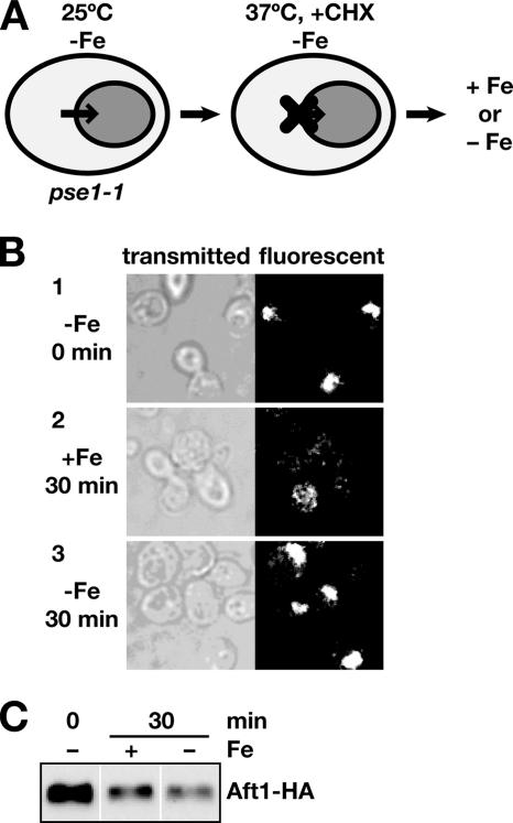
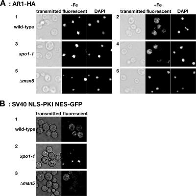
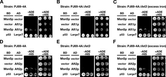
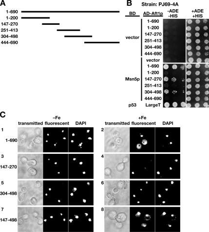
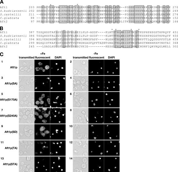
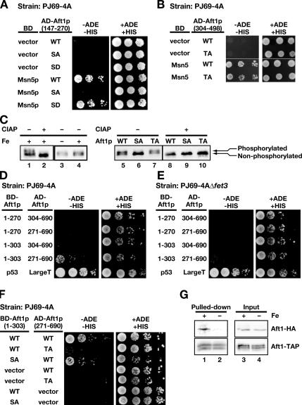
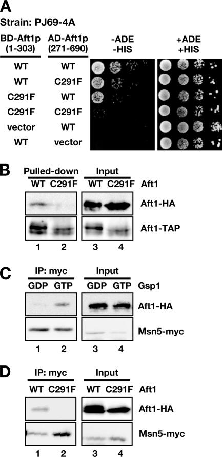
Aft1p is an iron-responsive transcriptional activator that plays a central role in maintaining iron homeostasis in Saccharomyces cerevisiae. Aft1p is regulated primarily by iron-induced shuttling of the protein between the nucleus and cytoplasm, but its nuclear import is not regulated by iron. Here, we have shown that the nuclear export of Aft1p is promoted in the presence of iron and that Msn5p is the nuclear export receptor (exportin) for Aft1p. Msn5p recognizes Aft1p in the iron-replete condition. Phosphorylation of S210 and S224 in Aft1p, which is not iron dependent, and the iron-induced intermolecular interaction of Aft1p are both essential for its recognition by Msn5p. Mutation of Cys291 of Aft1p to Phe, which causes Aft1p to be retained in the nucleus and results in constitutive activation of Aft1-target genes, disrupts the intermolecular interaction of Aft1p. Collectively, these results suggest that iron induces a conformational change in Aft1p, in which Aft1p Cys291 plays a critical role, and that, in turn, Aft1p is recognized by Msn5p and exported into the cytoplasm in an iron-dependent manner.


Figures

Figure 1.
Figure 1.
Nuclear export of Aft1p is promoted by iron. (A) Schematic diagram of the monitoring of Aft1p nuclear export. Shading represents the localization of Aft1p in the nucleus. (B) The nuclear export of Aft1p is promoted by iron. pse1-1 (PSY1201) cells expressing Aft1p-HA were grown to the logarithmic growth phase under conditions of iron starvation at 25°C, followed by cultivation at 37°C for 4 h to inactivate Pse1p. Cycloheximide was added to the cultures to inhibit further synthesis of Aft1p (1). Cells were further cultured at 37°C in the presence (2) or absence (3) of iron. At each of the indicated time points, cells were fixed, and the subcellular localization of Aft1p-HA was examined by indirect immunofluorescence microscopy. (C) The amount of Aft1 protein was not affected by the iron status of the cells. Lysates from yeast cells as treated described in B were separated by SDS-PAGE, and Aft1-HA was probed with an anti-HA antibody.
Figure 2.
Figure 2.
Aft1p accumulates in the nucleus of the Δmsn5 strain independently of iron status. (A) Wild-type (PSY580) (1 and 2), xpo1-1 (PSY1105) (3 and 4), or Δmsn5 (PSY1569) cells (5 and 6) were grown to the logarithmic growth phase in iron-starved (−Fe; 1, 3, and 5) or iron-replete (+Fe; 2, 4, and 6) conditions. After fixation, the subcellular localization of Aft1p-HA was examined by indirect immunofluorescence microscopy. Images taken with transmitted light and DAPI staining of nuclei are provided for comparison. (B) The subcellular localization of the SV40 NLS-PKI NES-GFP fusion protein was observed in wild-type (1), xpo1-1 (2), or Δmsn5 (3) cells. xpo1-1 cells were cultured at 37°C for 3 h before fixation (A and B).
Figure 3.
Figure 3.
Aft1p and Aft2p interact with Msn5p in an iron-dependent manner. PJ69-4A (A and D) or PJ69-4AΔfet3 (Y22; B, C, E, and F) cells were transformed with plasmids expressing AD-fused Aft1p (A–C), AD-fused Aft2p (D–F), or the corresponding empty vector, as well as a plasmid expressing BD-fused Msn5p or the corresponding empty vector. Cells were grown at 30°C for 4 d on medium containing adenine and histidine (+ADE +HIS), or lacking adenine and histidine but containing 10 mM 3-AT (−ADE −HIS). In C and F, 200 μM FeSO4 was added to the medium to create an excess iron condition. Cells were spotted using 10-fold serial dilutions beginning at an OD600 = 0.6. AD-fused SV40 Large T antigen (LargeT) and BD-fused p53 were used as a positive control.
Figure 4.
Figure 4.
The Msn5p-interacting domain within Aft1p. (A) Schematic presentation of the fragments of Aft1p tested for interaction with Msn5p. (B) Aft1p(147-270) and Aft1p(304-498) interact with Msn5p. Using PJ69-4A cells expressing the AD-fused Aft1p fragment indicated on the left and BD-Msn5p, the interaction between the fragments of Aft1p and Msn5p was tested as in Figure 3. (C) Subcellular localization of GFP fused Aft1p fragments. Y24 cells expressing GFP-Aft1p(1-690) (1 and 2), GFP-Aft1p(147-270) (3 and 4), GFP-Aft1p(304-498) (5 and 6), or GFP-Aft1p(147-498) (7 and 8) were cultured in the absence (−Fe; 1, 3, 5, and 7) or presence (+Fe; 2, 4, 6, and 8) of FOB, and the localization of GFP-fusion proteins was observed.
Figure 5.
Figure 5.
Two serine and four threonine residues are important for Aft1p-nuclear export. (A and B) Amino acid sequence of the Msn5p-interacting regions of Aft1p aligned with the relevant regions of predicted Aft1p orthologues in other fungal species and with that of Aft2p. The sequences outside amino acids 202-270 (A) and amino acids 387-451 (B) are not presented because there is little conservation in these sequences. Identical amino acids are boxed and similar residues are shaded. The serine and threonine residues that were mutated for the experiments shown in C and Figure 6 are indicated by asterisks. (C) Two serine and four threonine residues are involved in the cytoplasmic localization of Aft1p. Y24 cells expressing Aft1p-HA (1 and 2), Aft1p(SA)-HA (3 and 4), Aft1p(S170A)-HA (5 and 6), Aft1p(S240A)-HA (7 and 8), Aft1p(SD)-HA (9 and 10), Aft1p(TA)-HA (11 and 12), or Aft1p(STA)-HA (13 and 14) were cultured to the logarithmic growth phase in the presence (+Fe; 1, 3, 5, 7, 9, 11, and 13) or absence (−Fe; 2, 4, 6, 8, 10, 12, and 14) of FOB. Cells were fixed, and the subcellular localization of Aft1p-HA and indicated mutants was examined by indirect immunofluorescence microscopy.
Figure 6.
Figure 6.
Requirements for iron-dependent intermolecular interaction of Aft1p. (A) S210 and S224 are important for the interaction between Aft1p(147-270) and Msn5p. Interaction of Aft1p(147-270, SA) and Aft1p(147-270, SD) with Msn5p was examined with a yeast two-hybrid assay, as in Figure 3. (B) T421, T423, T431, and T435 are not important for the interaction between Aft1p(304-498) and Msn5p. Interaction of Aft1p(304-498, TA) with Msn5p was examined with a yeast two-hybrid assay, as in Figure 3. (C) S210 and S224 are involved in the phosphorylation of Aft1p. Proteins extracted from wild-type (BY4741) cells expressing Aft1p-HA (1–5 and 8), Aft1p(SA)-HA (6 and 9), or Aft1p(TA)-HA (7 and 10) were treated (+; 2 and 8–10) or not treated (−; 1 and 3–7) with CIAP and separated by SDS-PAGE. Aft1p-HA and its derivatives were detected by immunoblotting by using an anti-HA antibody. (D and E) Aft1p(1-303) and Aft1p(271-690) interact with each other in an iron-dependent manner. PJ69-4A (D) or PJ69-4AΔfet3 (E) cells were transformed with expression vectors for AD-fused Aft1p(271-690) or Aft1p(304-690) and BD-fused Aft1p(1-270) or Aft1p(1-303). Interactions between these Aft1p fragments were tested, as in Figure 3. (F) T421, T423, T431, and T435 are essential for the interaction between Aft1p(271-690) and Aft1p(1-303). The interactions between these Aft1p fragments were tested, as in Figure 3, using PJ69-4A cells expressing AD-fused Aft1p(271-690) or Aft1p(271-690, TA) and BD-fused Aft1p(1-303) or Aft1p(1-303, SA). (G) Aft1p interacts each other in iron-rich conditions. Δmsn5 cells expressing Aft1p-TAP and Aft1p-HA were cultured in the presence (1 and 3) or absence (2 and 4) of iron, and Aft1p-TAP was immunoprecipitated with IgG beads. Precipitates (lanes 1 and 2) and 1/20 of the precipitated lysates (3 and 4) were resolved by SDS-PAGE and analyzed by immunoblotting with an anti-HA antibody and an anti-TAP antibody (Open Biosystems).
Figure 7.
Figure 7.
Intermolecular interaction of Aft1p is required for Msn5p recognition and the mutation found in Aft1-1up disrupts the iron-mediated intermolecular interaction and Msn5p recognition of Aft1p. (A) Cys291 is important for the interaction between Aft1p(271-690) and Aft1p(1-303). PJ69-4A cells were transformed with expression vectors for AD-fused Aft1p(271-690) wild-type (WT) or C291F, and BD-fused Aft1p(1-303) WT or C291F, and the interactions between these Aft1p fragments were tested, as described in Figure 3. (B) Aft1pC291F failed to interact each other in iron-rich conditions. Δmsn5 cells expressing Aft1p-TAP and Aft1p-HA (lanes 1 and 3) or Aft1pC291F-TAP and Aft1pC291F-HA (lanes 2 and 4) were cultured in the presence of iron. Aft1p-TAP or Aft1pC291F-TAP was immunoprecipitated with IgG beads, and the precipitates were analyzed, as described in Figure 6G. (C) Aft1p was recognized by Msn5p in the presence of Ran-GTP, but not Ran-GDP. Lysates of yeast cells expressing Aft1p-HA were incubated with Msn5p-myc in the presence of either GTP-loaded His6-Gsp1G21V (lanes 2 and 4) or GDP-bound His6-Gsp1T26N (lanes 1 and 3). Anti-myc immunoprecipitates from the mixture (lanes 1 and 2) and 1/100 of the precipitated mixtures (lanes 3 and 4) were separated in SDS-PAGE and probed with an anti-HA antibody and an anti-myc antibody. (D) Aft1p, but not Aft1pC291F, was recognized by Msn5p in the presence of Ran-GTP. Lysates of yeast cells expressing Aft1p-HA (lanes 1 and 3) or Aft1pC291F-HA (lanes 2 and 4) were incubated with Msn5p-myc in the presence of GTP-loaded His6-Gsp1G21V. Anti-myc immunoprecipitates from the mixture (lanes 1 and 2) and 1/100 of the precipitated mixtures (lanes 3 and 4) were separated in SDS-PAGE and probed as described in C.

Similar articles

Cited by

References

    1. Akada R., Kallal L., Johnson D. I., Kurjan J. Genetic relationships between the G protein beta gamma complex, Ste5p, Ste20p and Cdc42p: investigation of effector roles in the yeast pheromone response pathway. Genetics. 1996;143:103–117. - PMC - PubMed
    1. Askwith C. C., de Silva D., Kaplan J. Molecular biology of iron acquisition in Saccharomyces cerevisiae. Mol. Microbiol. 1996;20:27–34. - PubMed
    1. Askwith C., Eide D., Van Ho A., Bernard P. S., Li L., Davis-Kaplan S., Sipe D. M., Kaplan J. The FET3 gene of S. cerevisiae encodes a multicopper oxidase required for ferrous iron uptake. Cell. 1994;76:403–410. - PubMed
    1. Beaudoin J., Labbe S. Copper induces cytoplasmic retention of fission yeast transcription factor cuf1. Eukaryot. Cell. 2006;5:277–292. - PMC - PubMed
    1. Bird A. J., McCall K., Kramer M., Blankman E., Winge D. R., Eide D. J. Zinc fingers can act as Zn2+ sensors to regulate transcriptional activation domain function. EMBO J. 2003;22:5137–5146. - PMC - PubMed

Publication types

MeSH terms