. 2007 Aug;18(8):2924-34.
doi: 10.1091/mbc.e06-09-0853. Epub 2007 May 30.

The nucleolar Net1/Cfi1-related protein Dnt1 antagonizes the septation initiation network in fission yeast

Affiliations

The nucleolar Net1/Cfi1-related protein Dnt1 antagonizes the septation initiation network in fission yeast

Quan-Wen Jin et al. Mol Biol Cell. 2007 Aug.

Abstract

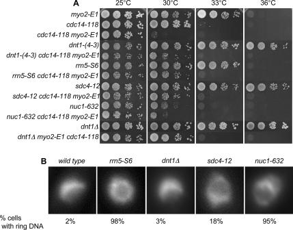
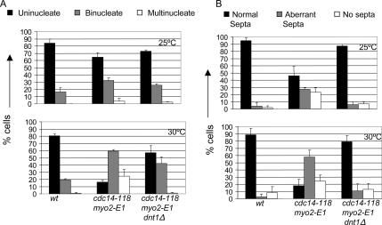
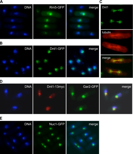
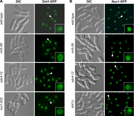
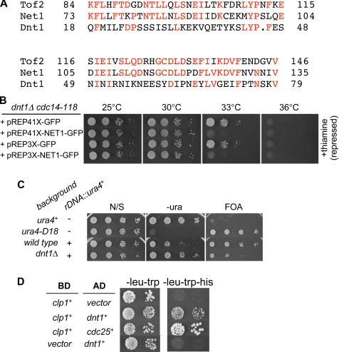
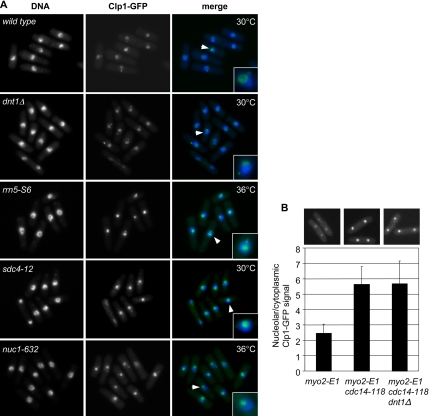
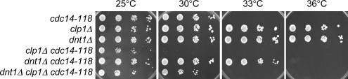
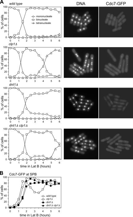
The septation initiation network (SIN) and mitotic exit network (MEN) signaling pathways regulate cytokinesis and mitotic exit in the yeasts Schizosaccharomyces pombe, and Saccharomyces cerevisiae, respectively. One function of these pathways is to keep the Cdc14-family phosphatase, called Clp1 in S. pombe, from being sequestered and inhibited in the nucleolus. In S. pombe, the SIN and Clp1 act as part of a cytokinesis checkpoint that allows cells to cope with cytokinesis defects. The SIN promotes checkpoint function by 1) keeping Clp1 out of the nucleolus, 2) maintaining the cytokinetic apparatus, and 3) halting the cell cycle until cytokinesis is completed. In a screen for suppressors of the SIN mutant cytokinesis checkpoint defect, we identified a novel nucleolar protein called Dnt1 and other nucleolar proteins, including Rrn5 and Nuc1, which are known to be required for rDNA transcription. Dnt1 shows sequence homology to Net1/Cfi1, which encodes the nucleolar inhibitor of Cdc14 in budding yeast. Like Net1/Cfi1, Dnt1 is required for rDNA silencing and minichromosome maintenance, and both Dnt1 and Net1/Cfi1 negatively regulate the homologous SIN and MEN pathways. Unlike Net1/Cfi1, which regulates the MEN through the Cdc14 phosphatase, Dnt1 can inhibit SIN signaling independently of Clp1, suggesting a novel connection between the nucleolus and the SIN pathway.


Figures

Figure 1.
Figure 1.
Suppressors of cdc14-118 myo2-E1 double mutant. (A) Selected suppressor single mutants and triple mutants with cdc14-118 myo2-E1 were grown in YE at 25°C, and then serial dilutions were spotted on YE plates. Plates were incubated at different temperatures as indicated for 3–5 d before photography. (B) Nuclear phenotype of the suppressors. Wild-type cells and suppressor mutants were first grown in YE at 25°C, and then they were shifted to 30°C for 4 h. Cells were fixed and stained with DAPI. Example nuclei from interphase cells are shown. Note the crescent-shaped nuclear DNA in wild type and dnt1Δ cells and ring-shaped nuclear DNA phenotype in rrn5-S6, sdc4-12, and nuc1-632 mutants. The percentage of cells with the ring-shaped nuclear DNA phenotype is indicated (n > 100).
Figure 2.
Figure 2.
dnt1Δ promotes proper completion of cytokinesis in the cdc14-118 myo2-E1 cells. Asynchronous cells of the indicated genotypes were grown at 25°C to log phase cell density, and portions were shifted to 30°C for 4 h. Cells were methanol fixed and stained simultaneously with DAPI and calcofluor to score for the number of nuclei per cell (A) and septum formation (B), respectively. Cells were scored for the presence of normal, aberrant, or lack of septa between nuclei. At least 100 cells were examined for each strain, and mean values (n = 3) were plotted.
Figure 3.
Figure 3.
Nucleolar localization of suppressor proteins. The indicated strains were grown at 30°C. (A) Cells transformed with pREP42-rrn5+-GFP were first grown in EMM with thiamine (repressed), and then they were induced for 18 h in EMM without thiamine. Cells were fixed and DAPI stained. Note that the GFP signal varies due to variation in the plasmid copy number between cells. (B) Cells carrying integrated dnt1-GFP were fixed and DAPI stained. (C) Cells carrying integrated dnt1-GFP and mCherry-tubulin expressed from a plasmid were imaged live by fluorescence microscopy. A montage of two late anaphase cells is shown. (D) Cells expressing both integrated dnt1-13myc and gar2-GFP were fixed, and then they were subjected to immunofluorescence with antibodies against Myc and GFP. Dnt1–13myc foci were concentrated in the nucleolus as marked by Gar2-GFP. (E) Cells carrying integrated nuc1-GFP were fixed and DAPI stained.
Figure 4.
Figure 4.
Nucleolar localization of Dnt1 and Nuc1 is disrupted in suppressor mutants. Wild-type and mutant cells with the indicated genotypes were first grown at 25°C, and then they were shifted to 30°C for 4 h before being visualized by fluorescence microscopy. Arrows indicate the nuclei enlarged in insets.
Figure 5.
Figure 5.
Relationship between S. pombe Dnt1 and S. cerevisiae Net1/Cfi1. (A) Sequence alignment between Dnt1, Net1, and Tof2 at the N termini of the three proteins. (B) Rescue of cdc14-118 by dnt1Δ can be reversed by expression of budding yeast NET1. dnt1Δ cdc14-118 cells transformed with either empty vectors (pREP41X-GFP or pREP3X-GFP) or the NET1-expressing plasmid (pREP41X-NET1-GFP or pREP3X-NET1-GFP) were first grown at 25°C in EMM medium plus thiamine, and then they were washed, diluted, and spotted on EMM plates with or without (data not shown) thiamine. Plates were incubated at different temperatures as indicated for 3–5 d. (C) Dnt1 is involved in rDNA silencing. Wild-type and dnt1Δ cells carrying ura4+ gene inserted at the rDNA repeats (rDNA::ura4+) and control wild-type cells carrying endogenous (ura4+) or deleted ura4+ gene (ura4-D18) were grown at 30°C and serially diluted and spotted onto the indicated plates. The plates were incubated at 30°C for 3–5 d before photography. (D) Dnt1 and Clp1 interact in the two-hybrid assay. Clp1 was fused with the DNA-binding domain of GAL4 (BD) and Dnt1 with the transcriptional activation domain of GAL4 (AD). Both fusion constructs were coexpressed and the growth on SD, −Leu, −Trp and SD, −Leu, −Trp, −His is shown. As controls, coexpressions of Clp1 and empty AD vector, and empty DB vector with Dnt1 (negative control) or an AD fusion with S. pombe Cdc25 (positive control) are shown. The two-hybrid interaction between Clp1 and Cdc25 has been described previously (Wolfe and Gould, 2004).
Figure 6.
Figure 6.
(A) Suppressors do not cause premature release of Clp1 from nucleolus. Wild-type and mutant cells with indicated genotypes were first grown at 25°C, and then they were shifted to 30 or 36°C for 4 h before being fixed and stained with DAPI. Arrows indicate the nuclei enlarged in insets. (B) Cells of the indicated genotypes were grown to log phase at 25°C, and then they were shifted to 30°C for 4 h. These cells were then methanol fixed and imaged for Clp1-GFP localization. The ratio of the mean average intensity of the Clp1-GFP signal in the nucleolus and cytoplasm is shown (n > 50). Representative images are shown. Only cells that were in telophase, as judged by nuclear positioning, were analyzed.
Figure 7.
Figure 7.
Dnt1 can regulate the SIN independently of Clp1. Wild-type and mutant cells with indicated genotypes were first grown in YE at 25°C, and then they were serially diluted and spotted on plates of YE. Plates were incubated at different temperatures as indicated for 3–5 d before photography.
Figure 8.
Figure 8.
dnt1Δ has a positive effect on SIN signaling even in the absence of Clp1. Wild-type and mutant cells with the indicated genotypes (all carrying Cdc7-GFP) were first grown in YE at 30°C, and then they were synchronized in early G2 by centrifugal elutriation and treated with 4 μM latrunculin B. Cells were withdrawn every 30 min for a period of 6 h, and then they were fixed and stained with DAPI. (A) Left, number of nuclei over time for each strain. Right, examples of DNA staining and Cdc7-GFP signal at the 4-h time point for each strain. (B) Quantification of Cdc7-GFP signals is shown. At least 200 cells were counted for each time point.

Similar articles

Cited by

References

    1. Allshire R. C., Nimmo E. R., Ekwall K., Javerzat J. P., Cranston G. Mutations derepressing silent centromeric domains in fission yeast disrupt chromosome segregation. Genes Dev. 1995;9:218–233. - PubMed
    1. Ayscough K. R., Stryker J., Pokala N., Sanders M., Crews P., Drubin D. G. High rates of actin filament turnover in budding yeast and roles for actin in establishment and maintenance of cell polarity revealed using the actin inhibitor latrunculin-A. J. Cell Biol. 1997;137:399–416. - PMC - PubMed
    1. Azzam R., Chen S. L., Shou W., Mah A. S., Alexandru G., Nasmyth K., Annan R. S., Carr S. A., Deshaies R. J. Phosphorylation by cyclin B-Cdk underlies release of mitotic exit activator Cdc14 from the nucleolus. Science. 2004;305:516–519. - PubMed
    1. Bahler J., Wu J. Q., Longtine M. S., Shah N. G., McKenzie A., 3rd, Steever A. B., Wach A., Philippsen P., Pringle J. R. Heterologous modules for efficient and versatile PCR-based gene targeting in Schizosaccharomyces pombe. Yeast. 1998;14:943–951. - PubMed
    1. Balasubramanian M. K., McCollum D., Gould K. L. Cytokinesis in fission yeast Schizosaccharomyces pombe. Methods Enzymol. 1997;283:494–506. - PubMed

Publication types

MeSH terms