. 2007 Jul;18(7):2037-45.
doi: 10.1681/ASN.2006121333. Epub 2007 May 30.

Role of gp91phox -containing NADPH oxidase in mediating the effect of K restriction on ROMK channels and renal K excretion

Affiliations

Role of gp91phox -containing NADPH oxidase in mediating the effect of K restriction on ROMK channels and renal K excretion

Elisa Babilonia et al. J Am Soc Nephrol. 2007 Jul.

Abstract

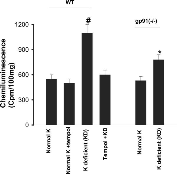
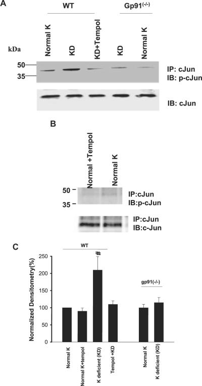
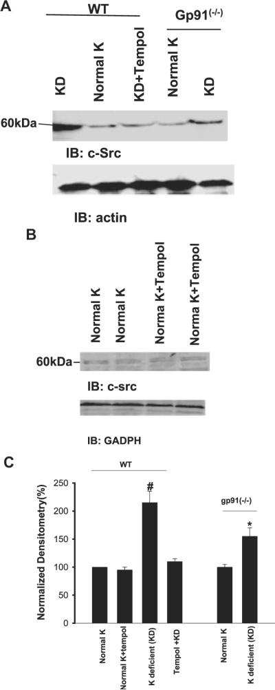
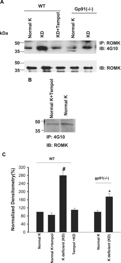
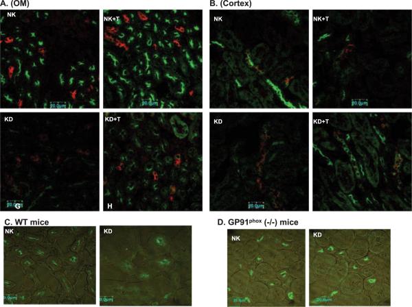
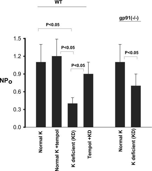
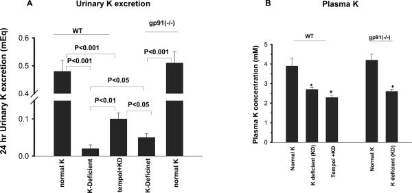
Previous study has demonstrated that superoxide and the related products are involved in mediating the effect of low K intake on renal K secretion and ROMK channel activity in the cortical collecting duct (CCD). This study investigated the role of gp91(phox)-containing NADPH oxidase (NOXII) in mediating the effect of low K intake on renal K excretion and ROMK channel activity in gp91(-/-) mice. K depletion increased superoxide levels, phosphorylation of c-Jun, expression of c-Src, and tyrosine phosphorylation of ROMK in renal cortex and outer medulla in wild-type (WT) mice. In contrast, tempol treatment in WT mice abolished whereas deletion of gp91 significantly attenuated the effect of low K intake on superoxide production, c-Jun phosphorylation, c-Src expression, and tyrosine phosphorylation of ROMK. Patch-clamp experiments demonstrated that low K intake decreased mean product of channel number (N) and open probability (P) (NP(o)) of ROMK channels from 1.1 to 0.4 in the CCD. However, the effect of low K intake on ROMK channel activity was significantly attenuated in the CCD from gp91(-/-) mice and completely abolished by tempol treatment. Immunocytochemical staining also was used to examine the ROMK distribution in WT, gp91(-/-), and WT mice with tempol treatment in response to K restriction. K restriction decreased apical staining of ROMK in WT mice. In contrast, a sharp apical ROMK staining was observed in the tempol-treated WT or gp91(-/-) mice. Metabolic cage study further showed that urinary K loss is significantly higher in gp91(-/-) mice than in WT mice. It is concluded that superoxide anions play a key role in suppressing K secretion during K restriction and that NOXII is involved in mediating the effect of low K intake on renal K secretion and ROMK channel activity.


Figures

Figure 1
Figure 1
Superoxide levels in the renal cortex and outer medulla from wild-type (WT) mice on a normal-K diet (1.1%), normal K + tempol treatment, K-deficient (KD) diet, and KD plus tempol treatment (left set of bars) and in gp91phox (-/-) mice on a control and/or KD diet (right set of bars). #The superoxide level in WT mice that were on a KD diet is significantly higher than that in every other group (P < 0.05). *Significant difference between control [gp91phox (-/-) mice on 1.1% K] and experimental group [gp91phox (-/-) mice on KD diet; P < 0.05].
Figure 2
Figure 2
(A) A Western blot demonstrating the effect of low K intake on cJun phosphorylation (top) and total c-Jun (bottom) in the renal cortex and outer medulla of WT mice on a normal-K diet, KD diet, and KD diet plus tempol treatment and gp91phox (-/-) mice that were fed a normal-K and/or a KD diet. (B) A Western blot showing the cJun phosphorylation in renal cortex and outer medulla in tempol-treated and non-tempol-treated WT mice that were fed a normal-K diet. (C) Bar graphs summarize the effect of K intake on cJun phosphorylation under different experimental conditions. #Phosphorylation of cJun in wt mice on KD diet is significantly higher than that in every other group (P < 0.05).
Figure 3
Figure 3
(A) A Western blot demonstrating the effect of low K intake on c-Src expression in the renal cortex and outer medulla of WT mice on a normal-K diet, KD diet, and KD diet plus tempol treatment and gp91phox (-/-) mice that were fed a normal-K and/or KD diet. (B) A Western blot showing the c-Src expression in renal cortex and outer medulla in WT mice on normal-K diet with or without tempol treatment. (C) Bar graph demonstrating the effect of dietary K intake on c-Src expression under different experimental conditions. #cSrc level in WT mice on KD diet is significantly higher than that in every other group (P < 0.05). *Significant difference between control [gp91phox (-/-] mice on 1.1% K) and experimental group [gp91phox (-/-) mice on KD diet; P < 0.05].
Figure 4
Figure 4
(A) Western blot showing the effect of low K intake on tyrosine phosphorylation of ROMK (top) and total ROMK expression (bottom) in the renal cortex and outer medulla in WT mice on a normal-K diet, KD diet, and KD diet plus tempol treatment and gp91phox (-/-) mice that were fed a normal-K and/or KD diet. (B)Western blot showing the tyrosine phosphorylation of ROMK in renal cortex and outer medulla in WT mice on normal-K diet with or without tempol treatment. (C) Bar graph showing the effect of K restriction on the tyrosine phosphorylation of ROMK. #Tyrosine phosphorylation of ROMK in WT mice on KD diet is significantly higher than that in every other group (P < 0.05). *Significant difference between control [gp91phox (-/-) mice on 1.1% K] and experimental group [gp91phox (-/-) mice on KD diet; P < 0.05].
Figure 5
Figure 5
Confocal images demonstrate the effect of dietary K intake on ROMK staining in wt mice (A through C) and gp91phox (-/-) mice (D). Double staining of aquaporin 2 (red) and ROMK (green) in outer medulla (A) or cortex (B) of tempol (T)-treated or non-tempol-treated mice on normal-K (NK, 1.1%) and KD diets, respectively. (C) ROMK staining in outer medulla of WT mice on NK or KD diet. (D) ROMK staining in outer medulla of gp91phox (-/-) mice on NK or KD diet.
Figure 6
Figure 6
The SK channel activity in the cortical collecting duct of WT mice on a normal-K diet, normal-K diet with tempol treatment, KD diet, and KD diet plus tempol treatment (left set of bars) and gp91phox (-/-) mice that were fed a normal-K and/or KD diet (right set of bars). The experiments were performed in cell-attached patches. Significance between different groups is indicated.
Figure 7
Figure 7
(A) Twenty-four-hour urinary K excretion in WT mice on normal-K diet (1.1%), WT mice on KD diet, tempol-treated WT mice on KD diet, gp91phox (-/-) mice on KD diet, and gp91phox (-/-) mice on normal-K diet. (B) Plasma K concentrations in WT mice on a normal-K diet, KD diet, or KD diet plus tempol for 7 d (left set of bars) and in gp91phox (-/-) mice on a normal-K or KD diet (right set of bars). *Significant difference from the corresponding control (normal K) and experimental groups.

References

    1. Stanton BA, Giebisch GH. Renal potassium transport. In: Windhager E, editor. Handbook of Physiology-Renal Physiology. Oxford University Press; Oxford: 1992. pp. 813–874.
    1. Giebisch G. Renal potassium transport: Mechanisms and regulation. Am J Physiol. 1998;274:F817–F833. - PubMed
    1. Rabinowitz L. Homeostatic regulation of potassium excretion. J Hypertens. 1989;7:433–442. - PubMed
    1. Babilonia E, Wei Y, Sterling H, Kaminski P, Wolin MS. Superoxide anions are involved in mediating the effect of low K intake on c-Src expression and renal K secretion in the cortical collecting duct. J Biol Chem. 2005;280:10790–10796. - PMC - PubMed
    1. Droge W. Free radicals in the physiological control of cell function. Physiol Rev. 2002;82:47–95. - PubMed

Publication types