

Functional cortical changes of the sensorimotor network are associated with clinical recovery in multiple sclerosis
- PMID: 17538952
- PMCID: PMC6870672
- DOI: 10.1002/hbm.20418
Functional cortical changes of the sensorimotor network are associated with clinical recovery in multiple sclerosis
Abstract
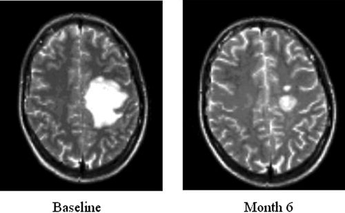
Objective: To assess the early cortical changes following an acute motor relapse secondary to a pseudotumoral lesion in MS patients, the longitudinal cortical functional correlates of clinical recovery, and the evolution over time of cortical reorganization.
Methods: FMRI during the performance of a simple motor task were obtained from 12 MS patients (after a clinical attack involving the motor system secondary to a pseudotumoral lesion) and 15 matched controls. In six patients and five controls, a longitudinal fMRI study was also performed.
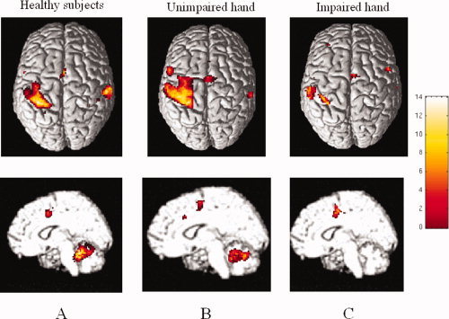
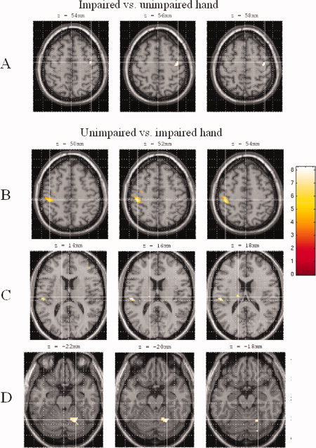
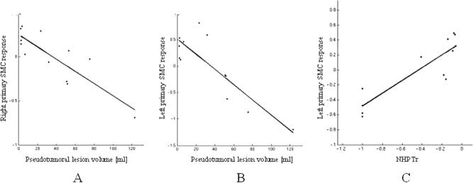
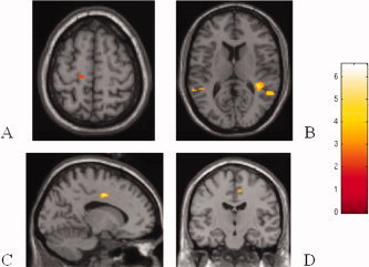
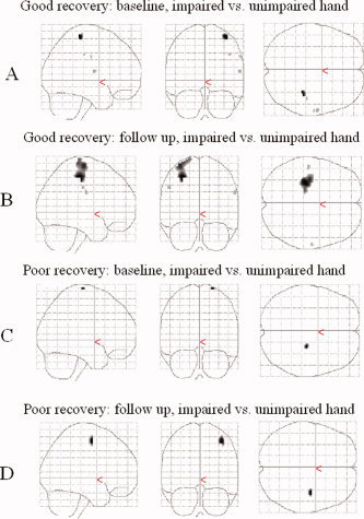
Results: In patients, at baseline, the primary sensorimotor cortex (SMC) of the ipsilateral (contralesional) hemisphere was significantly more active during task performance with the impaired than the unimpaired hand. During task performance with the unimpaired hand, the ipsilateral cerebellum and several motor areas in the contralateral hemisphere were significantly more active. Pseudotumoral lesion volume was correlated with activation of the primary SMC bilaterally (r = -0.86 and -0.85) and the nine-hole peg test score with activation of the primary SMC of the affected hemisphere (r = 0.88). A recovery of function of the primary SMC of the affected hemisphere was found in the four patients with clinical improvement. In the two patients without clinical recovery, there was a persistent recruitment of the primary SMC of the unaffected hemisphere.
Conclusions: Pseudotumoral MS lesions affecting the motor system can determine short-term cortical changes characterized by the recruitment of pathways in the unaffected hemisphere. The regain of function of motor areas of the affected hemisphere seems to be a critical factor for a favorable recovery.
(Copyright) 2006 Wiley-Liss, Inc.
Figures
References
-
- Barkhof F, Filippi M, Miller DH, Scheltens P, Campi A, Polman CH, Comi G, Ader HJ, Losseff N, Valk J ( 1997): Comparison of MRI criteria at first presentation to predict conversion to clinically definite multiple sclerosis. Brain 120: 2059–2069. - PubMed
-
- Calautti C, Baron JC ( 2003): Functional neuroimaging studies of motor recovery after stroke in adults: A review. Stroke 34: 1553–1566; Review. - PubMed
-
- Cifelli A, Matthews PM ( 2002): Cerebral plasticity in multiple sclerosis: Insights from fMRI. Mult Scler 8: 193–199; Review. - PubMed
-
- Dirnberger G, Duregger C, Lindinger G, Lang W ( 2004): Habituation in a simple repetitive motor task: A study with movement‐related cortical potentials. Clin Neurophysiol 115: 378–384. - PubMed
-
- Dobkin BH ( 2003): Functional MRI: A potential physiologic indicator for stroke rehabilitation interventions. Stroke 34: 23–28. - PubMed
MeSH terms
LinkOut - more resources
Full Text Sources
Medical
