. 2007 Jun 25;46(13):5260-70.
doi: 10.1021/ic0702926. Epub 2007 Jun 1.

Potentiometric and relaxometric properties of a gadolinium-based MRI contrast agent for sensing tissue pH

Affiliations

Potentiometric and relaxometric properties of a gadolinium-based MRI contrast agent for sensing tissue pH

Ferenc K Kalman et al. Inorg Chem. .

Abstract

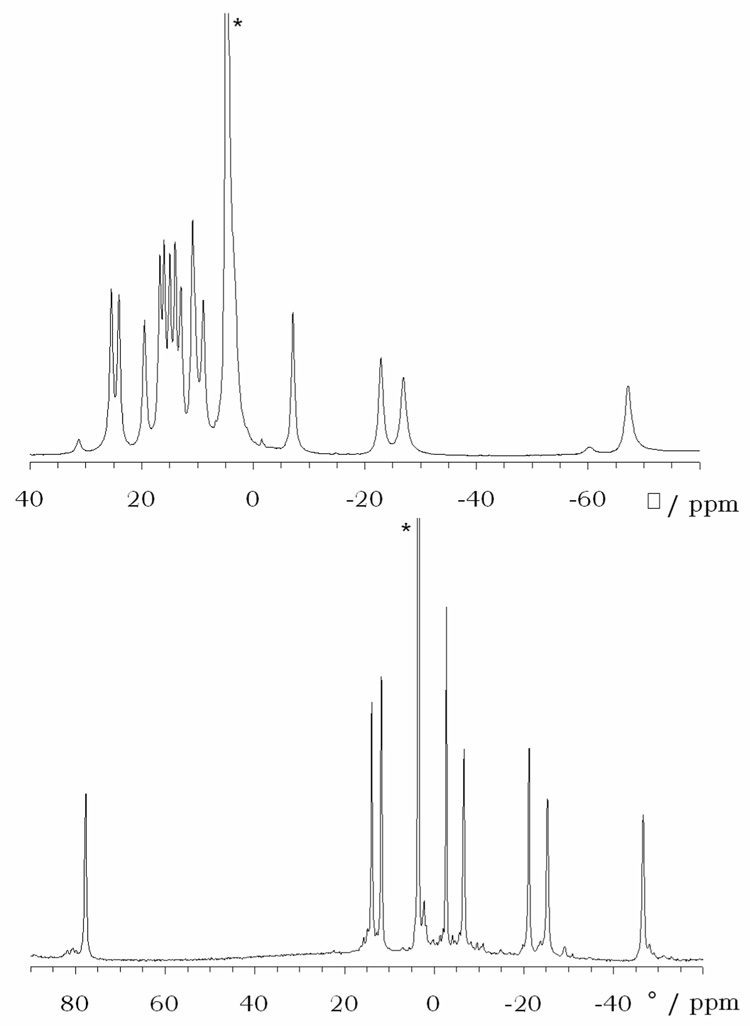
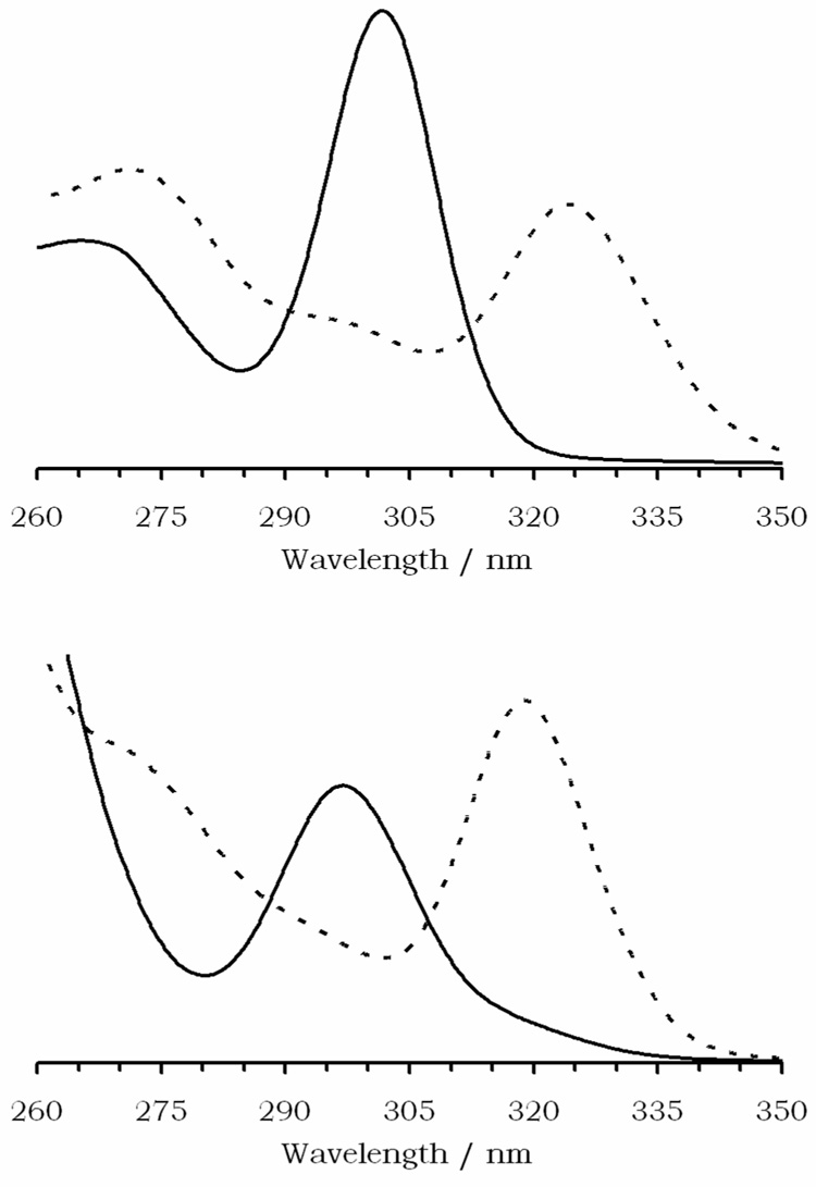
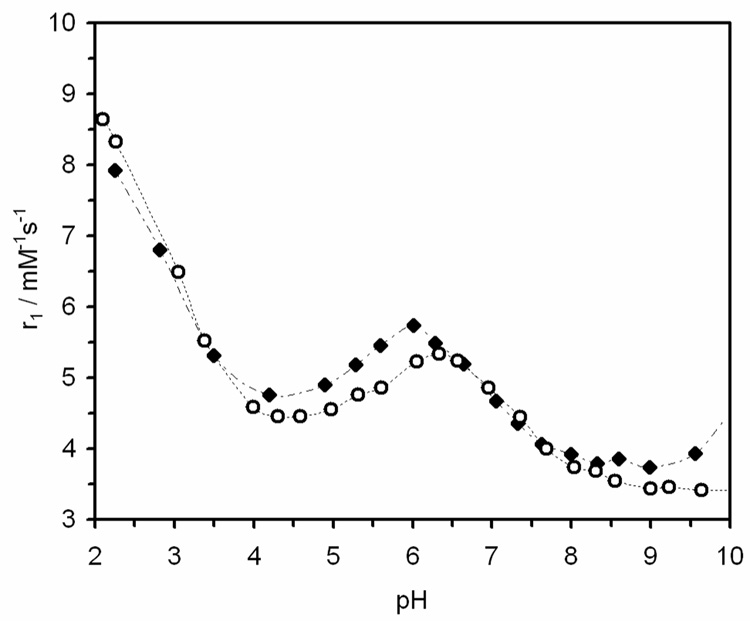
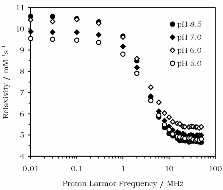
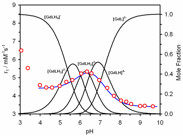
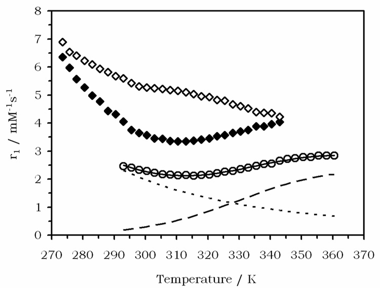
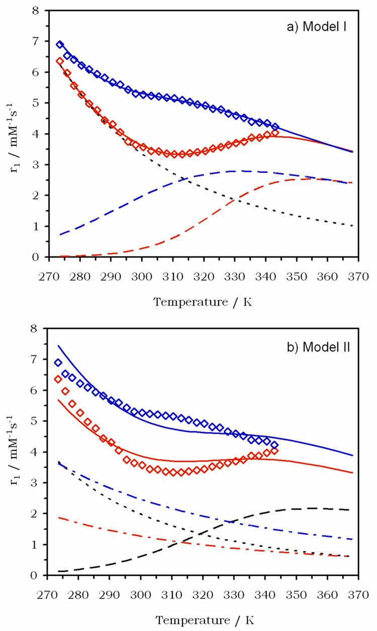
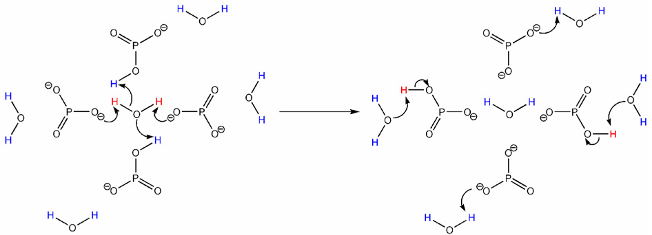
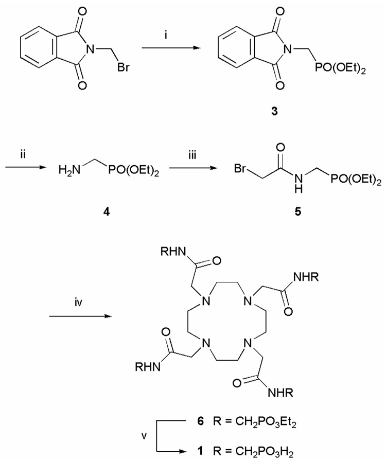
The pH-sensitive contrast agent, GdDOTA-4AmP (Gd1) has been successfully used to map tissue pH by MRI. Further studies now demonstrate that two distinct chemical forms of the complex can be prepared depending upon the pH at which Gd(3+) is mixed with ligand 1. The desired pH-sensitive form of this complex, referred to here as a Type II complex, is obtained as the exclusive product only when the complexation reaction is performed above pH 8. At lower pH values, a second complex is formed that, by analogy with an intermediate formed during the preparation of GdDOTA, we tentatively assign to a Type I complex where the Gd(3+) is coordinated only by the appended side-chain arms of 1. The proportion of Type I complex formed is largely determined by the pH of the complexation reaction. The magnitude of the pH-dependent change in the relaxivity of Gd1 was found to be less than earlier reported (Zhang, S.; Wu, K.; Sherry, A. D. Angew. Chem., Int. Ed. 1999, 38, 3192), likely due to contamination of the earlier sample by an unknown amount of Type I complex. Examination of the nuclear magnetic relaxation dispersion and relaxivity temperature profiles, coupled with information from potentiometric titrations, shows that the amphoteric character of the phosphonate side chains enables rapid prototropic exchange between the single bound water of the complex with the bulk water thereby giving Gd1 a unique pH-dependent relaxivity that is quite useful for the pH mapping of tissues by MRI.


Figures

Figure 1
Figure 1
The 1H NMR spectra of Yb1 complexes synthesized and recorded at a) pH 1, b) pH 9. The spectra were recorded in D2O at 296K and 270 MHz (the peaks arising from HOD are labelled with asterisks).
Figure 2
Figure 2
UV-visible absorption spectra of Ce1 (top) prepared at pH 1 (solid line) and pH 9 (dashed line). The absorption bands are similar to those observed for the Type I (solid line) and Type II (dashed line) complexes of CeDOTA (bottom).
Figure 3
Figure 3
The effect of solution pH on the relaxivity of Gd1 (open circles), recorded at 25 °C and 20 MHz ([Gd1] = 1.0 mM). The effect of solution pH on the relaxivity of Gd1 in the presence of 135 mM NaCl, 5 mM KCl and 2.5 mM CaCl2 ([Gd1] = 1 mM) is also shown (filled diamonds). Profiles recorded at 15 °C and 35 °C are provided as supplementary material.
Figure 4
Figure 4
Nuclear magnetic relaxation dispersion (NMRD) profiles for Gd1 at 25°C and pH 5 (open circles), pH 6 (open diamonds), pH 7 (closed diamonds) and pH 8.5 (closed circles). The pH for these experiments was maintained by using HEPES buffers.
Figure 5
Figure 5
The longitudinal relaxivity pH profile of Gd1 (25 °C, 20 MHz) (red circles) laid-over the speciation diagram of Gd1. The blue line is the overall relaxivity of the system calculated from the relaxivity of each species (Table 5) as determined by the regression analysis.
Figure 6
Figure 6
The temperature dependence of the relaxivity of Gd1 at pH 6.2 (open diamonds) and pH 8.3 (closed diamonds). The temperature dependence of the relaxivity of Gd2 is shown for comparison (open circles), the calculated relaxivity (solid line), the outer-sphere contribution (dotted line) and the inner-sphere contribution (dashed line) are also shown.
Figure 7
Figure 7
Fitting the temperature dependence of the relaxivity of Gd1 at pH 6.2 (blue) and pH 8.3 (red) to: a) a model that fixes the outer and second sphere contributions (dotted black line) and fits the data to changes in the inner sphere relaxivity (dashed lines). The fits are shown as solid lines. b) a model that assumes no change in inner-sphere relaxivity (dashed black line) and accounts for difference in relaxivity through changes in the second sphere contribution (dot-dash lines). The outer-sphere contribution (dotted black line) and fits (solid lines are also shown.
Figure 8
Figure 8
A schematic representation, viewed down the Gd-OH2 axis, of how the phosphonates in GdLH23− transfer protons between the coordinated water molecule and the bulk solvent. The relaxed protons of the coordinated water molecule (shown in red) are removed from the water molecule by the deprotonated phosphonates which act as bases. They are then replaced by unrelaxed protons from the bulk water (shown in blue) which are supplied by the monoprotonated phosphonates which are acting as acids.
Scheme 1
Scheme 1
Synthesis of 1. Reagents and conditions: i.) P(OEt)3 / Δ; ii.) N2H4 / EtOH; iii.) BrCH2COBr / K2CO3 / benzene; iv.) cyclen / K2CO3 / MeCN / 60°C; v.) 30% HBr / AcOH / RT.
Chart 1
Chart 1

References

    1. Sennoune Souad R, Luo D, Martinez-Zaguilan R. Cell Biochem. Biophys. 2004;40:185. - PubMed
    1. Tannock IF, Rotin D. Cancer Res. 1989;49:4373. - PubMed
    1. Vaupel P, Kallinowski F, Okunieff P. Cancer Res. 1989;49:6449. - PubMed
    1. Wike-Hooley JL, Haveman J, Reinhold JS. Radioter. Oncol. 1984;2:343. - PubMed
    1. Caravan P, Ellison JJ, McMurry TJ, Lauffer RB. Chem. Rev. 1999;99:2293. - PubMed

Publication types