. 2007 Jul 15;307(2):314-27.
doi: 10.1016/j.ydbio.2007.04.046. Epub 2007 May 3.

Cell type-autonomous and non-autonomous requirements for Dmrt1 in postnatal testis differentiation

Affiliations

Cell type-autonomous and non-autonomous requirements for Dmrt1 in postnatal testis differentiation

Shinseog Kim et al. Dev Biol. .

Abstract

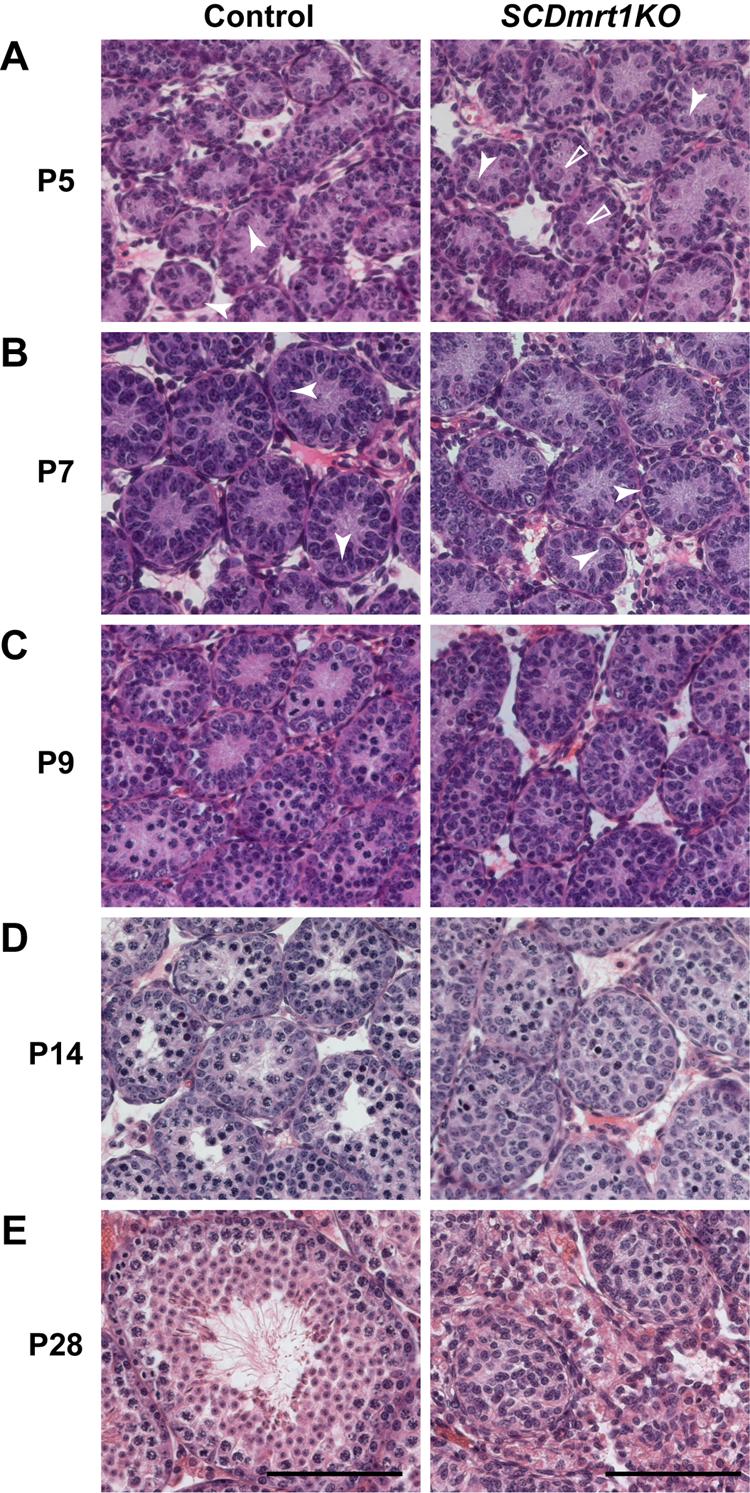
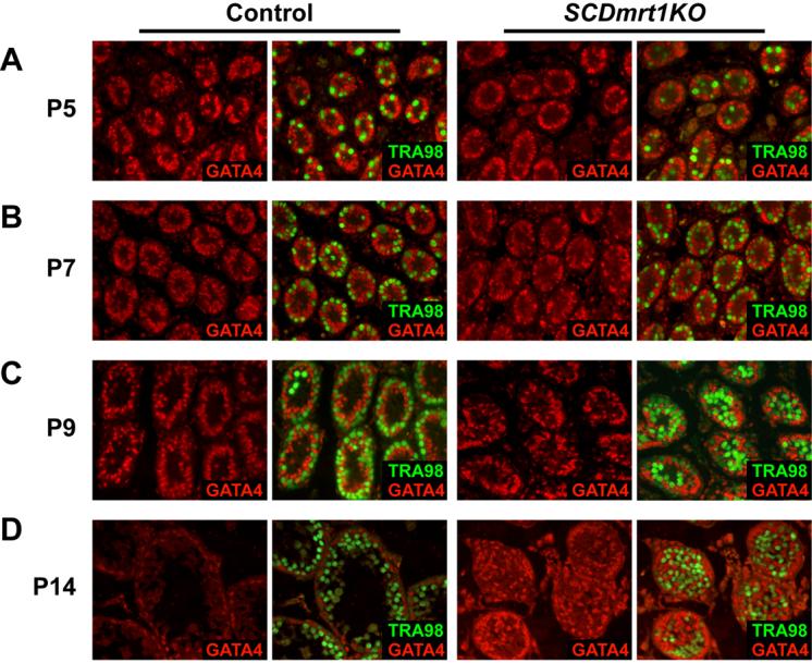
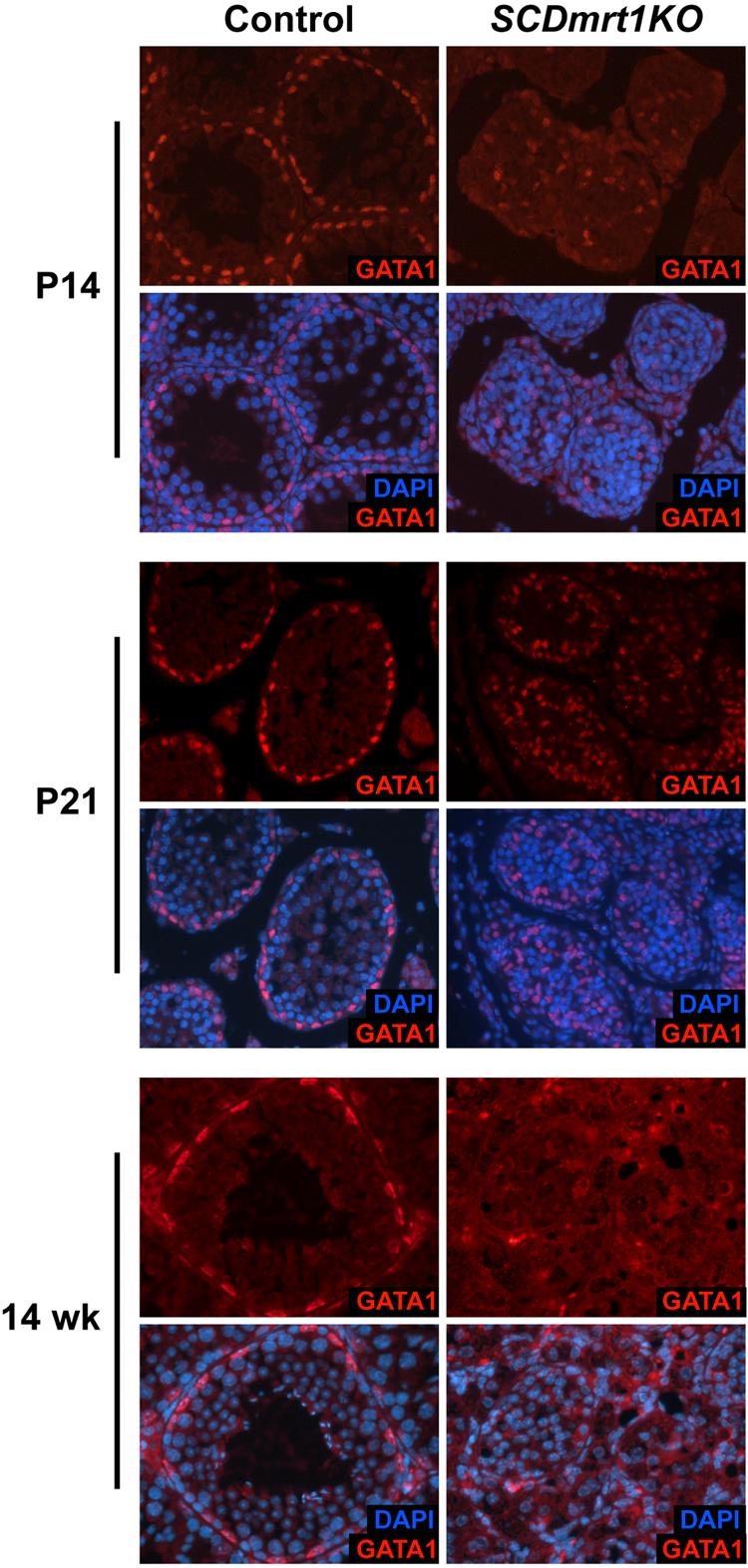
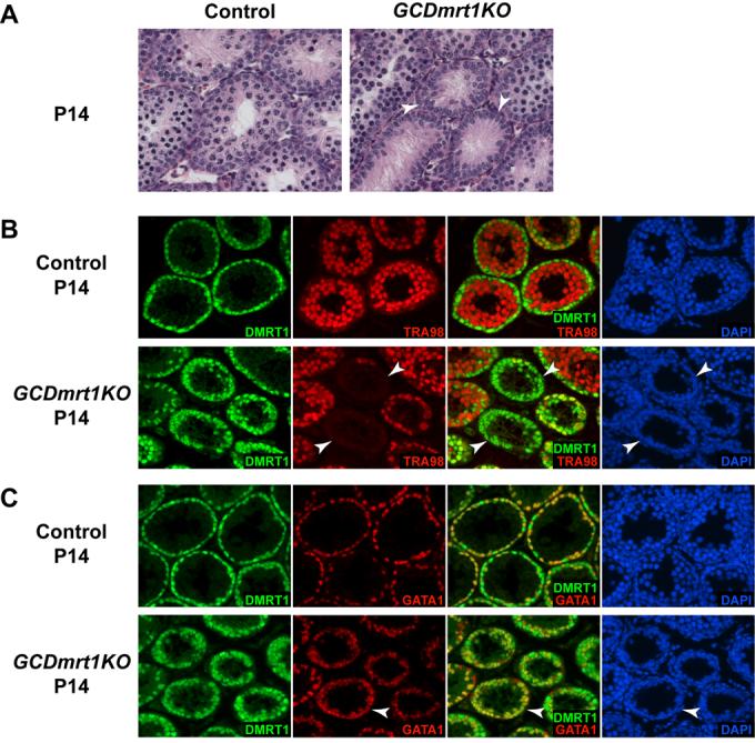
Genes containing the DM domain, a conserved DNA binding motif first found in Doublesex of Drosophila and mab-3 of Caenorhabditis elegans, regulate sexual differentiation in multiple phyla. The DM domain gene Dmrt1 is essential for testicular differentiation in vertebrates. In the mouse, Dmrt1 is expressed in pre-meiotic germ cells and in Sertoli cells, which provide essential support for spermatogenesis. Dmrt1 null mutant mice have severely dysgenic testes in which Sertoli cells and germ cells both fail to differentiate properly after birth. Here we use conditional gene targeting to identify the functions of Dmrt1 in each cell type. We find that Dmrt1 is required in Sertoli cells for their postnatal differentiation, and for germ line maintenance and for meiotic progression. Dmrt1 is required in germ cells for their radial migration to the periphery of the seminiferous tubule where the spermatogenic niche will form, for mitotic reactivation and for survival beyond the first postnatal week. Thus Dmrt1 activity is required autonomously in the Sertoli and germ cell lineages, and Dmrt1 activity in Sertoli cells is also required non-autonomously to maintain the germ line. These results demonstrate that Dmrt1 plays multiple roles in controlling the remodeling and differentiation of the juvenile testis.


Figures

Figure 1
Figure 1. Specific and efficient deletion of Dmrt1 in Sertoli cells of SCDmrt1KO mutants
(A) DMRT1 expression in Sertoli cells of control and SCDmrt1KO testes at P5. Double staining with antibodies to DMRT1 and the juvenile Sertoli cell marker GATA4. Merged image shows that DMRT1 is ablated from virtually all Sertoli cells in SCDmrt1KO testis. (B) DMRT1 expression in germ cells of control and SCDmrt1KO testes at P5. Double staining with antibodies to DMRT1 and the gonocyte and spermatogonial marker TRA98. Merged image shows that almost all germ cells express DMRT1 in SCDmrt1KO testis. The rare germ cells lacking DMRT1 may be entering meiosis and down-regulating DMRT1 expression, rather than mutant for Dmrt1, as they also were present in control animals (not shown).
Figure 2
Figure 2. Abnormal seminiferous tubule morphology in SCDmrt1KO testes
Hematoxylin/eosin staining of testis sections. (A) At P5 in both control and SCDmrt1KO testes most germ cells have migrated to the basal lamina at the periphery of the seminiferous tubule (closed arrowheads), although some germ cells remain in the center of the tubules (open arrowheads). (B) At P7, germ cell migration is complete in control and SCDmrt1KO testes (closed arrowheads) and cellular organization is normal in both. (C) At P9, control testes have Sertoli cells and spermatogonia at the periphery, meiotic cells in the adluminal compartment, and a developing central lumen. In SCDmrt1KO tubules Sertoli cells and spermatogonia are present at the periphery, but Sertoli cells also are found away toward the center of the tubules and minimal lumen formation is apparent. (D) At P14, control tubules are normally organized, with Sertoli cells and spermatogonia at the periphery, meiotic cells in the center, and an enlarging lumen forming. SCDmrt1KO tubules are disorganized with Sertoli cells and germ cells not properly localized and no lumen evident. (E) At P28, control tubule has abundant spermatogenesis, with spermatogonia established at the periphery, meiotic germ cells up to elongated spermatid stage, mature Sertoli cells and fully formed lumen. In SCDmrt1KO tubules germ cells are absent, and Sertoli cells retain immature morphology and fill the tubules. Scale bar: 100 μM. All images are at same magnification.
Figure 3
Figure 3. Germ cell mitotic reactivation does not require Dmrt1 in Sertoli cells
Double staining for the mitotic marker phospho-histone H3 (P-H3) and germ cell marker TRA98 reveals similar numbers of mitotic germ cells in control and SCDmrt1KO testes. In merged panels at far right, yellow cells are mitotic germ cells, and green cells are mitotic Sertoli cells. In these experiments, staining with the early meiotic prophase marker BC7 was used to confirm that the P-H3 positive germ cells were not meiotic (not shown).
Figure 4
Figure 4. Dmrt1 is not required in Sertoli cells for germ cell radial migration and meiotic initiation
(A) At P5, most germ cells (TRA98-positive) are at the periphery in both control and SCDmrt1KO tubules. (B) At P7 tubules in SCDmrt1KO testes appear normal, with most germ cells interspersed with Sertoli cells (GATA4-positive) at the periphery. (C) At P9, abundant germ cells are present in control and SCDmrt1KO tubules, and brightly staining TRA98-positive meiotic cells are present in both, but germ cells and Sertoli cells are beginning to become abnormally organized. (D) At P14, SCDmrt1KO testes still have abundant meiotic germ cells, but germ cells and Sertoli cell organization is severely abnormal in many tubules compared with controls.
Figure 5
Figure 5. Progression through meiotic prophase requires Dmrt1 in Sertoli cells
(A) Leptotene and zygotene marker BC7 at P14, showing many BC7-positive cells in both control and SCDmrt1KO testes. (B) Pachytene to elongated spermatid marker TRA369, showing very few positive cells in SCDmrt1KO testis, indicating a disruption in meiosis between leptonema and pachynema. All images are at same magnification; differences in tubule morphology between panels A and B are due to different fixation protocols used for the two antibodies.
Figure 6
Figure 6. Dmrt1 is required for normal activation of androgen receptor in Sertoli cells
At P5, androgen receptor (AR) is expressed in peritubular myoid cells (at the tubule margins) and interstitial cells in control and SCDmrt1KO testes. At P9, AR has been activated in Sertoli cells of control testes but not in SCDmrt1KO. At P21, AR expression is present in Sertoli cells of SCDmrt1KO, but at lower level than in control.
Figure 7
Figure 7. Dmrt1 is required for normal activation and for maintenance of GATA1 expression in Sertoli cells
At P14, GATA1 is expressed at reduced level in SCDmrt1KO testes relative to control (top panels) and reaches near-normal levels in Sertoli cells of SCDmrt1KO testes by P21 (middle). At 14 weeks (bottom), GATA1 expression is maintained in Sertoli cells of control but not SCDmrt1KO testes.
Figure 8
Figure 8. Dmrt1 is required in germ cells for their radial migration
(A) Double staining with antibodies to DMRT1 and GATA4 showing tubules in GCDmrt1KO testes at P7 in which all germ cells lack DMRT1 (arrowheads). (B) Double staining with antibodies to DMRT1 and TRA98 at P7 showing that many Dmrt1 mutant germ cells in GCDmrt1KO testis have failed to migrate to the periphery (arrowhead), whereas the great majority of germ cells in control testis have done so.
Figure 9
Figure 9. Germ cell survival and meiosis require Dmrt1 in germ cells
(A) Histology of control and GCDmrt1KO testes at P14. In GCDmrt1KO some tubule sections lack germ cells and have Sertoli cells of normal polarized morphology (arrowheads). (B) Double-staining with DMRT1 and TRA98 antibodies at P14. Some tubules in GCDmrt1KO testis have few or no germ cells (arrowheads). Surviving germ cells all are DMRT1-positive. Tubules in which wild type spermatogonia (double-positive cells) are present have abundant meiotic germ cells. (C) Double-staining with DMRT1 and GATA1 antibodies at P14, showing GCDmrt1KO tubule in which germ cells are absent and DMRT1 is expressed in almost all Sertoli cells (arrowhead). Red cells in merged image of GCDmrt1KO mutant are Sertoli cells (GATA4 positive) in which Dmrt1 was deleted by TNAP-Cre.

References

    1. Adams IR, McLaren A. Sexually dimorphic development of mouse primordial germ cells: switching from oogenesis to spermatogenesis. Development. 2002;129:1155–64. - PubMed
    1. Anderson R, Copeland TK, Scholer H, Heasman J, Wylie C. The onset of germ cell migration in the mouse embryo. Mech Dev. 2000;91:61–8. - PubMed
    1. Bowles J, Knight D, Smith C, Wilhelm D, Richman J, Mamiya S, Yashiro K, Chawengsaksophak K, Wilson MJ, Rossant J, Hamada H, Koopman P. Retinoid signaling determines germ cell fate in mice. Science. 2006;312:596–600. - PubMed
    1. Bremner WJ, Millar MR, Sharpe RM, Saunders PT. Immunohistochemical localization of androgen receptors in the rat testis: evidence for stage-dependent expression and regulation by androgens. Endocrinology. 1994;135:1227–34. - PubMed
    1. Chang C, Chen YT, Yeh SD, Xu Q, Wang RS, Guillou F, Lardy H, Yeh S. Infertility with defective spermatogenesis and hypotestosteronemia in male mice lacking the androgen receptor in Sertoli cells. Proc Natl Acad Sci U S A. 2004;101:6876–81. - PMC - PubMed

Publication types