. 2007 Oct;83(2):169-76.
doi: 10.1016/j.yexmp.2007.04.003. Epub 2007 May 4.

Matrix metalloproteinase-3 (stromelysin-1) in acute inflammatory tissue injury

Affiliations

Matrix metalloproteinase-3 (stromelysin-1) in acute inflammatory tissue injury

Kamalakar C Nerusu et al. Exp Mol Pathol. 2007 Oct.

Abstract

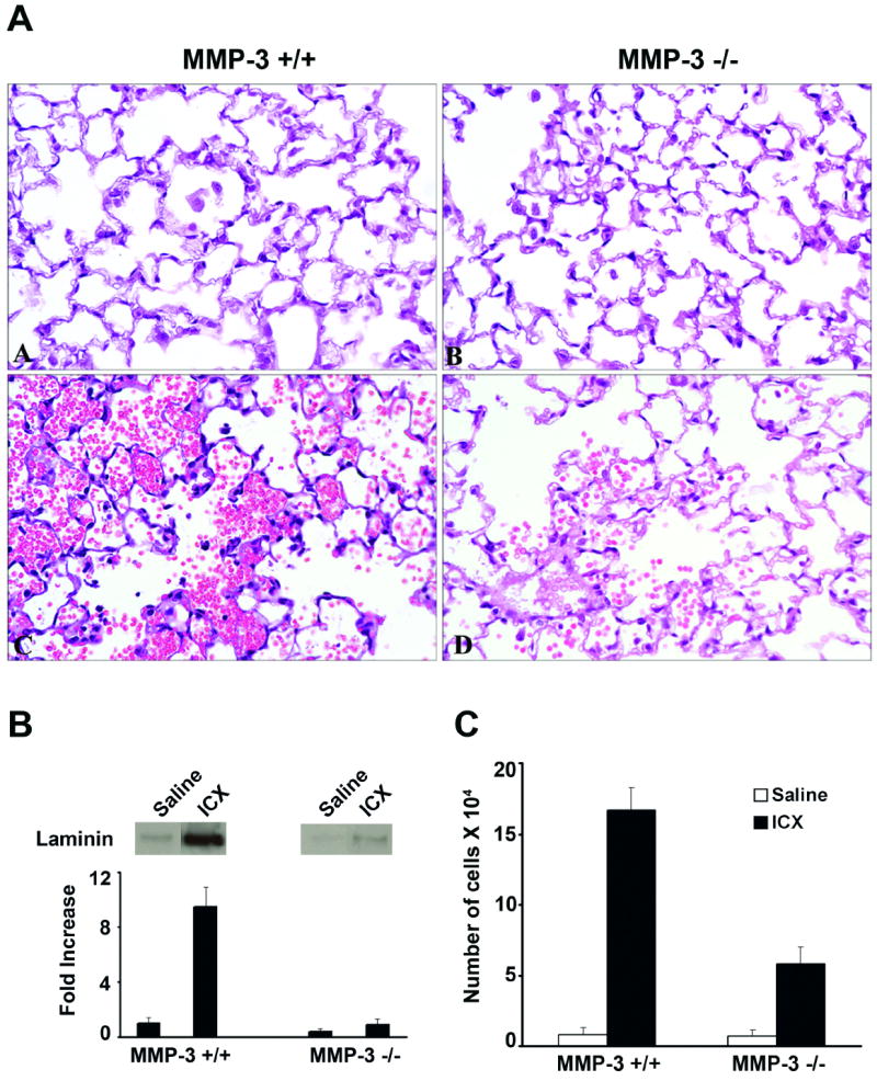
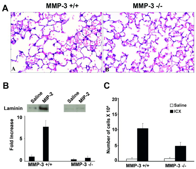
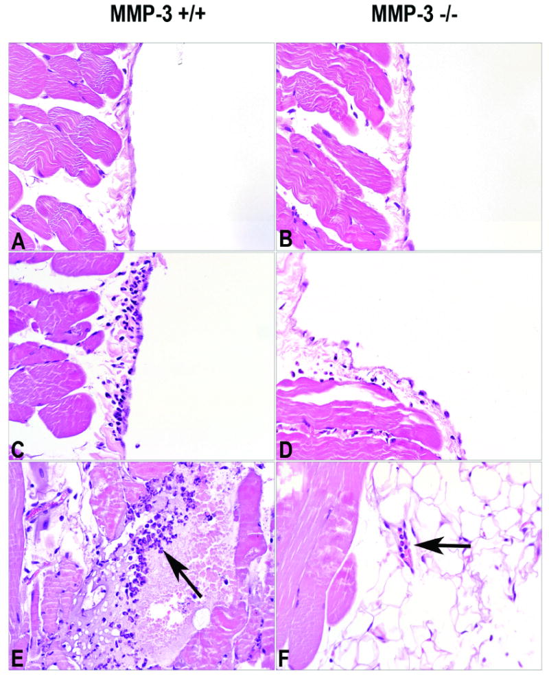
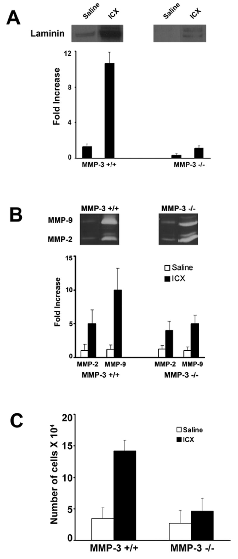
Mice lacking matrix metalloproteinase-3 (MMP-3; stromelysin-1) demonstrated significantly less injury than their normal counterparts following the formation of IgG-containing immune complexes in the alveolar wall or in the wall of the peritoneum. Likewise, mice lacking MMP-3 demonstrated less lung injury following intra-tracheal instillation of the chemotactic cytokine macrophage inhibitory protein-2 (MIP-2) than did mice with MMP-3. There was a relationship between tissue injury (evidenced histologically) and accumulation of anti-laminin 111 immunoreactive material in the bronchoalveolar lavage (BAL) or peritoneal lavage (PL) fluid. There was also a relationship between tissue injury and influx of neutrophils into the BAL or PL fluid. Taken together, these data demonstrate an important role for MMP-3 in acute inflammatory tissue injury.


Figures

Figure 1
Figure 1
Tissue injury, vascular wall damage and neutrophil accumulation in immune complex-challenged MMP-3 +/+ and MMP-3 -/- mice. Panel A: Histological evidence of lung injury in MMP-3 +/+ and MMP-3 -/- mice four hours after formation of IgG-containing immune complexes in the alveolar wall. Tissue damage is indicated by the presence of red blood cells and fibrin in the alveolar space. There is significantly more damage in the lungs of MMP-3 +/+ mice (section C) than in lungs of MMP-3 -/- mice (section D). There is essentially no lung damage in saline-challenged mice of either strain (sections A and B). Sections are hematoxylin and eosin stained (X160). Panel B: Anti-laminin 111 Immunoreactivity in BAL fluid of MMP-3 +/+ and MMP-3 -/- mice four hours after formation of IgG-containing immune complexes in the alveolar wall. Values represent means and standard deviations based on 8 animals per group. Statistical significance of the differences was assessed using ANOVA followed by paired-group comparisons. Differences between MMP-3 +/+ and corresponding MMP-3 -/- values were significant at p<0.05 level. Representative bands are shown in the inserts above the quantified material. Panel C: Neutrophils in BAL fluid of MMP-3 +/+ and MMP-3 -/- mice four hours after formation of IgG-containing immune complexes in the alveolar wall. Values represent means and standard deviations based on 8 animals per group. Statistical significance of the differences was assessed using ANOVA followed by paired-group comparisons. Differences between MMP-3 +/+ and corresponding MMP-3 -/- values were significant at p<0.05 level.
Figure 2
Figure 2
Tissue injury, vascular wall damage and neutrophil accumulation in MIP-2 - challenged MMP-3 +/+ and MMP-3 -/- mice. Panel A: Histological evidence of lung injury in MMP-3 +/+ and MMP-3 -/- mice four hours after intra-tracheal instillation of MIP-2. Tissue damage is indicated by the presence of red blood cells and fibrin in the alveolar space. There is significantly more damage in the lungs of MMP-3 +/+ mice (section A) than in lungs of MMP-3 -/- mice (section B). Sections are hematoxylin and eosin stained (X160). Panel B: Anti-laminin 111 Immunoreactivity in BAL fluid in BAL fluid of MMP-3 +/+ and MMP-3 -/- mice four hours after intra-tracheal instillation of MIP-2. Values represent means and standard deviations based on 8 animals per group. Statistical significance of the differences was assessed using ANOVA followed by paired-group comparisons. Differences between MMP-3 +/+ and corresponding MMP-3 -/- values were significant at p<0.05 level. Representative bands are shown in the inserts above the quantified material. Panel C: Neutrophils in BAL fluid of MMP-3 +/+ and MMP-3 -/- mice four hours after intra-tracheal instillation of MIP-2. Values represent means and standard deviations based on 8 animals per group. Statistical significance of the differences was assessed using ANOVA followed by paired-group comparisons. Differences between MMP-3 +/+ and corresponding MMP-3 -/- values were significant at p<0.05 level.
Figure 3
Figure 3
Histological evidence of peritoneal wall/mesentery injury in MMP-3 +/+ and MMP-3 -/- mice four hours after formation of IgG-containing immune complexes in the peritoneal wall/mesentery Sections A and B: Peritoneal wall from saline-injected animals. There is essentially no injury. Sections C and D: Peritoneal wall from immune complex-challenged animals. Focal areas of neutrophil accumulation along the peritoneal wall can be seen in both strains of mice. In MMP-3 +/+ mice, these areas are more wide spread and contain more neutrophils than similar areas in MMP-3 -/- mice. Section E: A neutrophil-rich lesion can be seen in the peritoneal wall of a MMP-3 +/+ mouse (arrow). Section F: In this MMP-3 -/- mouse neutrophils can be seen in a mesenteric vessel, but there is no evidence of extravascular neutrophils (arrow). Sections are hematoxylin and eosin stained (X160).
Figure 4
Figure 4
Panel A: Anti-laminin 111 Immunoreactivity in PL fluid of MMP-3 +/+ and MMP-3 -/- mice four hours after formation of IgG-containing immune complexes in the peritoneal wall/mesentery. Values represent means and standard deviations based on 6 animals per group. Statistical significance of the differences was assessed using ANOVA followed by paired-group comparisons. Differences between MMP-3 +/+ and corresponding MMP-3 -/- values were significant at p<0.05 level. Representative bands are shown in the inserts above the quantified material. Panel B: MMP-2 and MMP-9 levels in PL fluid of MMP-3 +/+ and MMP-3 -/- mice four hours after formation of IgG-containing immune complexes in the peritoneal wall/mesentery. Values represent means and standard deviations based on 3-4 animals per group. Representative bands are shown in the inserts. Panel C: Neutrophils in PL fluid of MMP-3 +/+ and MMP-3 -/- mice four hours after formation of IgG-containing immune complexes in the peritoneal wall/mesentery. Values represent means and standard deviations based on 6 animals per group. Statistical significance of the differences was assessed using ANOVA followed by paired-group comparisons. Differences between MMP-3 +/+ and corresponding MMP-3 -/- values were significant at p<0.05 level.

References

    1. Aumailley M, Bruckner-Tuderman L, Carter WG, et al. A simplified laminin nomenclature. Matrix Biol. 2005;24:326–332. - PubMed
    1. Betsuyaku T, Shipley JM, Liu Z, Senior RM. Neutrophil emigration in the lungs, peritoneum, and skin does not require gelatinase B. Am J Respir Cell Mol Biol. 1999;20:1303–1309. - PubMed
    1. Cederqvist K, Sorsa T, Tervahartiala T, Maisi P, Reunanen K, Lassus P, Andersson S. Matrix metalloproteinases-2, -8, and –9 and TIMP-2 in tracheal aspirates from preterm infants with respiratory distress. Pediatrics. 2001;108:686–692. - PubMed
    1. Choi KH, Lee HB, Jeong MY, Rhee YK, Chung MJ, Kwak YG, Lee YC. The role of matrix metalloproteinase-9 and tissue inhibitor of metalloproteinase-1 in crypogenic organizing pneumonia. Chest. 2002;121:1478–1485. - PubMed
    1. Conway JG, Wakefield JA, Brown RH, Marron BE, Sekut L, Stimpson SA, McElroy A, Menius A, Jeffreys JJ, Clark RL, McGeehan GM, Connolly KM. Inhibition of cartilage and bone destruction in adjuvant arthritis in the rat by a matrix metalloproteinase inhibitor. J Exp Med. 1995;182:449–457. - PMC - PubMed

Publication types

LinkOut - more resources