. 2008 Feb;209(2):426-45.
doi: 10.1016/j.expneurol.2007.03.029. Epub 2007 Apr 12.

Increased chondroitin sulfate proteoglycan expression in denervated brainstem targets following spinal cord injury creates a barrier to axonal regeneration overcome by chondroitinase ABC and neurotrophin-3

Affiliations

Increased chondroitin sulfate proteoglycan expression in denervated brainstem targets following spinal cord injury creates a barrier to axonal regeneration overcome by chondroitinase ABC and neurotrophin-3

James M Massey et al. Exp Neurol. 2008 Feb.

Abstract

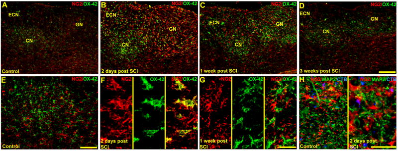
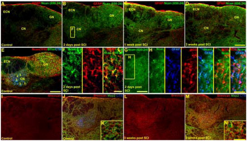
Increased chondroitin sulfate proteoglycan (CSPG) expression in the vicinity of a spinal cord injury (SCI) is a primary participant in axonal regeneration failure. However, the presence of similar increases of CSPG expression in denervated synaptic targets well away from the primary lesion and the subsequent impact on regenerating axons attempting to approach deafferented neurons have not been studied. Constitutively expressed CSPGs within the extracellular matrix and perineuronal nets of the adult rat dorsal column nuclei (DCN) were characterized using real-time PCR, Western blot analysis and immunohistochemistry. We show for the first time that by 2 days and through 3 weeks following SCI, the levels of NG2, neurocan and brevican associated with reactive glia throughout the DCN were dramatically increased throughout the DCN despite being well beyond areas of trauma-induced blood brain barrier breakdown. Importantly, regenerating axons from adult sensory neurons microtransplanted 2 weeks following SCI between the injury site and the DCN were able to regenerate rapidly within white matter (as shown previously by Davies et al. [Davies, S.J., Goucher, D.R., Doller, C., Silver, J., 1999. Robust regeneration of adult sensory axons in degenerating white matter of the adult rat spinal cord. J. Neurosci. 19, 5810-5822]) but were unable to enter the denervated DCN. Application of chondroitinase ABC or neurotrophin-3-expressing lentivirus in the DCN partially overcame this inhibition. When the treatments were combined, entrance by regenerating axons into the DCN was significantly augmented. These results demonstrate both an additional challenge and potential treatment strategy for successful functional pathway reconstruction after SCI.


Figures

Figure 1
Figure 1
Western blot analysis of GFAP, NG2, neurocan, brevican and aggrecan expression in the DCN of normal rats and rats following denervation by SCI. GFAP expression increases at 1 week and remains elevated at 3 weeks post injury. Most of the increases observed in NG2 and neurocan expression involve the 275Kd NG2 cleaved fragment and full length neurocan protein, both at nearly undetectable levels in normal rats. Both full length and C-terminal neurocan remain elevated at three weeks. Full length brevican protein expression increases at three weeks injury while there is only a small decrease in aggrecan expression at 1 week post injury. All optical densities (O.D.) for each target protein were divided by the O.D. of beta tubulin protein.
Figure 2
Figure 2
Quantification of real-time PCR and western blot analysis of GFAP (A, B), aggrecan (C, D), NG2 (E, F), neurocan (G–I) and brevican (J, K) expression in the DCN of normal rats (Ctr) and rats following denervation by SCI. Peak expression of mRNA precedes increases in GFAP, neurocan and brevican protein expression. *p<0.05; **p<0.01; ***p<0.001.
Figure 3
Figure 3
Low (A–D) and higher power (E–H) transverse images demonstrating the locations of changes in NG2 expression and activation of OX-42-IR microglia and probable O2A glia in the denervated dorsal column nuclei. In normal animals, NG2- (red) and OX-42-IR (green) labeled cells in the DCN have morphologies consistent with quiescent O2A glia and microglia, respectively (A, E). At two days following denervation by SCI when NG2 protein was highly expressed (see Figs. 1 & 2F), both OX-42- and NG2-IR were clearly increased when compared to normal rats (A, B). Also at this time, confocal microscopy revealed that each of these cell types displayed shortened thickened processes and some cells were co-labeled by both NG2- and OX-42-IR (F). By 1 week post injury the immunoreactivity of both antibodies was reduced (C) and had returned to control levels by 3 weeks post injury (D). Additionally at 1 week post SCI, NG2-IR no longer colocalized with OX-42-IR microglia and cell types labeled by either antibody had begun to revert to their normal morphology (G). Interestingly, in control animals, long varicose processes from NG2-IR O2A glia (red) were distributed through areas of dense CTB (blue) traced primary afferent terminals and MAP2-IR neurons (green) (H). This relationship was lost 2 days post injury (H). GN, gracile nucleus; CN, cuneate nucleus; ECN, external cuneate nucleus. Scale bars: A–D, 200μm; E, 100μm; F, G, 25μm; H, I, 25μm.
Figure 4
Figure 4
GFAP-IR astrocytes are sparsely distributed within the DCN of uninjured rats (A, C). Increases of GFAP protein beginning at 1 week and persisting through 3 weeks following the SCI (see Figs. 1, 2B) were localized to a distinct astrogliotic scar present within denervated areas of the DCN (B, D). GN, gracile nucleus; CN, cuneate nucleus; ECN, external cuneate nucleus. Scale bars: A, B, 200μm; C, D, 100μm.
Figure 5
Figure 5
Low (A–E, I, J, L, M) and high (F–H, K, N) power images demonstrating the locations of increased neurocan- and brevican-IR in the DCN following denervation by SCI. In uninjured animals, neurocan-IR (Ncan) was detected by antibodies directed against both the C-terminal (650.24, green, A) and N-terminal isoforms (1F6, red, E). Low levels of C-terminal and full length neurocan-IR were diffusely distributed in the normal DCN (A). In contrast, N-terminal neurocan-IR was stronger overall but low in the highly somatotopically organized areas of the cuneate and gracile nucleus identified by beta-tubulin III-IR neurons (BTiii, green) and traced CTB-IR (blue) primary afferents (E). Coinciding with beginning of increased expression of full length neurocan protein at 2 days post denervation by SCI (see Figs. 1, 2F), a clear increase in neurocan-IR (green) was observed in the DCN (B). Under higher power, much of the neurocan-IR (green) localized along and centered around the surfaces of GFAP-IR astrocyte cell bodies and processes (red, F; blue, H). Neurocan-IR did not localize with NG2-IR cells (red, G, H). Likewise, brevican-IR (red) also increased throughout the DCN 3 weeks following denervation (I–N) and localized along the surface of MAP2-IR (green) neurons (K, N) in areas of degenerating CTB-IR (blue) primary afferent terminals. GN, gracile nucleus; CN, cuneate nucleus; ECN, external cuneate nucleus. Scale bars: A–E, I, J, L, M, 200μm; F, 25μm; G, 100μm; H, 50μm; K, N, 25μm.
Figure 6
Figure 6
Low (A–F) and high power (G–N) photomicrographs of CSPGs constitutively expressed within the perineuronal nets detected by WFA cytochemistry and aggrecan-IR. WFA cytochemistry (red) prominently labeled perineuronal nets and extracellular matrix in all three DCN of uninjured animals identified by MAP2-IR (green) neurons and traced CTB-IR (blue) primary afferent terminals (A, D, G). This WFA labeling persisted throughout the 3 week period following denervation by SCI (B, C, E, F). Under higher power examination, WFA labeled structures (red, G; green H) could be seen surrounding MAP2- (green, G) and parvalbumin-IR (red, H) neurons. Aggrecan-IR (red, I–N) detected by Cat301 often colocalized with WFA labeling (green, I) labeling and surrounded BTiii-IR (green, J) neurons, but not GFAP-IR (green; K) astrocytes at all time points examined. No apparent changes in aggrecan-IR perineuronal net profiles were observed following denervation by SCI (L–N). GN, gracile nucleus; CN, cuneate nucleus; ECN, external cuneate nucleus. Scale bars: A–F, 200μm; G, L–N, 100μm; H–K, 25μm.
Figure 7
Figure 7
Images of sagittal sections demonstrating that axons from GFP (green) expressing DRG neurons microtransplanted into the gracile fasciculus (GF) regenerated through degenerating white matter but most were unable to enter the CSPG rich (see Figs. 5 & 6) denervated gracile nucleus (GN) containing NeuN-IR (red) neurons (A, C). Cytochrome oxidase (CO) cytochemistry identifies the location of the gracile nucleus and gracile fasciculus (B). Under higher magnification, regenerating axons were observed turning abruptly (arrow) at the border of the denervated gracile nucleus (C). In contrast, microtransplanted adult DRG neurons were capable of extending a few axons into portions of the denervated gracile nucleus after application of chABC (D). Scale bars: A, 100μm; B, 200; C, 50μm; D, 100μm.
Figure 8
Figure 8
Fewer (p<0.001) beta tubulin-IR (red) E15 rat DRG cells survived when cultured in conditioned media from mock transduced astrocytes (A) than from astrocytes infected by NT-3 lentivirus (B). Expression of NT-3 increased within the DCN following injection of NT-3 lentivirus (C) and resulted in increased axonal regeneration of GFP (green) expressing adult DRG neurons microtransplanted into the denervated gracile nucleus (GN) containing NeuN-IR (red) neurons (D). GF, gracile fasciculus. *p<0.05. Scale Bars: A,B, 100μm, D, 50μm.
Figure 9
Figure 9
Combined chABC and NT-3 lentivirus injections significantly increased axonal regeneration of GFP (green) expressing microtransplanted adult DRG neurons (A). The axons extended unimpeded into the denervated gracile nucleus (GN; A). Higher magnification examination revealed densely distributed GFP axons that ended with morphologies suggestive of synaptic boutons near NeuN-IR (red) neurons (B). Quantification revealed that treatment with either chABC or NT-3 lentivirus significantly increased axonal regeneration into the denervated gracile nucleus (C). Combined treatment of chABC and NT-3 lentivirus increased axonal entry into the denervated gracile nucleus nearly 10 fold over either treatment used alone (C). GF, gracile fasciculus. *p<0.05; **p<0.01; ***p<0.001. Scale bar: A, 100μm, B, 50μm.

Similar articles

Cited by

References

    1. Abdellatif AA, Pelt JL, Benton RL, Howard RM, Tsoulfas P, Ping P, Xu XM, Whittemore SR. Gene delivery to the spinal cord: comparison between lentiviral, adenoviral, and retroviral vector delivery systems. J Neurosci Res. 2006;84:553–567. - PMC - PubMed
    1. Aldskogius H, Kozlova EN. Central neuron-glial and glial-glial interactions following axon injury. Prog Neurobiol. 1998;55:1–26. - PubMed
    1. Asher RA, Morgenstern DA, Fidler PS, Adcock KH, Oohira A, Braistead JE, Levine JM, Margolis RU, Rogers JH, Fawcett JW. Neurocan is upregulated in injured brain and in cytokine-treated astrocytes. J Neurosci. 2000;20:2427–2438. - PMC - PubMed
    1. Barritt AW, Davies M, Marchand F, Hartley R, Grist J, Yip P, McMahon SB, Bradbury EJ. Chondroitinase ABC promotes sprouting of intact injured spinal systems after spinal cord injury. J Neurosci. 2006;26:10856–10867. - PMC - PubMed
    1. Biber K, Dijkstra I, Trebst C, De Groot CJ, Ransohoff RM, Boddeke HW. Functional expression of CXCR3 in cultured mouse and human astrocytes and microglia. Neuroscience. 2002;112:487–497. - PubMed

Publication types

MeSH terms