. 2007 Jul 27;370(5):826-36.
doi: 10.1016/j.jmb.2007.04.077. Epub 2007 May 10.

Cyclin box structure of the P-TEFb subunit cyclin T1 derived from a fusion complex with EIAV tat

Affiliations

Cyclin box structure of the P-TEFb subunit cyclin T1 derived from a fusion complex with EIAV tat

Kanchan Anand et al. J Mol Biol. .

Abstract

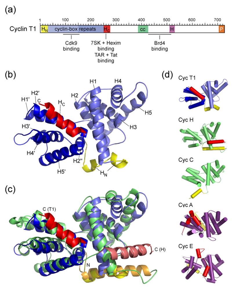
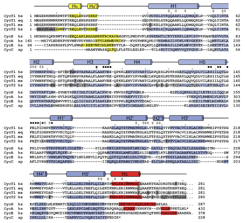
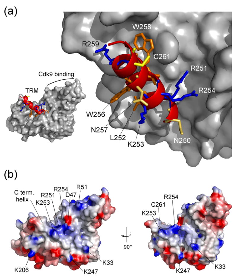
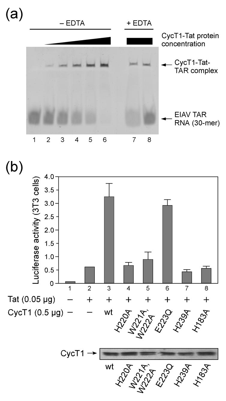
The positive transcription elongation factor b (P-TEFb) is an essential regulator of viral gene expression during the life cycle of human immunodeficiency virus type 1 (HIV-1). Its cyclin T1 subunit forms a ternary complex with the viral transcriptional transactivator (Tat) protein and the transactivation response (TAR) RNA element thereby activating cyclin dependent kinase 9 (Cdk9), which stimulates transcription at the level of chain elongation. We report the structure of the cyclin box domain of human cyclin T1 at a resolution of 2.67 A. The structure was obtained by crystallographic analysis of a fusion protein composed of cyclin T1 linked to the transactivator protein Tat from equine infectious anemia virus (EIAV), which is functionally and structurally related to HIV-1 Tat. The conserved cyclin box domain of cyclin T1 exhibits structural features for interaction with physiological binding partners such as Cdk9. A recognition site for Cdk/Cyclin substrates is partly covered by a cyclin T-specific insert, suggesting specific interactions with regulatory factors. The previously identified Tat/TAR recognition motif (TRM) forms a C-terminal helix that is partly occluded in the cyclin box repeat interface, while cysteine 261 is accessible to form an intermolecular zinc finger with Tat. Residues of the TRM contribute to a positively charged groove that may directly attract RNA molecules. The EIAV Tat protein instead appeared undefined from the electron density map suggesting that it is highly disordered. Functional experiments confirmed the TAR binding properties of the fusion protein and suggested residues on the second cyclin box repeat to contribute to Tat stimulated transcription.


Figures

Figure 1
Figure 1. Structure of the cyclin box domain of Cyclin T1
(a) Schematic diagram of the modular domain organization of human CycT1. The N-terminal cyclin domain contains the two canonical cyclin-box repeats and accessory N- and C-terminal helices. The central region is less conserved among different CycT’s but contains a proposed helical segment (cc, coiled coil) and a highly conserved histidine-rich segment (H). The C-terminal 20 residues are proline-rich (P). Proteins and RNA elements binding to CycT1 are indicated at their proposed interaction sites (Brd4, bromodomain-containing protein 4). (b) Overall structure of human CycT1 (8–263). Helices of the two repeats are denoted H1–H5 and H1′–H5′, coloured light and medium blue, respectively. N- and C-terminal helices (HN and HC) are shown in yellow and red, respectively. (c) Superimposition of CycT1 (blue) and human Cyclin H (green, AC: 1JKW). The two molecules were aligned on their cyclin box repeat structures (T1: 30–248; H: 49–262) resulting in an RMSD value of 2.2 Å. (d) Tube representation of A- and C-type cyclins. The orientation of all five molecules was aligned relative to helices H2 and H1′. N- and C-terminal helices of all cyclins are displayed in yellow and red, respectively. They are oriented differently in cyclins A (1FIN) and E (1W98) compared to cyclins C (1ZP2), H (1JKW) and T1 (2PK2, this study). Although CycT1 shares the closest structural similarity of the cyclin boxes to cyclin H, the orientation of its C-terminal helix aligns best to cyclin A.
Figure 2
Figure 2
Structure-based sequence alignment of the cyclin box repeats from various cyclin proteins. The alignment is based on the structures of human Cyclin H (1JKW, ref. 30), human Cyclin A (1FIN, ref. 8), human Cyclin E (1W98, ref. 12), Cyclin C from S. pombe (1ZP2, ref. 32) and human CycT1 (2PK2, this study). Secondary structure elements of CycT1 are indicated on top, helices of cyclins H, A, E and C are displayed by bars with the N- and C-terminal helices coloured yellow and red, respectively. Residues within the interface of the two repeats in CycT1 are indicated by diamonds. Residues of CycT1 that are proposed to interact with Cdk9 are marked by black squares.
Figure 3
Figure 3. The Tat-TAR recognition motif in Cyclin T1
(a) Conformation of the C-terminal region from residue 247 to 263 as ribbon plot within the CycT1 structure. Positively charged residues K253 and R254, polar asparagines N250 and N257, and the aromatic W256 are accessible on the surface of the TRM structure. In contrast, side chains of R251, L252, I255 and W258 connect the C-terminal helix to the cyclin box repeats. Cystein 261 which might form an intermolecular zinc-finger with Tat is fully exposed on the surface. (b) Electrostatic surface potential of CycT1 (displayed from −8 kBT to +8 kBT). Basic residues of the TRM form a combinatorial surface with the first cyclin box repeat in between both cyclin boxes that could attract negatively charged RNA molecules of 7SK or TAR.
Figure 4
Figure 4. TAR-binding of the CycT1-Tat fusion protein and functional analysis of mutant CycT1
(a) Electrophoretic mobility shift assay of CycT1-eqTat with a 30-mer equine TAR RNA. Increasing concentrations from 1 to 5 μM of the fusion protein in a buffer solution containing equal concentrations of zinc led to a complete shift of the RNA, which was used at 2 μM concentration, indicating the complex formation with the protein (lanes 2 to 6). Addition of EDTA in four- and twenty-fold concentration over the zinc ion concentration diminished the complex formation (lanes 7 and 8). (b) Functional analysis of mutant CycT1 proteins in cells. Mutation of aromatic residues H220, W221 and W222 to alanine in the Cyclin T specific region around helices H3′ and H4′ of the second cyclin box repeat reduced transcriptional activity, as does H239 on helix H5′ adjacent to the TRM. Mutation of H183 which forms the core interface between the first and second cyclin box repeat similarly diminished the CycT1 activity. The lower panel presents expression levels of CycT1 proteins as indicated by the arrow.

References

    1. Jones KA, Peterlin BM. Control of RNA initiation and elongation at the HIV-1 promoter. Annu Rev Biochem. 1994;63:717–743. - PubMed
    1. Wei P, Garber ME, Fang SM, Fischer WH, Jones KA. A novel CDK9-associated C-type cyclin interacts directly with HIV-1 Tat and mediates its high-affinity, loop-specific binding to TAR RNA. Cell. 1998;92:451–462. - PubMed
    1. Garber ME, Wei P, KewalRamani VN, Mayall TP, Herrmann CH, Rice AP, Littman DR, Jones KA. The interaction between HIV-1 Tat and human cyclin T1 requires zinc and a critical cysteine residue that is not conserved in the murine CycT1 protein. Genes Dev. 1998;12:3512–3527. - PMC - PubMed
    1. Murray AW. Recycling the cell cycle: cyclins revisited. Cell. 2004;116:221–234. - PubMed
    1. Shilatifard A, Conaway RC, Conaway JW. The RNA polymerase II elongation complex. Annu Rev Biochem. 2003;72:693–715. - PubMed

Publication types

MeSH terms

Associated data

LinkOut - more resources