. 2007 Sep;37(3):255-63.
doi: 10.1165/rcmb.2006-0289SM. Epub 2007 May 31.

Akt-mediated activation of HIF-1 in pulmonary vascular endothelial cells by S-nitrosoglutathione

Affiliations

Akt-mediated activation of HIF-1 in pulmonary vascular endothelial cells by S-nitrosoglutathione

D Jeannean Carver et al. Am J Respir Cell Mol Biol. 2007 Sep.

Abstract

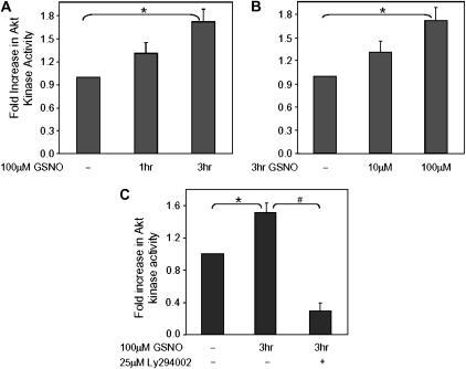
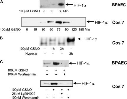
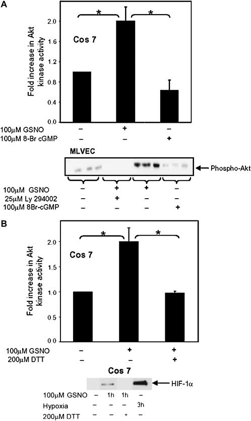
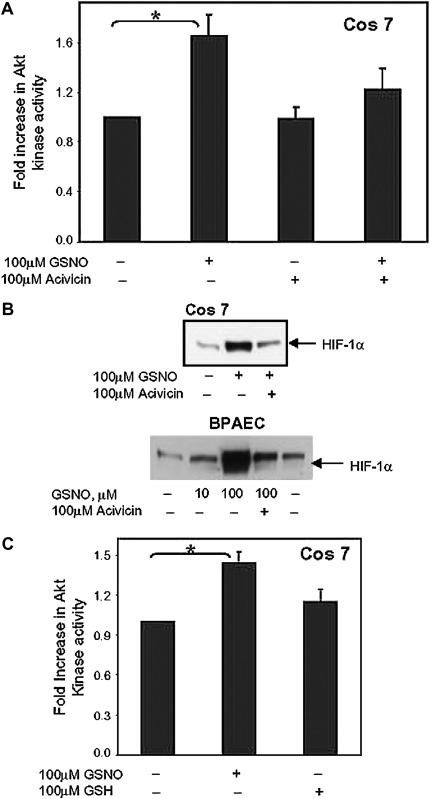
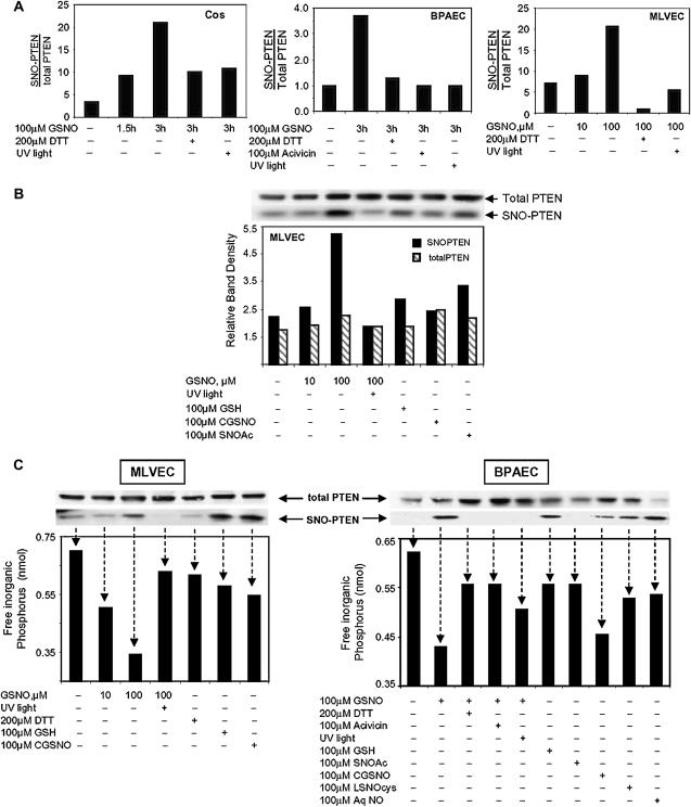
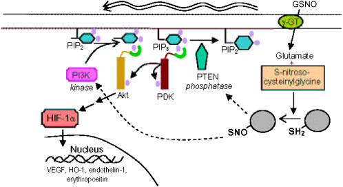
S-nitrosoglutathione (GSNO) stabilizes the alpha-subunit of hypoxia inducible factor-1 (HIF-1) in normoxic cells, but not in the presence of PI3K inhibitors. In this report, the biochemical pathway by which GSNO alters PI3K/Akt activity to modify HIF-1 expression was characterized in Cos cells and primary pulmonary vascular endothelial cells. GSNO increased Akt kinase activity--and downstream HIF-1alpha protein accumulation and DNA-binding activity--in a dose- and time-dependent manner. The PI3K inhibitors, wortmannin and LY294002, blocked these responses. Neither glutathione nor 8-bromo-cyclic GMP mimicked the GSNO-induced increases in Akt kinase activity. GSNO-induced Akt kinase activity and downstream HIF-1alpha stabilization were blocked by acivicin, an inhibitor of gamma-glutamyl transpeptidase (gammaGT), a transmembrane protein that can translate extracellular GSNO to intracellular S-nitrosocysteinylglycine. Dithiothreitol blocked GSNO-induced Akt kinase activity and HIF-1alpha stabilization. Moreover, the 3'-phosphatase of phosphoinositides, PTEN (phosphatase and tensin homolog deleted on chromosome ten) was S-nitrosylated by GSNO in pulmonary arterial endothelial cells, which was reversed by dithiothreitol and ultraviolet light. Interestingly, the abundance of S-nitrosylated PTEN also correlated inversely with PTEN activity. Taken together, these results suggest that GSNO induction of Akt appears to be mediated by S-nitrosylation chemistry rather than classic NO signaling through guanylate cyclase/cGMP. We speculate that gammaGT-dependent activation of Akt and subsequent activation of HIF-1 in vascular beds may be relevant to the regulation of HIF-1-dependent gene expression in conditions associated with oxyhemoglobin deoxygenation, as opposed to profoundly low Po(2), in the pulmonary vasculature.


Figures

<b>Figure 1.</b>
Figure 1.
GSNO increases Akt kinase activity in a time- and dose-dependent manner, an effect that is blocked by the phosphoinositol 3-kinase (PI3K) inhibitor, LY294002. Cos 7 cells were transiently co-transfected with plasmid DNA encoding HA-Akt and GFP-luciferase. Equal amounts of protein were immunoprecipitated with anti-HA antibody for each condition and Akt kinase activity assessed as described in Materials and Methods. Activity was corrected for transcription efficiency by luciferase activity. (A) Cells were treated with 100 μM GSNO for various times, or (B) were treated for 3 hours with varying concentrations of GSNO. (C) Additional Cos 7 cells were preincubated for 30 minutes with 100 μM Ly294002, a potent inhibitor of PI3K, then exposed to 100 μM GSNO for 3 hours. All cells were collected in duplicate or triplicate for each experiment and standard errors of the means were calculated (n = 7, *P = 0.002 for time course in A; n = 3, *P = 0.006 for dose response in B; n = 2, *P = 0.006 and #P = 0.006 for C).
<b>Figure 2.</b>
Figure 2.
GSNO causes an accumulation of HIF-1α protein and HIF-1α DNA binding in normoxia, but not in the presence of PI3K inhibitors. (A) BPAECs or Cos 7 cells were treated with 100 μM GSNO for various times. HIF-1α protein was detected in whole cell lysate in treated and untreated cells by Western blot analysis using anti–HIF-1α antibody. Blot is representative of several. (B) Cos 7 cells were treated with 100 μM GSNO or hypoxia for the times indicated. HIF-1 binding to DNA was determined by electromobility shift assay on whole cell lysate. (C) Cos 7 cells and BPAECs were treated with 100 μM GSNO in the presence or absence of the PI3K inhibitors 100nM wortmannin or 25 μM Ly 294002. HIF-1α protein was detected in whole cell lysate by Western blot analysis using anti–HIF-1α antibody.
<b>Figure 3.</b>
Figure 3.
Akt and HIF-1α activation by GSNO occurs through a thiol-dependent reaction. (A) Cos 7 cells were transfected as described in Figure 1, then treated with 100 μM GSNO or 100 μM 8-Br cGMP and Akt activity assessed. Results were corrected for transcription efficiency using luciferase activity (n = 3; *P = 0.007). Primary MLVEC were treated with 100 μM GSNO, ± Ly294002, or 8-Br cGMP for the times stated. Phosphorylated Akt protein was detected in whole cell lysate in treated and untreated cells by Western blot analysis using an anti-phospho(serine) Akt antibody. (B) Cos 7 cells were treated with GSNO, washed, then treated with 200 μM DTT. Akt kinase assays were preformed (n = 3; *P = 0.006), and HIF-1α protein was detected in whole cell lysate by Western blot analysis using anti–HIF-1α antibody.
<b>Figure 4.</b>
Figure 4.
GSNO activation of Akt requires the GSNO membrane transporter, γGT, and is not duplicated by reduced glutathione. (A) Cos 7 cells were transfected as in Figure 1. Cells were preincubated with 100 μM acivicin in the absence or presence of GSNO. Akt kinase activity was assessed as described (n = 7, *P = 0.005). (B) BPAEC and Cos 7 cells were treated with GSNO, with or without acivicin, and HIF-1α protein was detected in whole cell lysate in treated and untreated cells by Western blot analysis using anti–HIF-1α antibody. (C) Akt-transfected Cos 7 cells were treated with 100 μM GSNO or 100 μM GSH and Akt kinase activity assessed (n = 5, *P = 0.003).
<b>Figure 5.</b>
Figure 5.
S-nitroso-PTEN accumulates in the presence of GSNO and is inactivated in a time- and dose-dependent manner. (A) BPAEC, Cos 7, or MLVEC were treated with GSNO, GSH, S-nitroso-cysteinylglycine (CGSNO), S-nitroso-N'-acetylcysteine (SNOAc), or aqueous nitric oxide (AqNO) for the times and doses stated. Some cells were treated with DTT during the final 30 minutes of GSNO treatment or were preincubated with 100 μM acivicin. Lysate from some GSNO-treated cells was treated with ultraviolet light before analysis. Total PTEN protein was then detected in the lysate samples by Western blot analysis using anti-PTEN antibody. MAPK protein levels were assessed using anti-MAPK antibody to control for protein loading. S-nitrosylated PTEN (SNO-PTEN) was isolated from whole cell lysate using the biotin switch method. SNO-PTEN protein was detected by Western blot analysis using anti-PTEN antibody. Relative protein densities of the SNO-PTEN bands were compared with that of total PTEN. The data shown are single experiments. Similar patterns of results were seen in at least two trials for each cell line. (B) MLVEC cells were treated as stated. Data shown are Western blots representing total PTEN from whole cell lysate, and SNO-PTEN isolated from the same lysate using the biotin switch method. The graph represents the ratio of SNO-PTEN: total PTEN and is representive of several experiments illustrating the same pattern of SNO-PTEN: total PTEN ratios. (C) BPAEC and MLVEC cells were treated as described above. Western blots show bands representing total PTEN and SNO-PTEN isolated from the same lysate. The graph below represents the relative PTEN phosphatase activity on phosphatidylinositol 3,4,5-trisphosphate (PIP3). Arrows associate enzyme activity with the correlating SNO-PTEN and total PTEN bands detected by Western blot.
<b>Figure 6.</b>
Figure 6.
Proposed pathway by which GSNO can activate HIF 1 through PI3K/Akt in normoxia. This schematic illustrates the proposed regulation of HIF-1 in normoxia by the endogenous S-nitrosothiol, GSNO, through PI3K/Akt activation. In pulmonary vascular endothelial cells and Cos 7 cells, this appears to occur not through classic NO· diffusion with activation of guanylyl cyclase, but through a thiol-based reaction requiring the membrane protein, γGT. One target of this –SNO modification is activation of PI3K. Another target is PTEN, the counter-regulatory phosphatase of the PI3K/Akt pathway.

Similar articles

Cited by

References

    1. Semenza GL. O2-regulated gene expression: transcriptional control of cardiorespiratory physiology by HIF-1. J Appl Physiol 2004;96:1173–1177. - PubMed
    1. Zhou J, Callapina M, Goodall GJ, Brune B. Functional integrity of nuclear factor kappa B, phosphatidylinositol 3′-kinase, and mitogen-activated protein kinase signaling allows tumor necrosis factor alpha-evoked Bcl-2 expression to provoke internal ribosome entry site-dependent translation of hypoxia-inducible factor 1alpha. Cancer Res 2004;64:9041–9048. - PubMed
    1. Qian D, Lin HY, Wang HM, Zhang X, Liu DL, Li QL, Zhu C. Normoxic induction of the hypoxic-inducible factor-1 alpha by interleukin-1 beta involves the extracellular signal-regulated kinase 1/2 pathway in normal human cytotrophoblast cells. Biol Reprod 2004;70:1822–1827. - PubMed
    1. Kasuno K, Takabuchi S, Fukuda K, Kizaka-Kondoh S, Yodoi J, Adachi T, Semenza GL, Hirota K. Nitric oxide induces hypoxia-inducible factor 1 activation that is dependent on MAPK and phosphatidylinositol 3-kinase signaling. J Biol Chem 2004;279:2550–2558. - PubMed
    1. Palmer LA, Gaston B, Johns RA. Normoxic stabilization of hypoxia-inducible factor-1 expression and activity: redox-dependent effect of nitrogen oxides. Mol Pharmacol 2000;58:1197–1203. - PubMed

Publication types

MeSH terms

Substances