. 2007 Jun 20;26(12):2832-42.
doi: 10.1038/sj.emboj.7601738. Epub 2007 May 31.

Increased tumor cell dissemination and cellular senescence in the absence of beta1-integrin function

Affiliations

Increased tumor cell dissemination and cellular senescence in the absence of beta1-integrin function

Angelika Kren et al. EMBO J. .

Abstract

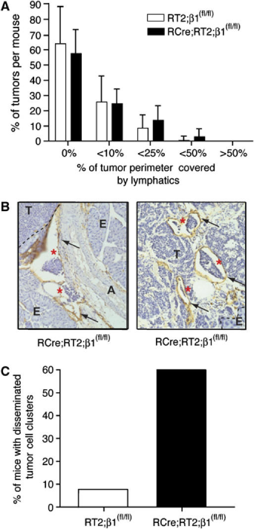
Integrins are transmembrane receptors that bind extracellular matrix proteins and enable cell adhesion and cytoskeletal organization, as well as transduction of signals into cells, to promote various aspects of cellular behavior, such as proliferation or survival. Integrins participate in many aspects of tumor biology. Here, we have employed the Rip1Tag2 transgenic mouse model of pancreatic beta cell carcinogenesis to investigate the role of beta(1)-integrin in tumor progression. Specific ablation of beta(1)-integrin function in pancreatic beta cells resulted in a defect in sorting between insulin-expressing beta cells and glucagon-expressing alpha cells in islets of Langerhans. Ablation of beta(1)-integrin in beta tumor cells of Rip1Tag2 mice led to the dissemination of tumor cell emboli into lymphatic blood vessels in the absence of ongoing lymphangiogenesis. Yet, disseminating beta(1)-integrin-deficient beta tumor cells did not elicit metastasis. Rather, primary tumor growth was significantly impaired by reduced tumor cell proliferation and the acquisition of cellular senescence by beta(1)-integrin-deficient beta tumor cells. The results indicate a critical role of beta(1)-integrin function in mediating metastatic dissemination and preventing tumor cell senescence.


Figures

Figure 1
Figure 1
Distorted sorting of α and β cells in β1-integrin-deficient islets of Langerhans. (A) Upper panel: immunohistochemical analysis by hematoxylin and eosin staining of histological sections of pancreatic islets from wild-type (C57) and β1-integrin-deficient (RCre;β1(fl/fl)) mice. Lower panel: immunofluorescence co-stainings for insulin (green), glucagon (red) and nuclei (DAPI, blue) of histological sections from wild-type (C57) and β1-integrin-deficient (RCre;β1(fl/fl)) mice. (B) FACS analysis of tumor cell suspensions from RT2;β1(fl/fl) control (left panel) and RCre;RT2;β1(fl/fl) experimental (right panel) mice stained for β1-integrin. Dashed black lines represent unstained cells, solid red line represents cells stained for β1-integrin.
Figure 2
Figure 2
Dissemination of tumor cell emboli but no lymphangiogenesis in β1-integrin-deficient tumors of RT2 mice. (A) Determination of peritumoral lymphatic density. White bars, tumors of RT2;β1(fl/fl) mice (12 mice, 162 tumors); black bars, tumors of RCre;RT2;β1(fl/fl) mice (15 mice, 205 tumors). Tumor sections were stained for LYVE-1 and categorized according to the degree of lymphatic coverage: not in contact with any lymphatic vessel (0%), tumors that were surrounded less than 10% (<10%), less than 25% (<25%), less than 50% (<50%) and more than 50% of (>50%). Statistical analysis (unpaired t-test) indicated that differences within groups are not significant. (B) LYVE-1 staining (brown, indicated by arrows) of histological sections of pancreata from RCre;RT2;β1(fl/fl) mice. Circulating tumor cell clusters are indicated by red asterisks. A, artery; E, exocrine pancreas; T, tumor. (C) Percentage of mice showing disseminated tumor cell clusters in RT2;β1(fl/fl) (white bars, n=12) and RCre;RT2;β1(fl/fl) mice (black bars; n=15).
Figure 3
Figure 3
Cell lines derived from RT2;β1(fl/fl) and RCre;RT2;β1(fl/fl) tumors. (A) Top panel: genotyping of cell lines derived from RT2 (βT2), RT2;β1(fl/fl) (βTi(fl/fl)), RCre;RT2;β1(fl/fl) (βTi(Δ/Δ)), and RT2;NCAM−/− (βTN2) tumors by PCR analysis. wt, wild-type β1-integrin alleles; fl, floxed alleles; Δ, deleted alleles. Bottom panel: immunoblotting analysis of NCAM expression in βT2, βTi(fl/fl), βTi(Δ/Δ) and βTN2 cells. (B) FACS analysis of β1-integrin surface expression levels of βTi(fl/fl) (black line) and βTi(Δ/Δ) (red line). Dashed lines represent controls (unstained cells). (C) Adhesion of the different genotype β tumor cells to collagen IV. Mock-transfected (clear bars) or NCAM140-transfected (dotted bars) βTi(fl/fl) and βTi(Δ/Δ) β tumor cell lines were seeded on either uncoated (gray bars) or collagen IV-coated (red bars) culture dishes. *P<0.003, unpaired t-test.
Figure 4
Figure 4
β1-integrin-deficient β tumor cells do not form primary tumors or colonize distant organs in transplantation experiments. (A) Average volumes of tumors arising from transplanted β tumor cell lines. A total of 106 each of wild-type β tumor cells (βT2), cells carrying floxed alleles of the β1-integrin gene (βTi(fl/fl)), cells deficient for β1-integrin expression (βTi(Δ/Δ)) and cells deficient for NCAM expression (βTN2) were injected into the flanks of C57Bl/6 mice. (B) Tumor incidence in C57Bl/6 mice injected with the cell lines described in (A). Sixteen sites in eight mice were injected per genotype cell line, and the percentage of sites with tumors is presented.
Figure 5
Figure 5
Reduced tumor cell proliferation and apoptosis in β1-integrin-deficient RT2 tumors. Tumor incidences (A) and tumor volumes (B) of pancreata derived from RT2;β1(fl/fl) control and RCre;RT2;β1(fl/fl) mice. Tumor cell proliferation was visualized by BrdU staining (C) and apoptotic cells were visualized by TUNEL reaction (D). BrdU- or TUNEL-positive cells were counted per × 40 microscopic field. *P>0.1; **P<0.05; ***P<0.01, unpaired t-test; n, number of analyzed mice. (E) Tumors of hematoxylin and eosin-stained sections were classified according to their histological grading. HYP, hyperplastic islets; AD, adenoma; G1, carcinoma grade1; G2, carcinoma grade 2; G3, carcinoma grade 3. (F) MTT growth assay of βTi(fl/fl) and βTi(Δ/Δ) tumor cell lines. Linear regression curves were calculated and are displayed for each cell line. *P<0.001, unpaired t-test of linear regression curves.
Figure 6
Figure 6
Impaired growth and filopodia formation of β1-integrin-deficient β tumor cells in a 3D culture system. (A) Phase-contrast micrographs of control (βTi(fl/fl)) and β1-integrin-deficient βTi(Δ/Δ) β tumor cells 2 days after seeding in Matrigel™. (B) Quantification of protrusion formation of βTi(fl/fl) and βTi(Δ/Δ) cells grown as in (A).
Figure 7
Figure 7
The lack of β1-integrin function induces cellular senescence. (A) Cell cycle analysis of tumors derived from RT2;β1(fl/fl) and RCre;RT2;β1(fl/fl) mice, as indicated. Three animals were analyzed per genotype. The percentages of cells in G0 are plotted. *P<0.05, unpaired t-test. (B) Examples of tumor cells of RCre;RT2;EII mice exhibiting expression of SA-β-Gal (turquoise, left and middle panels). Control tumors (RT2;EII, expressing β1-integrin) do not show any detectable signal for SA-β-Gal (right panels). Scale bar, 50 μm. T, tumor; E, exocrine pancreas; (C) Percentages of SA-β-Gal-positive and SA-β-Gal-negative tumors of RT2;EII (white bars) and RCre;RT2;EII (black bars) mice. Large, medium and small refer to tumor sizes of >3 mm, >1 mm and <1 mm in diameter, respectively.
Figure 8
Figure 8
Increased p21Cip1 levels in β1-integrin-deficient tumors. (A) Immunoblot for actin and p21Cip1 levels in lysates from control (RT2;β1(fl/fl) and RT2;EII) and β1-integrin-deficient (RCre;RT2;β1(fl/fl) and RCre;RT2;EII) tumors. M, molecular weight marker. (B) Quantification of relative p21Cip1 levels as determined by fluorescent quantitative immunoblotting shown in (A). *P<0.05, unpaired t-test.

Similar articles

Cited by

References

    1. Ahlgren U, Jonsson J, Jonsson L, Simu K, Edlund H (1998) Beta-cell-specific inactivation of the mouse Ipf1/Pdx1 gene results in loss of the beta-cell phenotype and maturity onset diabetes. Genes Dev 12: 1763–1768 - PMC - PubMed
    1. Alcorta DA, Xiong Y, Phelps D, Hannon G, Beach D, Barrett JC (1996) Involvement of the cyclin-dependent kinase inhibitor p16 (INK4a) in replicative senescence of normal human fibroblasts. Proc Natl Acad Sci USA 93: 13742–13747 - PMC - PubMed
    1. Banerji S, Ni J, Wang SX, Clasper S, Su J, Tammi R, Jones M, Jackson DG (1999) LYVE-1, a new homologue of the CD44 glycoprotein, is a lymph-specific receptor for hyaluronan. J Cell Biol 144: 789–801 - PMC - PubMed
    1. Bloch W, Forsberg E, Lentini S, Brakebusch C, Martin K, Krell HW, Weidle UH, Addicke K, Fassler R (1997) Beta 1 integrin is essential for teratoma growth and angiogenesis. J Cell Biol 139: 265–278 - PMC - PubMed
    1. Brooks PC, Clark RA, Cheresh DA (1994) Requirement of vascular integrin alpha v beta 3 for angiogenesis. Science 264: 569–571 - PubMed

Publication types