Review
. 2007;8(5):215.
doi: 10.1186/gb-2007-8-5-215.

The integrins

Affiliations
Review

The integrins

Yoshikazu Takada et al. Genome Biol. 2007.

Abstract

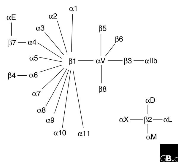
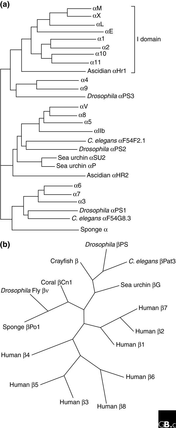
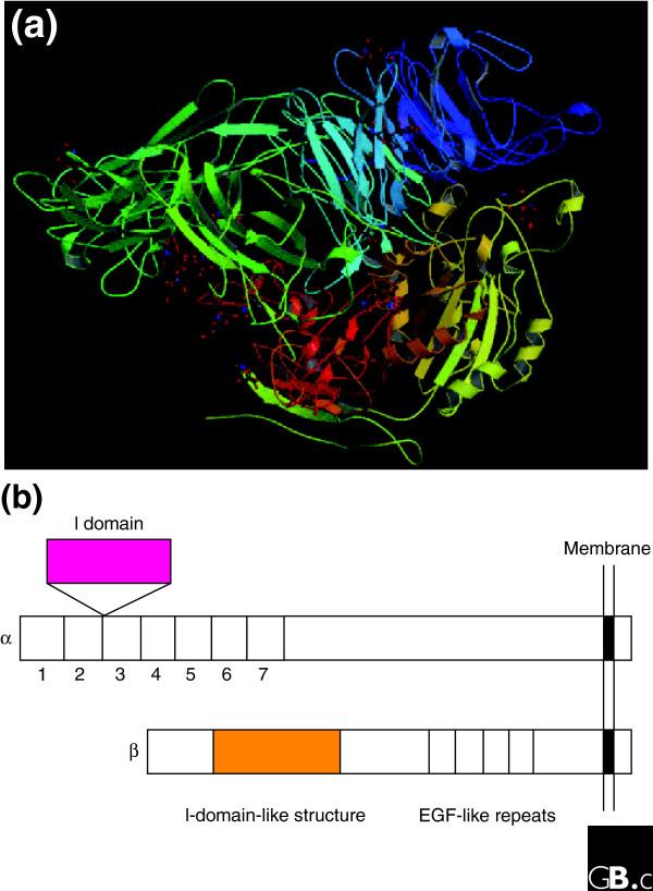
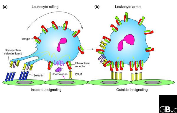
The integrins are a superfamily of cell adhesion receptors that bind to extracellular matrix ligands, cell-surface ligands, and soluble ligands. They are transmembrane alphabeta heterodimers and at least 18 alpha and eight beta subunits are known in humans, generating 24 heterodimers. Members of this family have been found in mammals, chicken and zebrafish, as well as lower eukaryotes, including sponges, the nematode Caenorhabditis elegans (two alpha and one beta subunits, generating two integrins) and the fruitfly Drosophila melanogaster (five alpha and one beta, generating five integrins). The alpha and beta subunits have distinct domain structures, with extracellular domains from each subunit contributing to the ligand-binding site of the heterodimer. The sequence arginine-glycine-aspartic acid (RGD) was identified as a general integrin-binding motif, but individual integrins are also specific for particular protein ligands. Immunologically important integrin ligands are the intercellular adhesion molecules (ICAMs), immunoglobulin superfamily members present on inflamed endothelium and antigen-presenting cells. On ligand binding, integrins transduce signals into the cell interior; they can also receive intracellular signals that regulate their ligand-binding affinity. Here we provide a brief overview that concentrates mostly on the organization, structure and function of mammalian integrins, which have been more extensively studied than integrins in other organisms.


Figures

Figure 1
Figure 1
The members of the human integrin superfamily and how they combine to form heterodimeric integrins. At least 18 α subunits and eight β subunits have been identified in humans, which are able to generate 24 different integrins. Integrin subunits that bind to each other to form a heterodimer are connected by solid lines. Each integrin has distinct ligand-binding specificity and tissue and cell distribution.
Figure 2
Figure 2
Phylogenetic trees of integrin subunits. Trees for (a) integrin α and (b) integrin β subunits are adapted from [58] and [59], respectively.
Figure 3
Figure 3
The extracellular region of a human integrin. (a) The crystal structure represents a net form of integrin αVβ3 with no bound RGD peptide (Protein Data Bank (PDB) code 1JV2) [3,4]. See PBD code 1L5G for the RGD-bound form. (b) The I (inserted or interactive) domain is present in seven human α subunits between β-propeller repeats 2 and 3, and is involved in ligand binding. An I-like domain is present in all human integrin β subunits along with four EGF-like repeats. Both the I and I-like domains have a Rossmann fold.
Figure 4
Figure 4
Leukocyte recruitment to the endothelial surface. (a) Binding of glycoprotein selectin ligands (yellow and purple) on the leukocyte to selectins (blue) on the endothelial surface, and weak binding of low-affinity leukocyte integrins (green) to ICAMs (pale yellow) on the endothelium facilitates cell tethering and rolling. This binding, together with signals from chemokines (pink), generates inside-out signals (yellow arrows) that shift the bound integrins to a high-affinity ligand-binding state. (b) Leukocyte arrest is mediated by clusters of high-affinity integrins (red) binding to ICAMs on the endothelial cells. These focal clusters can themselves signal outside-in to affect functions such as cell polarization and migration.

References

    1. Hynes RO. Integrins: bidirectional, allosteric signaling machines. Cell. 2002;110:673–687. doi: 10.1016/S0092-8674(02)00971-6. - DOI - PubMed
    1. Shimaoka M, Springer TA. Therapeutic antagonists and conformational regulation of integrin function. Nat Rev Drug Discov. 2003;2:703–716. doi: 10.1038/nrd1174. - DOI - PubMed
    1. Huhtala M, Heino J, Casciari D, de Luise A, Johnson MS. Integrin evolution: insights from ascidian and teleost fish genomes. Matrix Biol. 2005;24:83–95. doi: 10.1016/j.matbio.2005.01.003. - DOI - PubMed
    1. Xiong JP, Stehle T, Diefenbach B, Zhang R, Dunker R, Scott DL, Andrzej J, Goodman SL, Arnaout MA. Crystal structure of the extracellular segment of integrin αvβ3. Science. 2001;294:339–345. doi: 10.1126/science.1064535. - DOI - PMC - PubMed
    1. Xiong JP, Stehle T, Zhang R, Joachimiak A, Frech M, Goodman SL, Arnaout MA. Crystal structure of the extracellular segment of integrin alpha Vbeta3 in complex with an Arg-Gly-Asp ligand. Science. 2002;296:151–155. doi: 10.1126/science.1069040. - DOI - PubMed