. 2007 Jun;12(6):957-71.
doi: 10.1016/j.devcel.2007.04.001.

Functional redundancy of GSK-3alpha and GSK-3beta in Wnt/beta-catenin signaling shown by using an allelic series of embryonic stem cell lines

Affiliations

Functional redundancy of GSK-3alpha and GSK-3beta in Wnt/beta-catenin signaling shown by using an allelic series of embryonic stem cell lines

Bradley W Doble et al. Dev Cell. 2007 Jun.

Abstract

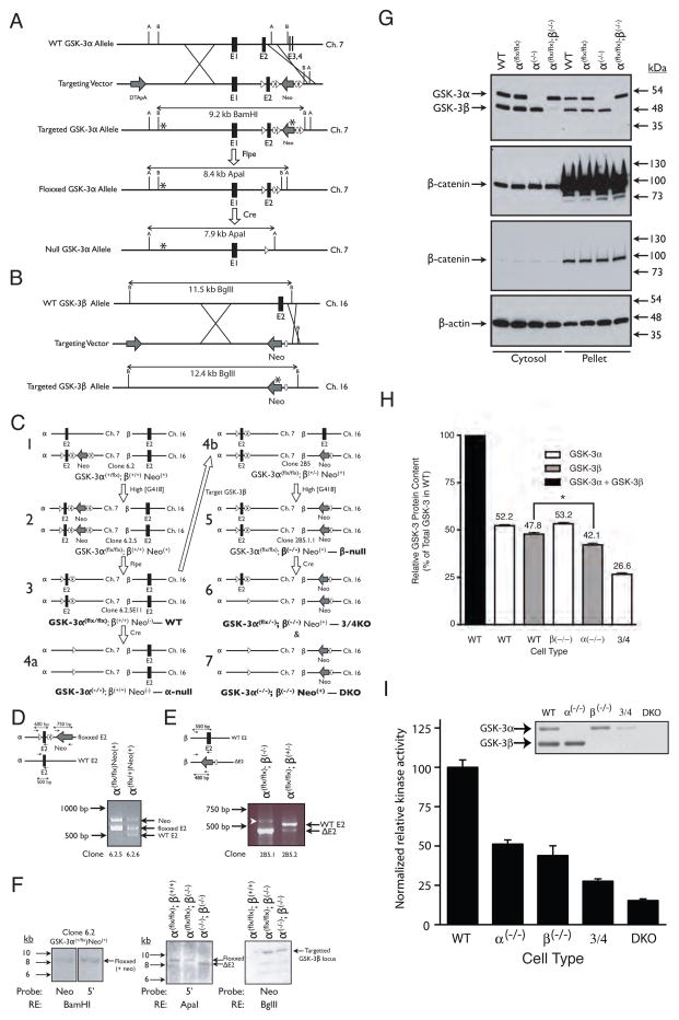
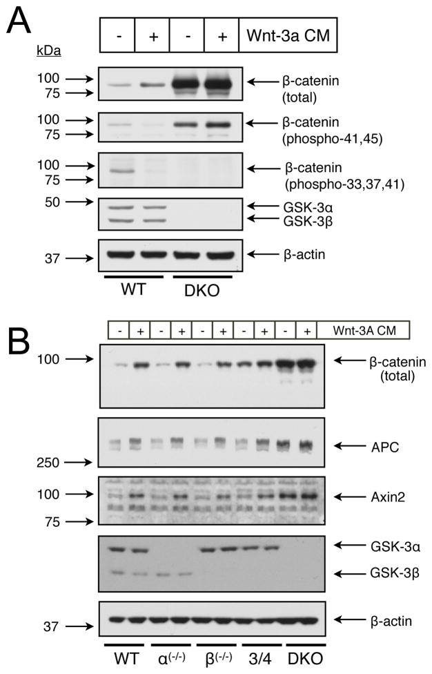
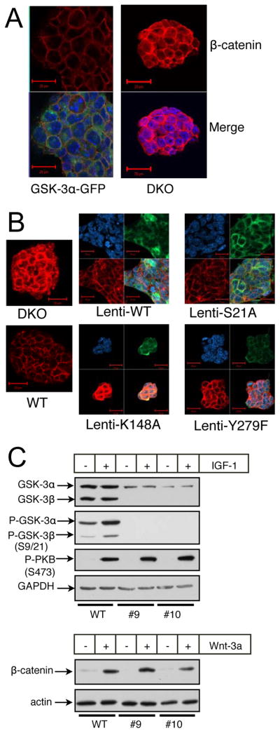
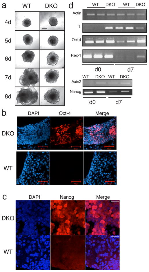
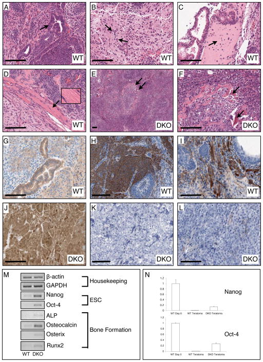
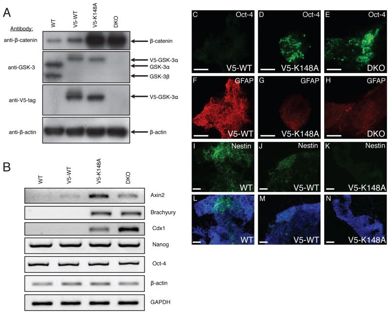
In mammalian cells, glycogen synthase kinase-3 (GSK-3) exists as two homologs, GSK-3alpha and GSK-3beta, encoded by independent genes, which share similar kinase domains but differ substantially in their termini. Here, we describe the generation of an allelic series of mouse embryonic stem cell (ESC) lines with 0-4 functional GSK-3 alleles and examine GSK-3-isoform function in Wnt/beta-catenin signaling. No compensatory upregulation in GSK-3 protein levels or activity was detected in cells lacking either GSK-3alpha or GSK-3beta, and Wnt/beta-catenin signaling was normal. Only in cells lacking three or all four of the alleles was a gene-dosage effect on beta-catenin/TCF-mediated transcription observed. Indeed, GSK-3alpha/beta double-knockout ESCs displayed hyperactivated Wnt/beta-catenin signaling and were severely compromised in their ability to differentiate, but could be rescued to normality by re-expression of functional GSK-3. The rheostatic regulation of GSK-3 highlights the importance of considering the contributions of both homologs when studying GSK-3 functions in mammalian systems.


Figures

Figure 1
Figure 1
Strategy used to create compound knockouts of GSK-3α and GSK-3β in ES cells. (A) Targeting strategy for GSK-3α. After homologous recombination with the targeting vector, exon 2 was replaced with a LoxP-flanked (floxxed) exon 2 and FRT-flanked (flrted) neomycin resistance cassette. B = BamHI; A = ApaI. (B) Targeting strategy for GSK-3β. After homologous recombination, exon 2 was replaced with a neomycin resistance cassette and a single FRT site. B = BglII. (C) Overview of the steps required to generate the various ESC lines used in this study (bold type). (D) Multiplex PCR analysis used to screen ESC clones for G418-mediated conversion from GSK-3α(+/flx) to GSK-3α (flx/flx) genotype (step 1–2 in panel C). (E) PCR analysis used to screen ESC clones for G418-mediated conversion from GSK-3α (flx/flx);GSK-3β(+/−) to GSK-3α(flx/flx);GSK-3β(−/−) genotype (step 4b-5 in panel C). White arrowhead indicates a non-specific band. In A-E, ovals represent FRT recombination sites, triangles represent LoxP recombination sites and asterisks represent hybridization sites for Southern blot probes. (F) Southern blots confirming proper targeting of GSK-3 loci. (G) Wild-type, GSK-3α(flx/flx), GSK-3α (−/−) and GSK-3β(−/−) ES cells all display the same level of cytosolic and membrane-associated (pellet) β–catenin. Two different exposures of the same β-catenin immunoblots are presented. (H) Quantitation of GSK-3 in WT and knockout cell lines. Three lysates, each from a different plate of cells, were analyzed per cell type. Error bars = SEM. * p <0.05 (ANOVA with Tukey’s post-hoc test). (I) Consequence of reduced GSK-3 gene dosage on kinase activity. Each bar represents the average from 2 independent kinase assay experiments with each assay performed in triplicate. Error bars = SEM. Inset – immunoblot of the GSK-3 band profiles in the eluates used in the kinase assays (normalized to total protein content).
Figure 2
Figure 2
Consequences of reduced GSK-3 gene dosage with respect to cytosolic β-catenin levels and β-catenin TCF-transactivation activity. (A) Immunoblot analysis of cytosolic GSK-3, β-catenin and GAPDH levels. Results from three independent clones with DKO or 3/4KO genotypes are presented. (B) Quantitative RT-PCR analysis of Axin2 transcript levels in WT and GSK-3 knockout ES cells. Error bars = SEM. (C) TCF reporter assay. Each bar represents the average from 4 independent samples. Error bars = SEM. * p < 0.05; ** p < 0.001 (ANOVA with Newman-Keuls post-hoc test). (D) Immunofluorescence analysis of a WT and DKO ES cell clone stained for β-catenin and DAPI (nuclei). Bar = 10 μm.
Figure 3
Figure 3
Immunoblot analysis of GSK-3 gene dosage effects on Wnt signaling intermediates. (A) Examination of β-catenin phosphorylation in WT and DKO ESCs treated 30 min. with Wnt-3a conditioned medium (CM). (B) Levels of key Wnt signaling molecules after long-term (20 hour) treatment with Wnt-3a CM.
Figure 4
Figure 4
Rescue of DKO ES cells with variants of GSK-3α. (A) Immunofluorescent images of DKO ES cells and DKO ES cells reconstituted with a GSK-3α-GFP fusion protein. β-catenin staining is red, GFP staining is green and nuclear staining is blue (DAPI). (B) Immunofluorescent images of WT and DKO ES cells as well as DKO ES cells rescued with various V5-epitope-tagged mutants of GSK-3α through lentiviral transduction. V5 staining is green, β-catenin staining is red and nuclear staining is blue. All images were obtained with identical PMT, resolution and scan-rate settings. (C) DKO ES cells rescued with a GSK-3α S21A mutant still respond to Wnt-3a. #9 and #10 are independent clones of DKO ES cells reconstituted with S21A-GSK-3α. Treatments with IGF-1 were for 15 minutes, while treatments with Wnt-3a conditioned medium were overnight.
Figure 5
Figure 5
GSK-3 double null ES cells (DKO) display a limited potential for differentiation. (A) Embryoid bodies derived from the same number (800) of WT or DKO ES cells at various time points after the initiation of differentiation. Bar = 200 μm (B). Oct-4 immunfluorescence staining of WT and DKO EBs differentiated for 14 days in the absence of LIF. Bar = 50 μm. (C) Nanog immmunofluorescence staining of WT and DKO EBs differentiated for 12 days in the absence of LIF. Bar = 20 μm. (D) Semi-quantitative RT-PCR analysis WT and DKO EBs at day 0 (d0) and day 7 (d7) of differentiation.
Figure 6
Figure 6
Histological, immunohistochemical, and PCR examination of teratomas generated from WT (GSK-3α(flx/flx)) and DKO ES cells. (A) The WT teratomas possess numerous examples of glandular epithelial structures indicative of endoderm (arrow indicates gut-like pseudostratified epithelium). (B) WT tumours also had large regions of neuronal tissue indicating differentiation into ectoderm (arrows point to neuronal nuclei). (C) Differentiation of WT ESCs into mesoderm was confirmed by observation of bone (arrow points to region of woven bone) and (D) muscle (inset shows magnification of striated skeletal muscle). (E, F) DKO teratomas contain tightly packed cells in undifferentiated tissue reminiscent of a carcinoma with scattered spicules of bone (arrows). (G) Immunohistochemical staining for β-catenin revealed normal membranous staining in epithelial cells of WT teratomas. H, I) Staining for the neuronal progenitor marker Nestin and the astrocytic marker GFAP was clearly observed in WT teratomas. (J) DKO teratomas displayed very high levels of β-catenin immunoreactivity in all cells in membrane, cytosol and nuclear compartments. (K, L) There was no detectable staining for Nestin or GFAP in DKO teratoma tissue sections. (M) Semi-quantitative RT-PCR analyses of WT vs. DKO gene expression. Bar = 100 μm.
Figure 7
Figure 7
The properties of DKO ESCs can be reverted to those of WT ESCs upon stable expression of WT, but not kinase-dead, GSK-3. (A) Immunoblot analyses of cytosolic lysates prepared from WT and DKO ESCs as well as DKO ESC cell lines stably expressing V5-tagged WT- or K148A-GSK-3α. (B) Semi-quantitative RT-PCR analysis of transcripts expressed in the same cell lines examined in panel A. (C–N) Immunofluorescence images of EBs maintained for 14 days in LIF-free medium. EBs derived from (C, F) V5-WT-GSK-3α-expressing DKO ESCs; (D, G) V5-K148A-GSK-3α expressing DKO ESCs; and (E, H) DKO ESCs double-stained for Oct-4 and GFAP, respectively. EBs derived from (I, L) WT ESCs; (J, M) V5-WT-GSK-3α-expressing DKO ESCs and (K, N) V5-K148A-GSK-3α-expressing DKO ESCs stained for Nestin and Nestin/DAPI (nuclear stain) merge, respectively. Bar = 100 μm in all micrographs.

References

    1. Amit S, Hatzubai A, Birman Y, Andersen JS, Ben-Shushan E, Mann M, Ben-Neriah Y, Alkalay I. Axin-mediated CKI phosphorylation of beta-catenin at Ser 45: a molecular switch for the Wnt pathway. Genes Dev. 2002;16:1066–1076. - PMC - PubMed
    1. Arnold SJ, Stappert J, Bauer A, Kispert A, Herrmann BG, Kemler R. Brachyury is a target gene of the Wnt/beta-catenin signaling pathway. Mech Dev. 2000;91:249–258. - PubMed
    1. Bain J, McLauchlan H, Elliott M, Cohen P. The specificities of protein kinase inhibitors: an update. Biochem J. 2003;371:199–204. - PMC - PubMed
    1. Ben-Shushan E, Thompson JR, Gudas LJ, Bergman Y. Rex-1, a gene encoding a transcription factor expressed in the early embryo, is regulated via Oct-3/4 and Oct-6 binding to an octamer site and a novel protein, Rox-1, binding to an adjacent site. Mol Cell Biol. 1998;18:1866–1878. - PMC - PubMed
    1. Bugeon L, Syed N, Dallman MJ. A fast and efficient method for transiently transfecting ES cells: application to the development of systems for conditional gene expression. Transgenic Res. 2000;9:229–232. - PubMed

Publication types

Grants and funding