Review
. 2007 Jun;45 Suppl A(6S):A15-24.
doi: 10.1016/j.jvs.2007.02.061.

Smooth muscle cell signal transduction: implications of vascular biology for vascular surgeons

Affiliations
Review

Smooth muscle cell signal transduction: implications of vascular biology for vascular surgeons

Akihito Muto et al. J Vasc Surg. 2007 Jun.

Abstract

Vascular smooth muscle cells exhibit varied responses after vessel injury and surgical interventions, including phenotypic switching, migration, proliferation, protein synthesis, and apoptosis. Although the source of the smooth muscle cells that accumulate in the vascular wall is controversial, possibly reflecting migration from the adventitia, from the circulating blood, or in situ differentiation, the intracellular signal transduction pathways that control these processes are being defined. Some of these pathways include the Ras-mitogen-activated protein kinase, phosphatidylinositol 3-kinase-Akt, Rho, death receptor-caspase, and nitric oxide pathways. Signal transduction pathways provide amplification, redundancy, and control points within the cell and culminate in biologic responses. We review some of the signaling pathways activated within smooth muscle cells that contribute to smooth muscle cell heterogeneity and development of pathology such as restenosis and neointimal hyperplasia.


Figures
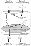
Figure 1
Figure 1
Signaling during SMC phenotype switching. The left side of the figure denotes pathways involved in signaling during SMC differentiation to the contractile phenotype. IGF-I causes expression of genes associated with the contractile, differentiated phenotype through the PI3K-Akt pathway, while at the same time blocks the Ras-MAPK pathway with the IPS-I/SHP2 complex. The right side of the figure denotes pathways involved in signaling during SMC phenotype switching to the synthetic phenotype. Several growth factors stimulate SMC phenotype switching by stimulating MAPK directly as well as by cleaving the IPS-I/SHP2 complex. MAPK transposition to the nucleus inhibits transcription of genes associated with the contractile phenotype and stimulates expression of genes associated with growth. Signals from each cascade inhibit the opposite cascade. IGF-I: insulin like growth factor-I; PDGF: platelet derived growth factor; EGF: epidermal growth factor; FGF: fibroblast growth factor; PI3K: phosphoinositide 3-kinase; IRS-I: insulin receptor substrate-I; SHP2: Scr homology protein 2; MAPK: mitogen-activated protein kinase. p: phosphorylation
Figure 2
Figure 2
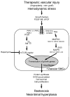
Proliferative signaling during SMC response to injury. The figure shows convergent signaling pathways resulting in protein synthesis and cell proliferation, leading to restenosis and neointimal hyperplasia. Implications of vascular intervention may include inducing growth factors that are SMC mitogens and chemoattractants. These factors stimulate SMC signal transduction pathways including the Ras-MAPK and the PI3K-Akt-mTOR pathways for growth gene transcription. PDGF-BB: platelet derived growth factor-BB; bFGF: basic fibroblast growth factor; PI3K: phosphoinositide 3-kinase; PDK1: 3-phosphoinositide-dependent kinase 1; mTOR: mammalian target of rapamycin ; MAPK: mitogen-activated protein kinase.
Figure 3
Figure 3
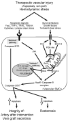
Apoptotic signaling during SMC response to injury. The figure shows divergent pathways resulting in control of apoptosis, leading to different outcomes. The extrinsic apoptosis pathway is stimulated by signals external to the SMC, whereas the intrinsic apoptosis pathway is stimulated by signals internal to the SMC nucleus and /or mitochondria. In the extrinsic pathway, apoptosis ligands activate caspases −8 and −10 or JNK. The intrinsic pathway is activated by DNA damage or genetic programs and induces the Apaf1-caspase−9 complex directly or through the release of cytochrome c. Both the internal and external pathways activate caspases−3, −5, and −7 to effect apoptosis. TNFα: tumor necrosis factorα; JNK: c-Jun N-terminal kinase; PKA: protein kinase A ; PI3K: phosphoinositide 3-kinase ; ERK: extracellular signal-regulated kinase; Cyto c: cytochrome c; Apaf1: apoptosis activating factor 1
Figure 4
Figure 4
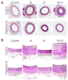
Relevance of mTOR signal transduction pathway in the response to vascular injury. The right carotid artery of New Zealand white rabbits was subjected to sham operation (control), balloon injury (B), outflow branch ligation to reduce flow (LF), or both balloon injury and reduction in flow (B+LF), and harvested after 21 days. Either rapamycin (5 mg/kg) or saline was orally administered daily from 48 hours prior to the procedure until harvest; in animals given rapamycin, serum levels (day 7) were therapeutic (mean 10.9 ± 0.5 ng/mL; therapeutic range 4–12 ng/mL; n=21). Animals treated with rapamycin demonstrated significant inhibition of neointimal thickening in balloon-injured arteries (B), including arteries treated with low flow (B+LF; p<0.0001). Negative remodeling was evident in all vessels in the low flow groups (LF, B+LF), and rapamycin did not affect this reduction in vessel size due to low flow. A, low power magnification; B, high power magnification. Reprinted from Vascular Pharmacology, Vol number, Paszkowiak et al, Evidence supporting changes in Nogo-B levels as a marker of neointimal expansion but not adaptive arterial remodeling, Pages No., Copyright (Year), with permission from Elsevier.

References

    1. Owens GK. Regulation of differentiation of vascular smooth muscle cells. Physiol Rev. 1995;75:487–517. - PubMed
    1. Kockx MM, Knaapen MW. The role of apoptosis in vascular disease. J Pathol. 2000;190:267–280. - PubMed
    1. Ross R. Atherosclerosis--an inflammatory disease. N Engl J Med. 1999;340:115–126. - PubMed
    1. Schwartz SM, Heimark RL, Majesky MW. Developmental mechanisms underlying pathology of arteries. Physiol Rev. 1990;70:1177–1209. - PubMed
    1. Mitra AK, Gangahar DM, Agrawal DK. Cellular, molecular and immunological mechanisms in the pathophysiology of vein graft intimal hyperplasia. Immunol Cell Biol. 2006;84:115–124. - PubMed

Publication types

MeSH terms

Substances