Review
. 2007 Jun;45 Suppl A(Suppl A):A99-103.
doi: 10.1016/j.jvs.2007.02.046.

Tissue engineering applications to vascular bypass graft development: the use of adipose-derived stem cells

Affiliations
Review

Tissue engineering applications to vascular bypass graft development: the use of adipose-derived stem cells

Paul DiMuzio et al. J Vasc Surg. 2007 Jun.

Abstract

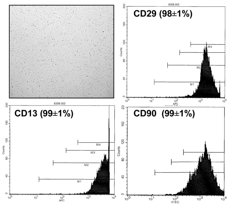
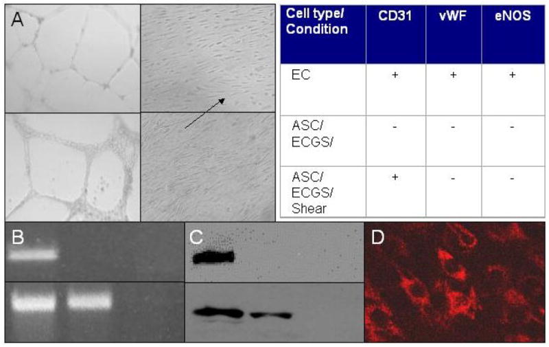
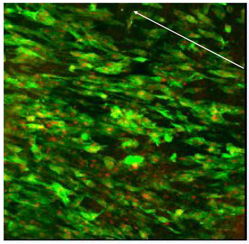
The burgeoning field of vascular tissue engineering holds promise for the creation of a practical and successful small-diameter arterial bypass graft. Many creative combinations of autologous cells and scaffolds exist along with an equally long list of microenvironmental cues used to create a functional arterial conduit. This review outlines our work using abdominal wall fat as a source of autologous stem cells for vascular tissue engineering, focusing specifically on this stem cell's availability and potency to differentiate into endothelial-like cells. In a series of 49 patients undergoing elective peripheral vascular surgery, an abundant quantity of adult stem cells was harvested from fat obtained by liposuction. The efficacy of the isolation did not appear influenced by advanced age, obesity, renal failure, or vascular disease, although fat from diabetic patients yielded significantly less stem cells. In addition, these adipose-derived stem cells acquired several morphologic and molecular endothelial phenotypes when exposed to growth factors (endothelial cell growth supplement and vascular endothelial growth factor) and physiologic shear stress in vitro. Taken together, these studies suggest that fat appears to be a viable source of autologous stem cells for use in vascular tissue engineering.


Figures

Figure 1
Figure 1. Morphological and molecular characterization of adipose-derived stem cells (ASCs)
Inverted phase light micrograph (40x, unstained) of ASCs grown in culture demonstrating spindle-like morphology. After negative selection of cells for CD31 and CD45 using magnetic cell sorting, representative fluorescent activated cell sorting of ASCs grown in culture (passage four) demonstrate a homogeneous population of cells positive for CD13, 29 and 90 surface markers.
Figure 2
Figure 2. Differentiation of ASCs in response to endothelial cell growth supplement (ECGS) and shear stress
A. Morphological studies. Inverted phase light micrographs (40x, unstained) of human endothelial cells (upper two panels) forming cords on Matrigel (left) and aligning in the direction of shear stress (right). Similar findings are noted in human ASCs cultured in ECGS for a minimum of three weeks (lower two panels). B. Molecular studies (CD31 message). RT-PCR detecting CD31 message in human endothelial (left lane), ASCs (middle lane) and smooth muscle (right lane) cells before (upper row) and after (lower row) exposure to 12dynes shear for four days. ASCs grown in ECGS and exposed to shear express CD31 message, an endothelial cell phenotype marker. C. Molecular studies (CD31 protein). In studies analogous to those in B, Western blot for CD31 protein demonstrates that ASCs cultured in ECGS and exposed to shear stress express CD31 at the protein level. D. Immunohistochemical study (CD31). Laser confocal micrograph (40x) of ASCs grown in ECGS, exposed to shear stress and stained with human CD31 Mab confirm the presence of CD31 protein within the differentiated ASCs. Table. Summary of molecular studies of ASCs grown in ECGS and exposed to shear stress. Neither vWF nor eNOS was demonstrated by RT-PCR or Western blot.
Figure 3
Figure 3. Attachment and retention of ASCs to vascular scaffolding
Laser confocal micrograph (100x, phalloidin and propidium iodide stain) of human stem cells seeded onto decellularized vein allograft. After 24h of static seeding, the graft was exposed to gradually increasing shear force (from 0 to 9 dynes) over 72h. The arrow indicates direction of flow. The stem cells aligned with flow and maintained a confluent monolayer of cells under physiological shear force.

Similar articles

Cited by

References

    1. Riha GM, Lin PH, Lumsden AB, Yao Q, Chen C. Application of stem cells for vascular tissue engineering. Tissue Engineering. 2005;11:1535–52. - PubMed
    1. Herring MB, Dilley R, Jersild RA, Jr, Boxer L, Gardner A, Glover J. Seeding arterial prostheses with vascular endothelium. The nature of the lining. Ann Surg. 1979;190:84–90. - PMC - PubMed
    1. Weinberg CB, Bell E. A blood vessel model constructed from collagen and cultured vascular cells. Science. 1986;87:397–400. - PubMed
    1. Isenberg BC, Williams C, Tranquillo RT. Small-diameter artificial arteries engineered in vitro. Circ Res. 2006;98:25–35. - PubMed
    1. Barrilleaux B, Phinney DG, Prockop Darwin, O’Connor KC. Review: Ex vivo engineering of living tissues with adult stem cells. Tissue Engineering. 2006;12:1–13. - PubMed

MeSH terms