. 2007 Oct 1;93(7):2457-71.
doi: 10.1529/biophysj.107.108068. Epub 2007 Jun 1.

Folding and unfolding of gammaTIM monomers and dimers

Affiliations

Folding and unfolding of gammaTIM monomers and dimers

Brijesh Patel et al. Biophys J. .

Abstract

Kinetic simulations of the folding and unfolding of triosephosphate isomerase (TIM) from yeast were conducted using a single monomer gammaTIM polypeptide chain that folds as a monomer and two gammaTIM chains that fold to the native dimer structure. The basic protein model used was a minimalist Gō model using the native structure to determine attractive energies in the protein chain. For each simulation type--monomer unfolding, monomer refolding, dimer unfolding, and dimer refolding--thirty simulations were conducted, successfully capturing each reaction in full. Analysis of the simulations demonstrates four main conclusions. First, all four simulation types have a similar "folding order", i.e., they have similar structures in intermediate stages of folding between the unfolded and folded state. Second, despite this similarity, different intermediate stages are more or less populated in the four different simulations, with 1), no intermediates populated in monomer unfolding; 2), two intermediates populated with beta(2)-beta(4) and beta(1)-beta(5) regions folded in monomer refolding; 3), two intermediates populated with beta(2)-beta(3) and beta(2)-beta(4) regions folded in dimer unfolding; and 4), two intermediates populated with beta(1)-beta(5) and beta(1)-beta(5) + beta(6) + beta(7) + beta(8) regions folded in dimer refolding. Third, simulations demonstrate that dimer binding and unbinding can occur early in the folding process before complete monomer-chain folding. Fourth, excellent agreement is found between the simulations and MPAX (misincorporation proton alkyl exchange) experiments. In total, this agreement demonstrates that the computational Gō model is accurate for gammaTIM and that the energy landscape of gammaTIM appears funneled to the native state.


Figures

FIGURE 1
FIGURE 1
Triosephosphate isomerase dimer from PDB structure 1YPI. One monomer is colored blue with interface residues in cyan. The other monomer is colored red with interface residues in yellow.
FIGURE 2
FIGURE 2
All atom coordinates (left) and Cα model (right) of the γTIM monomer.
FIGURE 3
FIGURE 3
Schematic for folding (right arrows) and unfolding (left arrows) for (top) γTIM Gō model monomer and (bottom) γTIM Gō model dimer.
FIGURE 4
FIGURE 4
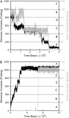
Sample trajectories of γTIM dimer (A) unfolding and (B) refolding. The left y axis shows the total number of intramolecular contacts (black lines) for the two protein chains (587 maximum for each chain = 1174 total). The right y axis shows the number of intermolecular contacts (gray lines) at the dimer interface between the two chains (108 maximum).
FIGURE 5
FIGURE 5
Intermediate stages of γTIM monomer unfolding (dashed gray lines), dimer unfolding (dashed black lines), monomer refolding (solid gray lines), and dimer refolding (solid black lines). (A) α-helix (black) and β-sheet (gray) regions of γTIM. (B) Probability of residue folding in group B: β2β3. (C) Probability of residue folding in group C: β2β4. (D) Probability of residue folding in group D: β1β5. (E) Probability of residue folding in group E: β1β8.
FIGURE 6
FIGURE 6
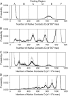
(A) Probability of populating folding groups A–F during monomer unfolding, (B) monomer refolding, (C) dimer unfolding, and (D) dimer refolding. Error boundaries are indicated in gray.
FIGURE 7
FIGURE 7
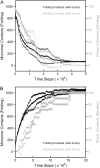
Average number of intramolecular (black lines) or intermolecular (gray lines) contacts obtained from 30 trajectories of (A) dimer unfolding and (B) dimer refolding. Error boundaries are indicated with black points for intramolecular contacts and gray points for intermolecular contacts.
FIGURE 8
FIGURE 8
Residues folded in early-folding intermediate 2 (gray points) and late-folding intermediate 1 (black points) for 47 residues studied with alkyl-exchange experiments.
FIGURE 9
FIGURE 9
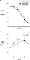
Percent match of simulation folding groups A–F (Fig. 5) at predicting experimental intermediates (A) 2 and (B) 1 from Fig. 8. Data points from simulations of monomer refolding, monomer unfolding, dimer refolding, and dimer unfolding are slightly offset to aid the eye.

Similar articles

Cited by

References

    1. Bryngelson, J. D., and P. G. Wolynes. 1987. Spin glasses and the statistical mechanics of protein folding. Proc. Natl. Acad. Sci. USA. 84:7524–7528. - PMC - PubMed
    1. Onuchic, J. N., Z. Luthey-Schulten, and P. G. Wolynes. 1997. Theory of protein folding: the energy landscape perspective. Annu. Rev. Phys. Chem. 48:545–600. - PubMed
    1. Leopold, P. E., M. Montal, and J. N. Onuchic. 1992. Protein folding funnels: a kinetic approach to the sequence-structure relationship. Proc. Natl. Acad. Sci. USA. 89:8721–8725. - PMC - PubMed
    1. Garel, T., and H. Orland. 1988. Mean-field model for protein folding. Europhys. Lett. (Switzerland). 6:307–310.
    1. Shakhnovich, E. I., and A. M. Gutin. 1989. The nonergodic (spin-glass-like) phase of heteropolymer with quenched disordered sequence of links. Europhys. Lett. (Switzerland). 8:327–332.

Publication types

LinkOut - more resources