. 2007 Aug;189(15):5738-48.
doi: 10.1128/JB.00395-07. Epub 2007 Jun 1.

Structure and function of cold shock proteins in archaea

Affiliations

Structure and function of cold shock proteins in archaea

Laura Giaquinto et al. J Bacteriol. 2007 Aug.

Abstract

Archaea are abundant and drive critical microbial processes in the Earth's cold biosphere. Despite this, not enough is known about the molecular mechanisms of cold adaptation and no biochemical studies have been performed on stenopsychrophilic archaea (e.g., Methanogenium frigidum). This study examined the structural and functional properties of cold shock proteins (Csps) from archaea, including biochemical analysis of the Csp from M. frigidum. csp genes are present in most bacteria and some eucarya but absent from most archaeal genome sequences, most notably, those of all archaeal thermophiles and hyperthermophiles. In bacteria, Csps are small, nucleic acid binding proteins involved in a variety of cellular processes, such as transcription. In this study, archaeal Csp function was assessed by examining the ability of csp genes from psychrophilic and mesophilic Euryarchaeota and Crenarchaeota to complement a cold-sensitive growth defect in Escherichia coli. In addition, an archaeal gene with a cold shock domain (CSD) fold but little sequence identity to Csps was also examined. Genes encoding Csps or a CSD structural analog from three psychrophilic archaea rescued the E. coli growth defect. The three proteins were predicted to have a higher content of solvent-exposed basic residues than the noncomplementing proteins, and the basic residues were located on the nucleic acid binding surface, similar to their arrangement in E. coli CspA. The M. frigidum Csp was purified and found to be a single-domain protein that folds by a reversible two-state mechanism and to exhibit a low conformational stability typical of cold-adapted proteins. Moreover, M. frigidum Csp was characterized as binding E. coli single-stranded RNA, consistent with its ability to complement function in E. coli. The studies show that some Csp and CSD fold proteins have retained sufficient similarity throughout evolution in the Archaea to be able to function effectively in the Bacteria and that the function of the archaeal proteins relates to cold adaptation. The initial biochemical analysis of M. frigidum Csp has developed a platform for further characterization and demonstrates the potential for expanding molecular studies of proteins from this important archaeal stenopsychrophile.


Figures

FIG. 1.
FIG. 1.
Complementation of cold sensitivity in E. coli BX04 cells expressing M. frigidum csp. The pIN-csp plasmid and the pIN vector (control) were transformed into cold-sensitive strain BX04 and streaked onto LB plates containing ampicillin with (A, C, E) and without (B, D) 2 mM IPTG, and the plates were incubated at 30°C (A), 23°C (B, C), or 15°C (D, E).
FIG. 2.
FIG. 2.
Primary-structure analysis of Csp homologs. Amino acid sequence alignments of Csp homologs from E. coli (CspA), M. frigidum, uncultured Crenarchaeota, “C. symbiosum,” H. marismortui, Halobacterium sp. strain NRC-1 (genes 101 and 1836), and H. lacusprofundi (genes 846 and 1547). Aromatic residues, negatively charged residues, and positively charged residues are highlighted in yellow, blue, and red, respectively. β-Strands in E. coli CspA are indicated by bars, and the residues composing the RNA binding motifs, RNP1 and RNP2, are boxed. The lengths of the proteins are shown on the right in amino acids.
FIG. 3.
FIG. 3.
Three-dimensional structure models of Csp homologs. The nuclear magnetic resonance structure of E. coli CspA (A) and homology models of M. frigidum Csp (B), uncultured Crenarchaeota Csp (C), “C. symbiosum” Csp (D), and Csps from H. marismortui (E), Halobacterium sp. strain NRC-1 (genes 101 and 1836) (F and G), H. lacusprofundi (genes 846 and 1547) (H and I), and M. burtonii CSD (J) are shown. Positively charged residues exposed on the surface are in red writing. E. coli Csp β-sheets 1 to 5 are in black writing. E. coli Csp loops 1 to 4 are in white writing. M. burtonii CSD cysteine residues typical of Zn ribbons are in green writing. The models are presented alphabetically from left to right, with A in the top left corner and J at the bottom.
FIG. 4.
FIG. 4.
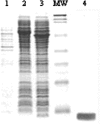
Expression and purification of M. frigidum Csp. SDS-gel electrophoresis analysis of the purification of M. frigidum Csp by affinity chromatography on chitin beads. M. frigidum Csp was purified from crude lysate of BL21(DE3) cells expressing the M. frigidum Csp-intein-CBD fusion. Shown are the insoluble fraction (lane 1), the soluble fraction in sample buffer without DTT (lane 2), the soluble fraction in sample buffer with 70 mM DTT (lane 3), the purified M. frigidum Csp (lane 4), and a broad-range protein molecular size standard (lane MW; Bio-Rad) corresponding to 224, 122, 90, 51.5, 35.3, 28.7, 21, and 7.2 kDa.
FIG. 5.
FIG. 5.
TUG-GE of M. frigidum Csp. Unfolding (N⇆U) (A) and refolding (U⇆N) (B) transition curves of M. frigidum Csp in a TUG (0 to 7 M) perpendicular to the direction of electrophoresis is shown. The urea gradient is counterbalanced by an inverse acrylamide gradient (15 to 11%). The free energy of unfolding at 0 M urea was calculated by graphically extrapolating from the transition region, and the concentration of urea at ΔG = 0 is [urea]1/2. Fu, fraction of unfolded molecules; ΔG, free-energy difference between folded and unfolded states; [urea]1/2, concentration of urea at equilibrium; R, universal gas constant (8.314 kJ mol−1); T, absolute temperature.
FIG. 6.
FIG. 6.
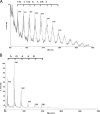
Mass spectra of M. frigidum Csp. Shown are the positive-ion MALDI-TOF mass spectra of M. frigidum Csp incubated in subtilisin (A) and M. frigidum Csp incubated in subtilisin and RNase A (B) determined by C18 ZipTip extraction and a 3-HPA matrix. The ribonucleotide bases corresponding to the mass difference between consecutive peaks are indicated. The average residue mass for each of the ribonucleotide bases is as follows: adenosine, 329.21; cytidine, 305.18; uridine, 306.17; guanosine, 345.21.
FIG. 7.
FIG. 7.
Phylogenetic tree of Csp homologs. The tree was constructed with the PHYLIP server.

Similar articles

Cited by

References

    1. Aravind, L., and E. V. Koonin. 1999. DNA-binding proteins and evolution of transcription regulation in the archaea. Nucleic Acids Res. 27:4658-4670. - PMC - PubMed
    1. Bae, W., S. Phadtare, K. Severinov, and M. Inouye. 1999. Characterization of Escherichia coli cspE, whose product negatively regulates transcription of cspA, the gene for the major cold shock protein. Mol. Microbiol. 31:1429-1441. - PubMed
    1. Béjà, O., E. V. Koonin, L. Aravind, L. T. Taylor, H. Seitz, J. L. Stein, D. C. Bensen, R. A. Feldman, R. V. Swanson, and E. F. DeLong. 2002. Comparative genomic analysis of archaeal genotypic variants in a single population and in two different oceanic provinces. Appl. Environ. Microbiol. 68:335-345. - PMC - PubMed
    1. Berquist, B. R., J. Soneja, and S. DasSarma. 2005. Comparative genomic survey of information transfer systems in two diverse extremely halophilic archaea, Halobacterium sp. strain NRC-1 and Haloarcula marismortui, p. 148-182. In N. Gunde-Cimerman, A. Oren, and A. Plemenitas (ed.), Adaptation to life at high salt concentrations in Archaea, Bacteria, and Eukarya. Springer, Dordrecht, The Netherlands.
    1. Bradford, M. M. 1976. A rapid and sensitive method for the quantitation of microgram quantities of protein utilizing the principle of protein-dye binding. Anal. Biochem. 72:248-254. - PubMed

Publication types

MeSH terms