. 2007 Aug;189(16):5987-95.
doi: 10.1128/JB.00049-07. Epub 2007 Jun 1.

Crystal structures of the receiver domain of the response regulator PhoP from Escherichia coli in the absence and presence of the phosphoryl analog beryllofluoride

Affiliations

Crystal structures of the receiver domain of the response regulator PhoP from Escherichia coli in the absence and presence of the phosphoryl analog beryllofluoride

Priti Bachhawat et al. J Bacteriol. 2007 Aug.

Abstract

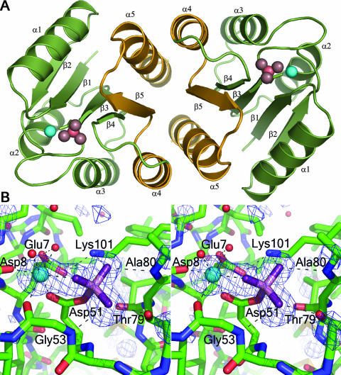
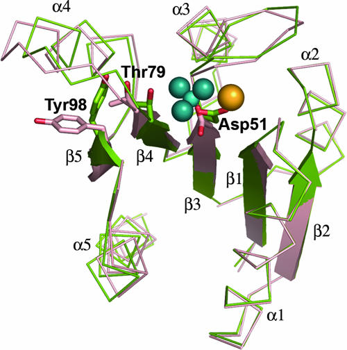
The response regulator PhoP is part of the PhoQ/PhoP two-component system involved in responses to depletion of extracellular Mg(2+). Here, we report the crystal structures of the receiver domain of Escherichia coli PhoP determined in the absence and presence of the phosphoryl analog beryllofluoride. In the presence of beryllofluoride, the active receiver domain forms a twofold symmetric dimer similar to that seen in structures of other regulatory domains from the OmpR/PhoB family, providing further evidence that members of this family utilize a common mode of dimerization in the active state. In the absence of activating agents, the PhoP receiver domain crystallizes with a similar structure, consistent with the previous observation that high concentrations can promote an active state of PhoP independent of phosphorylation.


Figures

FIG. 1.
FIG. 1.
BeF3-activated PhoPN. (A) The regulatory-domain dimer. Activated PhoPN dimerizes with twofold rotational symmetry, forming an α4-β5-α5 interface. A ribbon depiction of the dimer is shown in green with the α4-β5-α5 dimer interface highlighted in gold and repetitive secondary-structure elements labeled. The noncovalent BeF3 complex and Mg2+ within the active site are shown as pink and cyan spheres, respectively. (B) Stereo view of the active site. A difference electron density map (FoFc) calculated at 1.9 Å with omission of the Mg2+ and BeF3 complex from the model is shown in blue, contoured at 3σ. Carbons, nitrogens, and oxygens of the protein are shown as green, blue, and red sticks, respectively. The noncovalent BeF3 complex is shown in stick format with beryllium and fluorides in light and dark purple, respectively. Water molecules and Mg2+ are shown as red and cyan spheres, respectively. The network of hydrogen bonds that coordinates BeF3 and Mg2+ is shown as dashed lines. All molecular images were generated using Pymol (http://www.pymol.org).
FIG. 2.
FIG. 2.
Differences in the backbone conformations of activated versus unactivated PhoPN. The conformational changes of the switch residues Thr79 and Tyr98, when BeF3 is bound at the active site Asp51, are correlated with minor differences in the overall positioning of secondary-structural elements relative to the unactivated protein. The unactivated protomer (chain A) is shown in red, and the activated protein is shown in green. The BeF3 complex is shown in blue, and Mg2+ is shown in yellow. Tyr98 has an outward orientation in the unactivated molecule and an inward orientation in the activated molecule.
FIG. 3.
FIG. 3.
Electron density in the β4-α4-β5 region of activated and unactivated PhoPN proteins. Stereo views of the σA-weighted 2FoFc electron density maps contoured at 2.5σ and calculated at 1.9 Å for activated PhoPN (A) and at 2.54 Å for (B) chain A and (C) chain B of unactivated PhoPN. The protein model is shown in stick representation (carbon, blue; nitrogen, dark blue; oxygen, red), and the relevant secondary-structural elements and tyrosine 98 are labeled in panel A for reference.
FIG. 4.
FIG. 4.
Comparison of B factors of unactivated and activated PhoPN. (A) Average main-chain B factors for unactivated PhoPN chain A (pink), unactivated PhoPN chain B (orange), and activated PhoPN (green). (B) Differences between B factors of activated PhoPN and unactivated PhoPN chain A (pink) and unactivated PhoPN chain B (orange). The secondary-structural elements are shown in blue at the top.
FIG. 5.
FIG. 5.
Size exclusion chromatography of unphosphorylated and phosphorylated PhoPN and PhoP. (A) Elution profiles of PhoPN (solid line) and phosphorylated PhoPN (dotted line) on a TosoHaas G2000SW column. (B) Elution profiles of full-length PhoP (solid line) and phosphorylated PhoP (dotted line) on a TosoHaas G3000SWXL column. The conditions for chromatography and the protein standards used for calibration of the column are described in Materials and Methods.

References

    1. Bachhawat, P., G. V. Swapna, G. T. Montelione, and A. M. Stock. 2005. Mechanism of activation for transcription factor PhoB suggested by different modes of dimerization in the inactive and active states. Structure 13:1353-1363. - PMC - PubMed
    1. Bent, C. J., N. W. Isaacs, T. J. Mitchell, and A. Riboldi-Tunnicliffe. 2004. Crystal structure of the response regulator 02 receiver domain, the essential YycF two-component system of Streptococcus pneumoniae in both complexed and native states. J. Bacteriol. 186:2872-2879. - PMC - PubMed
    1. Bijlsma, J. J., and E. A. Groisman. 2005. The PhoP/PhoQ system controls the intramacrophage type three secretion system of Salmonella enterica. Mol. Microbiol. 57:85-96. - PubMed
    1. Birck, C., Y. Chen, F. M. Hulett, and J. P. Samama. 2003. The crystal structure of the phosphorylation domain in PhoP reveals a functional tandem association mediated by an asymmetric interface. J. Bacteriol. 185:254-261. - PMC - PubMed
    1. Brünger, A. T., P. D. Adams, G. M. Clore, W. L. DeLano, P. Gros, R. W. Grosse-Kunstleve, J. S. Jiang, J. Kuszewski, M. Nilges, N. S. Pannu, R. J. Read, L. M. Rice, T. Simonson, and G. L. Warren. 1998. Crystallography and NMR system: a new software suite for macromolecular structure determination. Acta Crystallogr. D 54:905-921. - PubMed

Publication types

MeSH terms

LinkOut - more resources