. 2007 Aug;189(15):5482-94.
doi: 10.1128/JB.00647-07. Epub 2007 Jun 1.

Cysteine scanning mutagenesis and topological mapping of the Escherichia coli twin-arginine translocase TatC Component

Affiliations

Cysteine scanning mutagenesis and topological mapping of the Escherichia coli twin-arginine translocase TatC Component

Claire Punginelli et al. J Bacteriol. 2007 Aug.

Abstract

The TatC protein is an essential component of the Escherichia coli twin-arginine (Tat) protein translocation pathway. It is a polytopic membrane protein that forms a complex with TatB, together acting as the receptor for Tat substrates. In this study we have constructed 57 individual cysteine substitutions throughout the protein. Each of the substitutions resulted in a TatC protein that was competent to support Tat-dependent protein translocation. Accessibility studies with membrane-permeant and -impermeant thiol-reactive reagents demonstrated that TatC has six transmembrane helices, rather than the four suggested by a previous study (K. Gouffi, C.-L. Santini, and L.-F. Wu, FEBS Lett. 525:65-70, 2002). Disulfide cross-linking experiments with TatC proteins containing single cysteine residues showed that each transmembrane domain of TatC was able to interact with the same domain from a neighboring TatC protein. Surprisingly, only three of these cysteine variants retained the ability to cross-link at low temperatures. These results are consistent with the likelihood that most of the disulfide cross-links are between TatC proteins in separate TatBC complexes, suggesting that TatC is located on the periphery of the complex.


Figures

FIG. 1.
FIG. 1.
Positions of the single cysteine substitutions in TatC that were constructed in this study. Residues that were substituted to cysteine for topological labeling studies are shown in hatched ovals, while those located towards the center of each predicted transmembrane α-helix that were substituted to cysteine for the initial disulfide cross-linking analysis are represented as solid medium-gray ovals. Further residues in transmembrane helix one that were mutated to cysteine are depicted as solid light-gray ovals. Transmembrane helices four and five are shown in broken lines, since a study by Gouffi et al. (18) questioned their existence.
FIG. 2.
FIG. 2.
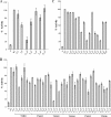
Periplasmic TMAO reductase activities of E. coli tat mutant strains producing TatA, TatB and cysteine-substituted TatC proteins that were used for sulfhydryl labeling (A) or disulfide cross-linking (B and C). Periplasmic TMAO reductase activities were measured from the ΔtatABCD ΔtatE pcnB derivative, DADE-P (Δ), or DADE-P carrying either pUNITAT2 (29) (wild type [WT]) and (A) pUNITATCC4H (Cys) or pUNITATCC4H encoding cysteine-substituted TatC proteins or (B and C) pUNITATCC4 (Cys) or pUNITATCC4 encoding cysteine-substituted TatC proteins. In each case, the amino acid position of the substitution is shown under each column. One hundred percent activity was taken to be that determined for E. coli strain DADE-P carrying pUNITAT1 and corresponds to an activity of typically 0.85 μmol benzyl viologen oxidized/min/mg protein. In panel B, the substitutions in transmembrane helix one (TMH1) to transmembrane helix six (TMH6) are shown. The error bars represent the standard errors of the means (n = 3 or 4).
FIG. 3.
FIG. 3.
TatC variant G144C forms a strong cross-linked dimer in untreated membranes. Western blot analysis of membranes isolated from the ΔtatABCD ΔtatE pcnB strain, E. coli strain DADE-P harboring plasmid producing TatA, TatB, and either wild-type TatCHis (pUNITAT2), the TatCHis cysteine-less derivative (pUNITATCC4H), or single cysteine substitutions as indicated. Samples were analyzed as isolated or were reduced by treatment with 10 mM DTT for 1 h (indicated by the symbols − and +, respectively, above each lane), and all samples were analyzed by SDS-PAGE under nonreducing conditions.
FIG. 4.
FIG. 4.
Sulfhydryl labeling of spheroplasts overproducing TatA, TatB, and cysteine variants of TatC. Spheroplasts were incubated with buffer alone (−), with MPB (lab), or with AMS followed by MPB (block), as described in Materials and Methods. Spheroplasts were subsequently lysed, and membranes were isolated, dispersed with SDS, and immunoprecipitated with anti-TatC antisera. Samples were separated by SDS-PAGE (12.5% acrylamide) and electroblotted, and MPB-labeled protein was detected with a 1:5,000 dilution of streptavidin-HRP (Strep). The same blot was then stripped and reprobed with a 1:5000 dilution of anti-tetra-His (α-His) antibody (QIAGEN) to detect TatC protein. (A) Labeling reactions with control samples expressing a cysteine-less TatC variant and the single cysteine substitutions L9C (cytoplasmic location) and G144C and S148C (both periplasmic [peri] location). (B) Labeling reactions with TatC cysteine variants designed to probe the cellular location of the loop region between transmembrane helices four and five: I183C, P186C, and I191C. The positions of these residues on a cartoon of TatC are shown at the right-hand side of each panel (cytoplasmic [cyto] or periplasmic [peri] location).
FIG. 5.
FIG. 5.
The TatC G144C, A26C, and Y36C variants form disulfide cross-links when oxidized at room temperature or on ice. Membrane samples were prepared from the ΔtatABCD ΔtatE pcnB strain, DADE-P, coexpressing single cysteine-substituted TatC variants G144C (A), A26C (B), and Y36C (C) together with TatA and TatB from pUNITATCC4. For membranes harboring the TatC G144C or A26C substitution, a sample as prepared was retained (lanes marked C), and the remainder of the membranes were treated with DTT to reduce preformed dimer as described in Materials and Methods. Samples (100 μg membrane protein) were then subjected to oxidizing conditions (by the addition of copper phenanthroline) at either room temperature (ORT) or on ice (O0°C). A sample of the DTT-treated membranes are also shown (lanes marked R). For membranes harboring the TatC Y36C variant (which shows no disulfide-bonded dimer in the membrane as isolated), samples (100 μg membrane protein) were incubated for one hour with either buffer alone at room temperature (C) or oxidant at either room temperature (ORT) or on ice (O0°C). Samples (10 μg membrane protein) were resolved by SDS-PAGE (12.5% acrylamide), and TatC proteins were visualized by immunoblotting with anti-TatC antisera.
FIG. 6.
FIG. 6.
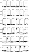
Immunoblot analysis of cross-linked E. coli TatC proteins containing cysteine substitutions in each transmembrane helix. Membrane samples were prepared from the ΔtatABCD ΔtatE pcnB strain, DADE-P, coexpressing single cysteine-substituted TatC variants together with TatA and TatB from pUNITATCC4. Samples (100 μg membrane protein) were subjected to either oxidizing conditions (O; shown in the leftmost lane for each sample) or reducing conditions (R; middle lane for each sample) or incubated with buffer alone (C; rightmost lane for each sample) each at room temperature as described in Materials and Methods. Samples (10 μg membrane protein) were resolved by SDS-PAGE (12.5% acrylamide), and TatC proteins were visualized by immunoblotting with anti-TatC antisera. The position of the cysteine substitution is shown above each panel. The positions of TatC monomer (*) and dimer (**) forms are indicated. (A) Substitutions in transmembrane helix one; (B) substitutions in transmembrane helix two; (C) substitutions in transmembrane helix three; (D) substitutions in transmembrane helix four; (E) substitutions in transmembrane helix five; (F) substitutions in transmembrane helix six.
FIG. 7.
FIG. 7.
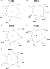
Helical wheel projections of portions of TatC transmembrane helix two (TMH2) to transmembrane helix six (TMH6) that were analyzed by disulfide cross-linking. The percentage next to each residue is the percentage of cross-linked dimer (expressed as a percentage of monomer plus dimer) formed after oxidation at room temperature for 1 h.
FIG. 8.
FIG. 8.
Immunoblot analysis of cross-linked E. coli TatC proteins containing further cysteine substitutions in transmembrane helix one. Membrane samples were prepared from the ΔtatABCD ΔtatE pcnB strain, DADE-P, coexpressing single cysteine-substituted TatC variants together with TatA and TatB from pUNITATCC4. Samples (100 μg membrane protein) were subjected to either oxidizing conditions (O; shown in the leftmost lane for each sample) or reducing conditions (R; middle lane for each sample) or incubated with buffer alone (C; rightmost lane for each sample), each at room temperature. Samples (10 μg membrane protein) were resolved by SDS-PAGE (12.5% acrylamide), and TatC proteins were visualized by immunoblotting with anti-TatC antisera. The position of the cysteine substitution is shown above each panel. The positions of TatC monomer (*) and dimer (**) forms are indicated.
FIG. 9.
FIG. 9.
Helical wheel projection for the entire TatC transmembrane helix one. For those residues that show cross-linking, the percentage of cross-linked dimer (expressed as a percentage of monomer plus dimer) formed after oxidation at room temperature for 1 h is indicated. Residues A26 and Y36 are shown boxed.

References

    1. Alami, M., I. Luke, S. Deitermann, G. Eisner, H. G. Koch, J. Brunner, and M. Müller. 2003. Differential interactions between a twin-arginine signal peptide and its translocase in Escherichia coli. Mol. Cell 12:937-946. - PubMed
    1. Allen, S. C., C. M. Barrett, N. Ray, and C. Robinson. 2002. Essential cytoplasmic domains in the Escherichia coli TatC protein. J. Biol. Chem. 277:10362-10366. - PubMed
    1. Banerjee, S., and D. G. Fraenkel. 1972. Glucose-6-phosphate dehydrogenase from Escherichia coli and from a “high-level” mutant. J. Bacteriol. 110:155-160. - PMC - PubMed
    1. Behrendt, J., K. Standar, U. Lindenstrauss, and T. Brüser. 2004. Topological studies on the twin-arginine translocase component TatC. FEMS Microbiol. Lett. 234:303-308. - PubMed
    1. Berks, B. C., T. Palmer, and F. Sargent. 2003. The Tat protein translocation pathway and its role in microbial physiology. Adv. Microb. Physiol. 47:187-254. - PubMed

Publication types

MeSH terms

LinkOut - more resources