

Sugar radicals formed by photoexcitation of guanine cation radical in oligonucleotides
- PMID: 17547448
- PMCID: PMC2526165
- DOI: 10.1021/jp071107c
Sugar radicals formed by photoexcitation of guanine cation radical in oligonucleotides
Abstract
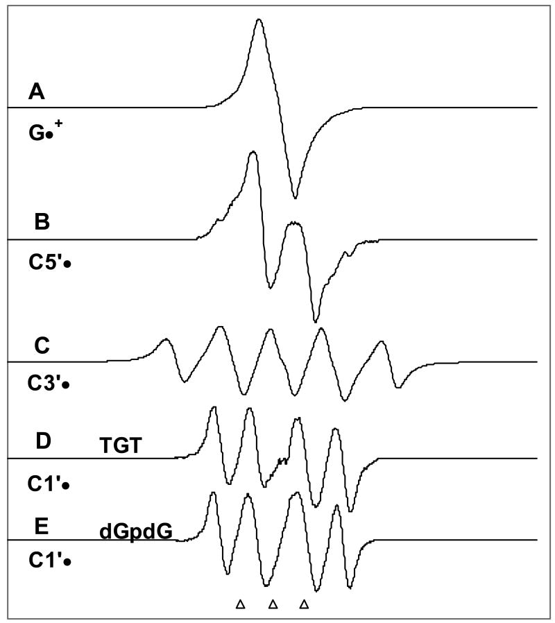
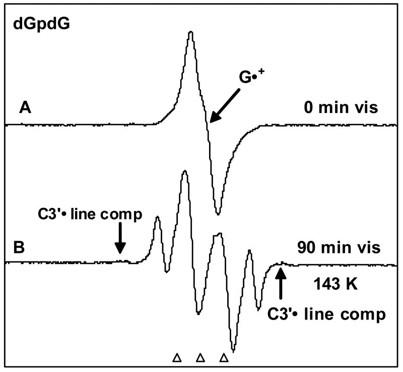
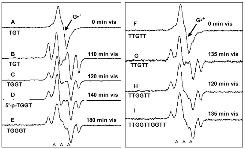
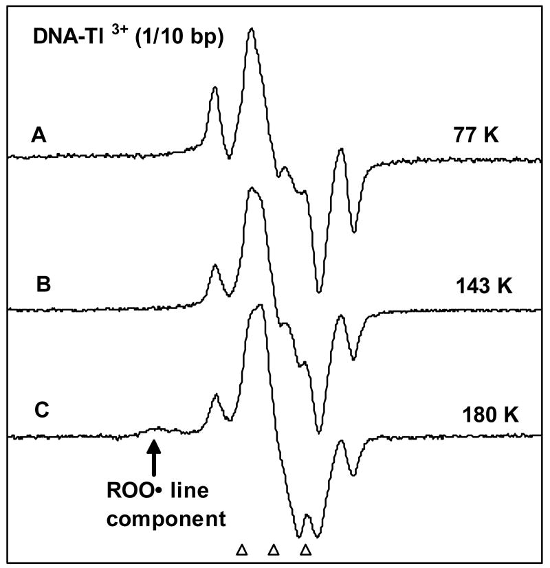
This work presents evidence that photoexcitation of guanine cation radical (G+*) in dGpdG and DNA-oligonucleotides TGT, TGGT, TGGGT, TTGTT, TTGGTT, TTGGTTGGTT, AGA, and AGGGA in frozen glassy aqueous solutions at low temperatures leads to hole transfer to the sugar phosphate backbone and results in high yields of deoxyribose radicals. In this series of oligonucleotides, we find that G+* on photoexcitation at 143 K leads to the formation of predominantly C5'* and C1'* with small amounts of C3'*. Photoconversion yields of G+* to sugar radicals in oligonucleotides decreased as the overall chain length increased. However, for high molecular weight dsDNA (salmon testes) in frozen aqueous solutions, substantial conversion of G+* to C1'* (only) sugar radical is still found (ca. 50%). Within the cohort of sugar radicals formed, we find a relative increase in the formation of C1'* with length of the oligonucleotide, along with decreases in C3'* and C5'*. For dsDNA in frozen solutions, only the formation of C1'* is found via photoexcitation of G+*, without a significant temperature dependence (77-180 K). Long wavelength visible light (>540 nm) is observed to be about as effective as light under 540 nm for photoconversion of G+* to sugar radicals for short oligonucleotides but gradually loses effectiveness with chain length. This wavelength dependence is attributed to base-to-base hole transfer for wavelengths >540 nm. Base-to-sugar hole transfer is suggested to dominate under 540 nm. These results may have implications for a number of investigations of hole transfer through DNA in which DNA holes are subjected to continuous visible illumination.
Figures
References
-
-
von Sonntag C. The Chemical Basis of Radiation Biology. Taylor & Francis; London: 1987. pp. 221–294.Lett JT. Prog Nuceic Acid Res Mol Biol. 1990;39:305–352.Becker D, Sevilla MD. Adv Radiat Biol. 1993;17:121–180.Becker D, Sevilla MD. Royal Society of Chemistry Specialist Periodical Report. In: Gilbert BC, Davies MJ, Murphy DM, editors. Electron Spin Resonance. Vol. 16. 1998. pp. 79–114.Weiland B, Hüttermann J. Int J Radiat Biol. 1998;74:341–358. and references therein. Weiland B, Hüttermann J. Int J Radiat Biol. 1999;75:1169–1175. and references therein. Ward JF. Cold Spring Harb Symp Quant Biol. 2000;65:377–382. and references therein. Sevilla MD, Becker D. Royal Society of Chemistry Specialist Periodical Report. In: Gilbert BC, Davies MJ, Murphy DM, editors. Electron Spin Resonance. Vol. 19. 2004. pp. 243–278.Bernhard WA, Close DM. In: Charged Particle and Photon Interactions with Matter Chemical, Physicochemical and Biological Consequences with Applications. Mozumdar A, Hatano Y, editors. Marcel Dekkar, Inc.; New York, Basel: 2004. pp. 431–470.von Sonntag C. Free-radical-induced DNA Damage and Its Repair. Springer-Verlag; Berlin, Heidelberg: 2006. pp. 335–447.Becker D, Adhikary A, Sevilla MD. In: Charge Migration in DNA Physics, Chemistry and Biology Perspectives. Chakraborty T, editor. Springer-Verlag; Berlin, Heidelberg: 2007. In Press.
-
-
- Becker D, Bryant-Friedrich A, Trzasko C, Sevilla MD. Radiat Res. 2003;160:174–185. - PubMed
-
- Boudaïffa B, Cloutier P, Hunting D, Huels MA, Sanche L. Science. 2000;287:1658–1660. - PubMed
-
- Sanche L. Mass Spectrometry Reviews. 2002;21:349–369. - PubMed
-
- Li X, Sevilla MD, Sanche L. J Am Chem Soc. 2003;125:13668–13669. - PubMed
Publication types
MeSH terms
Substances
Grants and funding
LinkOut - more resources
Full Text Sources
Miscellaneous
