. 2007 Aug;27(16):5686-98.
doi: 10.1128/MCB.01109-06. Epub 2007 Jun 4.

Nerve growth factor stimulates the concentration of TrkA within lipid rafts and extracellular signal-regulated kinase activation through c-Cbl-associated protein

Affiliations

Nerve growth factor stimulates the concentration of TrkA within lipid rafts and extracellular signal-regulated kinase activation through c-Cbl-associated protein

Allison S Limpert et al. Mol Cell Biol. 2007 Aug.

Abstract

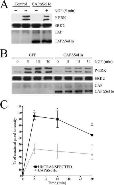
Nerve growth factor (NGF) acts through its receptor, TrkA, to elicit the neuronal differentiation of PC12 cells through the action of extracellular signal-regulated kinase 1 (ERK1) and ERK2. Upon NGF binding, TrkA translocates and concentrates in cholesterol-rich membrane microdomains or lipid rafts, facilitating formation of receptor-associated signaling complexes, activation of downstream signaling pathways, and internalization into endosomes. We have investigated the mechanisms responsible for the localization of TrkA within lipid rafts and its ability to activate ERK1 and ERK2. We report that NGF treatment results in the translocation of activated forms of TrkA to lipid rafts, and this localization is important for efficient activation of the ERKs. TrkA is recruited and retained within lipid rafts through its association with flotillin, an intrinsic constituent of these membrane microdomains, via the adapter protein, c-Cbl associated protein (CAP). Mutant forms of CAP that lack protein interaction domains block TrkA localization to lipid rafts and attenuate ERK activation. Importantly, suppression of endogenous CAP expression inhibited NGF-stimulated neurite outgrowth from primary dorsal root ganglion neurons. These data provide a mechanism for the lipid raft localization of TrkA and establish the importance of the CAP adaptor protein for NGF activation of the ERKs and neuronal differentiation.


Figures

FIG. 1.
FIG. 1.
MβCD treatment decreases ERK and TrkA activation. PC12 cells were treated with 10 mM MβCD, which depletes cholesterol from cell membranes, for 15 min prior to NGF stimulation (100 ng/ml) for the indicated times. (A) Western blots were probed for phospho-ERK1/2 (P-ERK). The blots were stripped and subsequently reprobed for ERK2 to control for protein loading. (B) The data are represented as mean pixel intensity normalized to the percentage of maximal phospho-ERK response. The data shown are an average (± standard error) of three independent experiments (*, P < 0.05). (C) Western blots were probed for phospho-TrkA (P-TrkA) and then stripped and reprobed for TrkA to control for protein loading.
FIG. 2.
FIG. 2.
TrkA, SOS, and CAP concentrate in the flotillin-containing membrane fraction following NGF treatment. PC12 cell lysates were fractionated using discontinuous sucrose gradients. Fraction 1 represents the top of the gradient. PC12 cells were left untreated or stimulated with NGF for 5 min and lysed, and the lysates were resolved on sucrose gradients. Gradients were fractionated, and equal amounts of protein from corresponding fractions were separated by SDS-PAGE. Western blots were probed for flotillin, TrkA, phospho-TrkA (P-TrkA), CAP, SOS, FRS2, and TfR1. Flotillin, which is a marker for lipid raft membranes, was used as a marker for the lipid raft fraction, while TfR1 was used to mark the heavy membrane fractions. These results are representative of three independent experiments.
FIG. 3.
FIG. 3.
Following NGF stimulation, CAP becomes associated with proteins important in lipid rafts and in MAPK activation. PC12 cells were stimulated with NGF for the indicated times, and cell lysates were prepared. (A) Flotillin was immunoprecipitated (IP) from cell lysates and the resulting Western blot (IB) was probed for CAP and flotillin. These data are representative of three independent experiments. (B) TrkA-CAP interactions were investigated by immunoprecipitation of TrkA followed by Western blotting with antibodies to CAP. The blots were stripped and reprobed for TrkA. (C) PC12 cells expressing Myc-tagged APS or GFP were stimulated with NGF. The tagged proteins were then immunoprecipitated from cell lysates with anti-Myc antibodies and then analyzed by Western blotting. The blots were probed with anti-CAP antibodies and then stripped and reprobed with anti-Myc antibodies to control for protein expression levels and immunoprecipitation efficiency. (D) SOS was immunoprecipitated from cell lysates. Immunoprecipitates were resolved by SDS-PAGE. Western blots were probed for CAP. The blots were then stripped and reprobed with anti-SOS antibodies to control for immunoprecipitation efficiency. (E) PC12 cells were stimulated with NGF for the indicated times and then lysed. Cbl was immunoprecipitated from cell lysates. Immunoprecipitates were analyzed using Western blotting and probed for CAP and then reprobed with anti-Cbl antibodies.
FIG. 4.
FIG. 4.
Both TrkA and CAP colocalize with flotillin on the cell surface following NGF stimulation. (A) PC12 cells were treated with NGF for 0, 5, and 10 min. Cells were fixed and stained for CAP (red) and flotillin (green). Cells were then examined by confocal microscopy. (B) PC12 cells were treated with NGF for 0, 5, and 10 min. Cells were fixed and stained for TrkA (red) and flotillin (green). Cells were examined by confocal microscopy. Arrows indicate areas of colocalization. Scale bar, 5 μm.
FIG. 5.
FIG. 5.
CAPΔSoHo but not full-length CAP inhibits the concentration of TrkA, SOS, and CAP in the lipid raft membrane fraction. PC12 cells expressing FLAG-CAP (A) or FLAG-CAPΔSoHo (B) were left untreated or stimulated with NGF for 5 min. The lipid raft membrane fractions were then isolated by sucrose gradient fractionation. Fraction 1 represents the top of the gradient. Lipid raft fractions were analyzed using Western blotting with antiflotillin, anti-Trk, anti-CAP, anti-SOS, anti-FLAG, anti-FRS2, and anti-TfR1 antibodies. The CAP antibody recognizes both the endogenous and the transfected CAP proteins. In panel A, FLAG antibodies are used to detect the expressed full-length CAP protein which is the same size as the endogenous protein. In panel B, the upper CAP band represents endogenous CAP levels, while the lower band shows the CAPΔSoHo-transfected protein. These results are representative of three independent experiments.
FIG. 6.
FIG. 6.
CAPΔSoHo inhibits colocalization of TrkA and flotillin following NGF stimulation. PC12 cells were transfected with FLAG-CAP or FLAG-CAPΔSoHo; 48 h later they were either left untreated or stimulated with NGF for 5 min. Cells were fixed and stained for TrkA (red), flotillin (green), or FLAG (blue) and examined by confocal microscopy. Arrows indicate points of colocalization. Open arrowheads denote transfected cells. Scale bar, 5 μm.
FIG. 7.
FIG. 7.
CAPΔSoHo inhibits ERK activation. (A) Purified FLAG-CAPΔSoHo protein was introduced into PC12 cells using the Chariot protein transduction reagent. Cells were incubated with or without NGF for 5 min and then lysed. Cell lysates were probed for phospho-ERK (P-ERK) to examine differences in ERK activation and for ERK2 to control for protein loading. Anti-CAP was used to probe for the incorporation of CAPΔSoHo into the cells. This antibody recognizes both endogenous (upper band) and mutant CAP proteins (lower band) which differ in their molecular weights. (B) PC12 cells were transfected with FLAG-CAPΔSoHo or GFP constructs using Lipofectamine 2000. Cells were stimulated with NGF for indicated times. Western blots were probed for phospho-ERK, ERK 2, and CAP. (C) Graphical analysis of data from three independent experiments. Data are represented as the average (± standard error) mean pixel intensity normalized to the percentage of maximal activation (*, P < 0.05).
FIG. 8.
FIG. 8.
CAP-mediated protein interactions are necessary for NGF but not EGF stimulation of the ERKs. (A) PC12 cells were transfected with constructs expressing full-length FLAG-CAP, FLAG-CAPΔSoHo, or GFP. Cells were incubated with or without NGF for 5 min. Cells were lysed, and the lysates were examined by Western blotting analysis. Western blots were probed for phospho-ERK (P-ERK), ERK2, and FLAG. (B) PC12 cells were transfected with constructs expressing FLAG-CAPΔSH3 or GFP. Cells were either treated with NGF for 5 min or left untreated. Cells were lysed and analyzed for protein concentration. Equal amounts of protein from each sample were separated by SDS-PAGE and analyzed by Western blotting. Blots were probed for phospho-ERK, ERK2, and FLAG. (C) PC12 cells were transfected with constructs expressing FLAG-CAP, FLAG-CAPΔSoHo, or GFP and then treated with EGF for 5 min or left untreated. Cells were lysed, and lysates were analyzed for protein concentration. Equal amounts of protein from each sample were separated by SDS-PAGE and analyzed by Western blotting. Blots were probed for phospho-ERK, ERK 2, and FLAG.
FIG. 9.
FIG. 9.
CAPΔSoHo inhibits TrkA phosphorylation and internalization. (A) PC12 cells were transfected with GFP, FLAG-CAP, or FLAG-CAPΔSoHo. Cells were left untreated or stimulated with NGF for 5 min. Cells were lysed, and lysates were probed with antibodies to phospho-Trk (P-Trk), TrkA, and FLAG. (B) PC12 cells were transfected with FLAG-CAPΔSoHo- or GFP-expressing constructs. Cells were left untreated or treated with NGF for 5, 15, or 30 min. Cells were then lysed, and lysates were analyzed for protein concentration using the Bradford protein assay. Equal amounts of protein from each sample were resolved using SDS-PAGE. Western blots were probed for phospho-TrkA, TrkA, and CAP. These results are representative of three independent experiments. Cells were transfected with FLAG-CAP (C) or FLAG-CAPΔSoHo (D) and were treated with NGF for 15 min or left untreated. Cells were incubated with biotin to label cell surface proteins. Cell lysates were then incubated with neutravidin beads to specifically isolate the biotin-labeled proteins. Western blotting was performed, and blots were probed for TrkA. Lysates were probed with anti-FLAG antibodies to detect expression of the transfected proteins.
FIG. 10.
FIG. 10.
CAPΔSoHo affects the activation of the small G proteins Ras and Rap1. PC12 cells were transfected with constructs expressing FLAG-CAP, FLAG-CAPΔSoHo, or GFP. Cells were left untreated or stimulated with NGF for 5 min. Lysates were incubated with agarose-immobilized Raf-RBD protein to specifically isolate activated forms of Ras and Rap1. The Raf-RBD associated proteins were analyzed using Western blots probed for Ras (A) and Rap1 (B). Lysates were probed for total Ras and Rap1 protein and for expression of the FLAG-tagged proteins. (C) Quantitative analysis of Rap1 activation following 5 min of NGF stimulation from three independent experiments. Data are represented as the average (± standard error) mean pixel intensity derived from Western blots of Rap1 bands normalized to the vector control.
FIG. 11.
FIG. 11.
CAP siRNA reduces neurite outgrowth in cultured primary DRG neurons. DRG neurons were cultured from E14 mouse embryos and transfected using Amaxa nucleofection with FAM-labeled control siRNA (A) or FAM-labeled CAP siRNA (B) and incubated in NGF-containing medium. (C and D) Cells transfected with FAM-labeled siRNA were counted and scored for longest individual neurite length (short neurites, <250 μm; long neurites, >250 μm). Statistical analysis was done to determine the percentage of total transfected neurons extending short neurites or long neurites. In >200 cells the ranges were as follows: control siRNA, 21.5% ± 5.5% short and 78% ± 6% long; CAP siRNA, 66.5% ± 1.5% short and 33.5% ± 1.5% long. (E) Lysates from DRG neurons transfected with control and CAP siRNAs were probed for CAP protein expression by Western blotting.

Similar articles

Cited by

References

    1. Ahn, M. Y., K. D. Katsanakis, F. Bheda, and T. S. Pillay. 2004. Primary and essential role of the adaptor protein APS for recruitment of both c-Cbl and its associated protein CAP in insulin signaling. J. Biol. Chem. 279:21526-21532. - PubMed
    1. Anderson, R. G. 1998. The caveolae membrane system. Annu. Rev. Biochem. 67:199-225. - PubMed
    1. Arispe, N., and M. Doh. 2002. Plasma membrane cholesterol controls the cytotoxicity of Alzheimer's disease AbetaP (1-40) and (1-42) peptides. FASEB J. 16:1526-1536. - PubMed
    1. Baumann, C. A., V. Ribon, M. Kanzaki, D. C. Thurmond, S. Mora, S. Shigematsu, P. E. Bickel, J. E. Pessin, and A. R. Saltiel. 2000. CAP defines a second signalling pathway required for insulin-stimulated glucose transport. Nature 407:202-207. - PubMed
    1. Bickel, P. E., P. E. Scherer, J. E. Schnitzer, P. Oh, M. P. Lisanti, and H. F. Lodish. 1997. Flotillin and epidermal surface antigen define a new family of caveolae-associated integral membrane proteins. J. Biol. Chem. 272:13793-13802. - PubMed

Publication types

MeSH terms

LinkOut - more resources