. 2007 Aug;27(16):5664-72.
doi: 10.1128/MCB.00602-07. Epub 2007 Jun 4.

Linear distance from the locus control region determines epsilon-globin transcriptional activity

Affiliations

Linear distance from the locus control region determines epsilon-globin transcriptional activity

Motoshi Shimotsuma et al. Mol Cell Biol. 2007 Aug.

Abstract

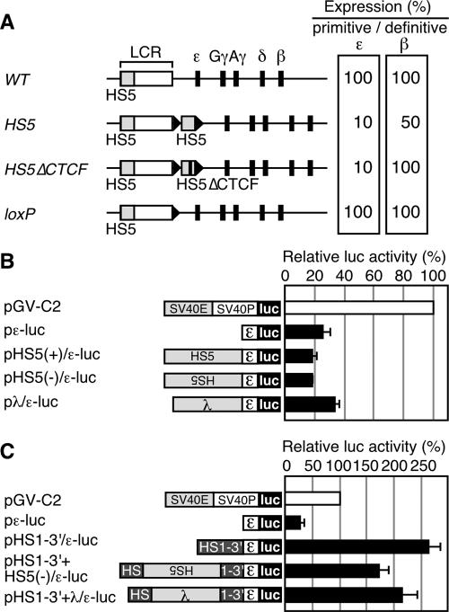
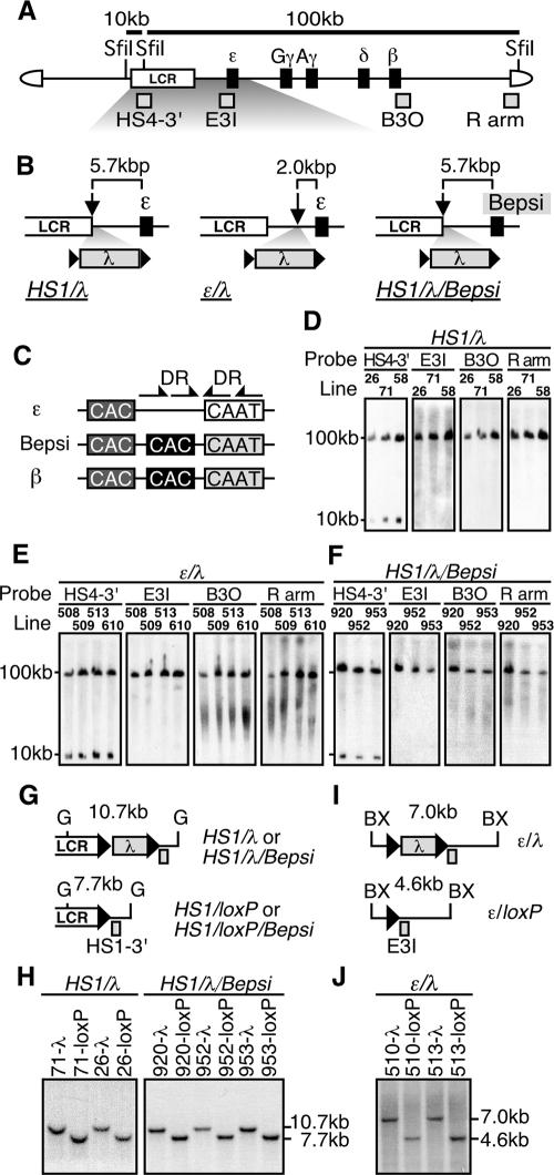
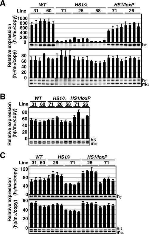
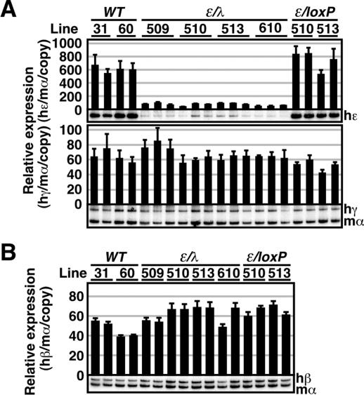
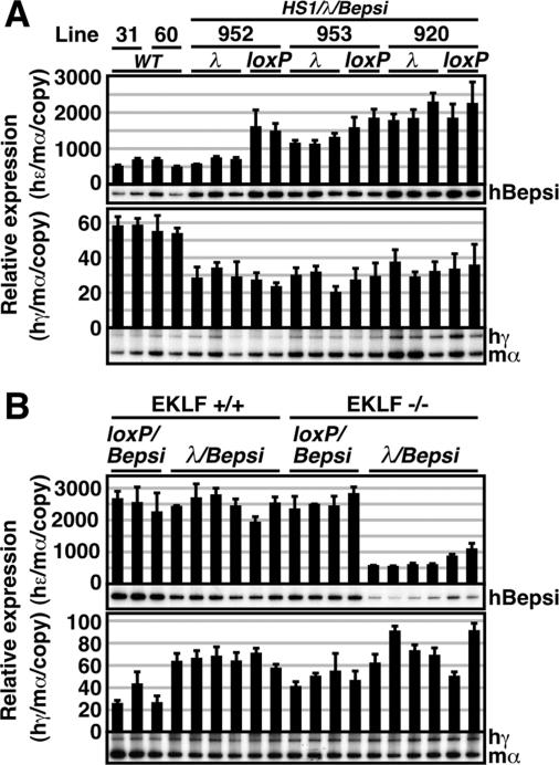
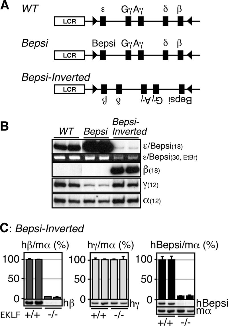
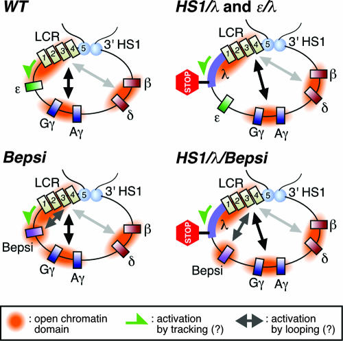
Enhancer elements modulate promoter activity over vast chromosomal distances, and mechanisms that ensure restrictive interactions between promoters and enhancers are critical for proper control of gene expression. The human beta-globin locus control region (LCR) activates expression of five genes in erythroid cells, including the proximal embryonic epsilon- and the distal adult beta-globin genes. To test for possible distance sensitivity of the genes to the LCR, we extended the distance between the LCR and genes by 2.3 kbp within the context of a yeast artificial chromosome, followed by the generation of transgenic mice (TgM). In these TgM lines, epsilon-globin gene expression decreased by 90%, while the more distantly located gamma- or beta-globin genes were not affected. Remarkably, introduction of a consensus EKLF binding site into the epsilon-globin promoter rendered its expression distance insensitive; when tested in an EKLF-null genetic background, expression of the mutant epsilon-globin gene was severely compromised. Thus, the epsilon-globin gene differs in its distance sensitivity to the LCR from the other beta-like globin genes, which is, at least in part, determined by the transcription factor EKLF.


Figures

FIG. 1.
FIG. 1.
(A) Relative expression of the primitive ɛ-globin or the definitive β-globin genes in YAC TgM. The values in the WT TgM are set at 100. Results from the WT (28), HS5 (containing a 2.6 kbp HS5 fragment inserted 3′ to HS1) (30), HS5▵CTCF (containing the HS5 fragment lacking CTCF binding sites), and loxP (same as the WT, except that a ∼40-bp loxP footprint (arrowhead) is retained) TgM are shown. (B) Analysis of potential silencer activity associated with HS5 in K562 cells. Structures of the reporter constructs used in the transfection assays are shown on the left. The human ɛ-globin promoter region (174 to +38) was subcloned into the luciferase reporter plasmid to generate pɛ-luc. The HS5 DNA fragment (2.6 kbp; same as the one used in the HS5 TgM) was inserted upstream of the ɛ-globin promoter in sense or antisense orientations to generate pHS5±/ɛ-luc. A 2.3-kbp fragment from λ DNA was inserted upstream of the ɛ-globin gene promoter to generate pλ/ɛ-luc. The reference reporter construct pGV-C2 is driven by the simian virus 40 (SV40) enhancer (E) and promoter (P). K562 cells were transfected with the luciferase reporter plasmids as well as the control pCMV-β-Gal. Luciferase activities were normalized to β-Gal activities to control for transfection efficiencies. The relative luciferase activity is expressed relative to that in the pGV-C2 (set at 100). Each value of luciferase activity represents the mean ± SD for at least three independent experiments. (C) HS5 DNA insertion does not attenuate the enhancer activity localized within the 3′ region of HS1 (HS1-3′). The HS1-3′ DNA fragment (∼1 kbp) was inserted upstream of the ɛ-globin promoter, within the context of pɛ-luc, to generate pHS1-3′/ɛ-luc. The HS5 (2.6 kbp) or the λ (2.3 kbp) DNA fragment was inserted in the HS1-3′ region (at the same position as the HS5 fragment was inserted in the HS5 TgM) of pHS1-3′/ɛ-luc to generate pHS1-3′+HS5(−)/ɛ-luc and pHS1-3′+λ/ɛ-luc, respectively. These reporter plasmids were transfected into K562 cells and assayed as described above.
FIG. 2.
FIG. 2.
(A) Structure of the 150-kbp human β-globin YAC. The positions of the β-like globin genes (solid rectangles) are shown relative to the LCR. SfiI restriction enzyme sites are located 5′ to HS5, between HS4 and HS3, and in the right arm of the YAC. Probes (gray rectangles) used for long-range fragment analyses shown in panels D, E, and F, and expected restriction enzyme fragments with their sizes are shown. (B) Schematic representation of the mutant β-globin loci. The λ DNA fragment (2.3 kbp) was floxed (solid triangles) and inserted 3′ to HS1 (HS1/λ and HS1/λ/Bepsi) or introduced between HS1 and the ɛ-globin gene (ɛ/λ). In the HS1/λ/Bepsi construct, the promoter sequence of the ɛ-globin gene was mutated to mimic that of the β-globin gene, in addition to the λ insertion. (C) The positions of the distal (dark gray) or proximal (solid) CAC boxes are shown relative to the CAAT box in the human ɛ-globin, β-globin, and Bepsi gene promoters. Arrows indicate direct repeat (DR) elements, present only in the ɛ-globin promoter (29). (D, E, and F) Long-range structural analyses of the human β-globin YAC in TgM. The whole β-globin locus is contained within two SfiI fragments (10 and 100 kbp) as described for panel A above. DNAs from thymus cells were digested with SfiI in agarose plugs, separated by pulsed-field gel electrophoresis, and hybridized separately to probes (shown in panel A). A schematic representation of the transgene loci around the inserted λ DNA (G and I) and Southern blot analyses for confirming Cre-loxP-mediated in vivo recombination (H and J) are shown. Cre-loxP recombination removes the 2.3-kbp λ DNA insert from each mutated locus, which creates a 7.7-kbp BglII (G, HS1/loxP, and HS1/loxP/Bepsi) or a 4.6-kbp BstXI (BX, ɛ/loxP) fragment in each locus. Tail DNAs from each mutant and loxP footprint TgM lines were digested with BglII (HS1/λ and HS1/λ/Bepsi series) or BstXI (ɛ/λ series), separated on agarose gels, and hybridized to probes HS1-3′ or E3I. Arrowheads, loxP sequences.
FIG. 3.
FIG. 3.
Expression of the human β-like globin genes in HS1/λ TgM. (A) Total RNA was prepared from the yolk sacs of more than two embryos (9.5 dpc) derived from the intercross of male transgenic and female WT animals. Samples were collected from two independent litters of each mutant line. Expression of human ɛ (hɛ)- and human γ (hγ)-globin compared to endogenous mouse α (mα)-globin genes was separately analyzed by semiquantitative RT-PCR. The signals for hɛ-globin at 18 cycles and hγ/mα-globin at 12 cycles were quantified by PhosphorImager, and the ratios of hɛ/mα (top) and hγ/mα (bottom) were calculated (the mα signal at 12 cycles was set at 100%, and the values are normalized by transgene copy numbers). (B) Total RNA was prepared from the spleens of 1-month-old anemic mice. Samples were collected from two individuals from each line of TgM. Expression of human β (hβ)-globin compared to endogenous mα-globin genes was analyzed by semiquantitative RT-PCR. The signal for hβ/mα-globin at 12 cycles was quantified, and the ratio of hβ/mα was calculated (the mα signal at 12 cycles was set at 100%). (C) Total RNA was prepared from the fetal livers (14.5 dpc) derived from the intercross of male transgenic and female WT animals. Samples were collected from two independent litters of each mutant line. Expression of hγ- and hβ-globin compared to endogenous mα-globin genes was analyzed separately by semiquantitative RT-PCR. The signals for hγ-globin at 18 cycles and hβ/mα-globin at 12 cycles were quantified, and the ratios of hγ/mα (top) and hβ/mα (bottom) were calculated (the mα signal at 12 cycles was set at 100%). The averages ± SD from at least three independent experiments was calculated and are graphically depicted. Representative results are shown below each panel.
FIG. 4.
FIG. 4.
Expression of the human β-like globin genes in ɛ/λ TgM. (A) Total RNA was prepared from the yolk sacs of more than two embryos (9.5 dpc) from two independent litters of each mutant line. Expression of human ɛ (hɛ)- and human γ (hγ)-globin compared to endogenous mouse α (mα)-globin genes was separately analyzed by semiquantitative RT-PCR. (B) Total RNA was prepared from the spleens of 1-month-old anemic mice. Samples were collected from two individuals from each line of TgM. Expression of human β (hβ)-globin compared to endogenous mα-globin genes was analyzed by semiquantitative RT-PCR as described in the legend to Fig. 3.
FIG. 5.
FIG. 5.
Expression of the human β-like globin genes in the HS1/λ/Bepsi TgM. (A) Total RNA was prepared from the yolk sacs of more than two embryos (9.5 dpc) from two independent litters of each mutant line. Expression of human Bepsi (hBepsi)- and human γ (hγ)-globin compared to endogenous mouse α (mα)-globin genes was separately analyzed by semiquantitative RT-PCR. λ, HS1/λ/Bepsi; loxP, HS1/loxP/Bepsi. (B) HS1/λ/Bepsi TgM (line 920) was bred with the EKLF-null mouse to analyze β-like globin gene expression in the presence (EKLF+/+) or absence (EKLF−/−) of endogenous EKLF activity. Total RNA was prepared from the yolk sacs of more than two embryos (9.5 dpc) from two independent litters of the mutant line. Expression of hBepsi- and hγ-globin compared to endogenous mα-globin genes was separately analyzed by semiquantitative RT-PCR. See the legend to Fig. 3 for details.
FIG. 6.
FIG. 6.
Expression of the human β-like globin genes in Bepsi-Inverted TgM. (A) Schematic representation of the Bepsi-Inverted locus. A pair of the loxP sites (arrowheads) was introduced into the human β-globin YAC in inverted orientation to generate a pseudo-WT locus (28). Then, the ɛ-globin promoter in the WT locus was mutated to generate the Bepsi locus. Following the establishment of TgM lines with the Bepsi YAC, the genes were inverted by mating with Cre-expressing TgM (Bepsi-Inverted). (B) Semiquantitative RT-PCR analysis of β-like globin genes expression in the yolk sacs of the mutant TgM. Amplified Bepsi transcript was also stained with the ethidium bromide (EtBr). PCR cycle numbers used are shown in parentheses. (C) Semiquantitative RT-PCR analysis of the β-like globin genes expression in the yolk sacs of the EKLF+/+ and EKLF−/− mice. The expression levels of the β-like globin genes in the EKLF+/+ background was set at 100. The average and SD and shown graphically for each animal. Representative RT-PCR results for human β (hβ)-, γ (hγ)-, Bepsi (hBepsi)-, and mouse α (mα)-globin in different EKLF mutant backgrounds (+/+ and −/−) are shown below each panel. See the legend to Fig. 3 for details.
FIG. 7.
FIG. 7.
Two modes of LCR-promoter communication (tracking and looping) may control developmental stage- and/or gene-specific activation within the human β-globin locus. The 5′ and 3′ HSs (light blue ovals and numbered rectangles) may be involved in forming higher order chromatin architecture within the β-globin locus, one part of which is a powerful enhancer (the LCR). In primitive erythroid cells, the ɛ-globin gene may be activated by the LCR via tracking mechanism (WT, green arrow) and therefore displays sensitivity to the linear distance from the LCR (HS1/λ and ɛ/λ). Introduction of the CAC (presumptive EKLF binding) site into and/or disruption of the direct repeat sites from the ɛ-globin promoter (Bepsi) may allow it to be activated by means of a looping mechanism (arrow). Thus, the Bepsi promoter is highly active even after the λ DNA is inserted (HS1/λ/Bepsi). Predicted open chromatin domains are indicated.

Similar articles

Cited by

References

    1. Bell, A. C., A. G. West, and G. Felsenfeld. 1999. The protein CTCF is required for the enhancer blocking activity of vertebrate insulators. Cell 98:387-396. - PubMed
    1. Bulger, M., T. Sawado, D. Schubeler, and M. Groudine. 2002. ChIPs of the beta-globin locus: unraveling gene regulation within an active domain. Curr. Opin. Genet. Dev. 12:170-177. - PubMed
    1. Carter, D., L. Chakalova, C. S. Osborne, Y. F. Dai, and P. Fraser. 2002. Long-range chromatin regulatory interactions in vivo. Nat. Genet. 32:623-626. - PubMed
    1. Choi, O. R., and J. D. Engel. 1988. Developmental regulation of beta-globin gene switching. Cell 55:17-26. - PubMed
    1. Dean, A. 2006. On a chromosome far, far away: LCRs and gene expression. Trends Genet. 22:38-45. - PubMed

Publication types

LinkOut - more resources