. 2007 Aug;27(15):5445-55.
doi: 10.1128/MCB.02127-06. Epub 2007 Jun 4.

Prion protein repeat expansion results in increased aggregation and reveals phenotypic variability

Affiliations

Prion protein repeat expansion results in increased aggregation and reveals phenotypic variability

Elizabeth M H Tank et al. Mol Cell Biol. 2007 Aug.

Abstract

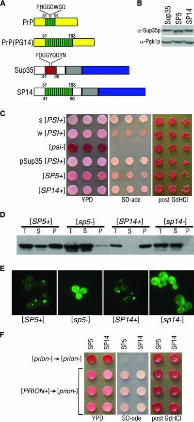
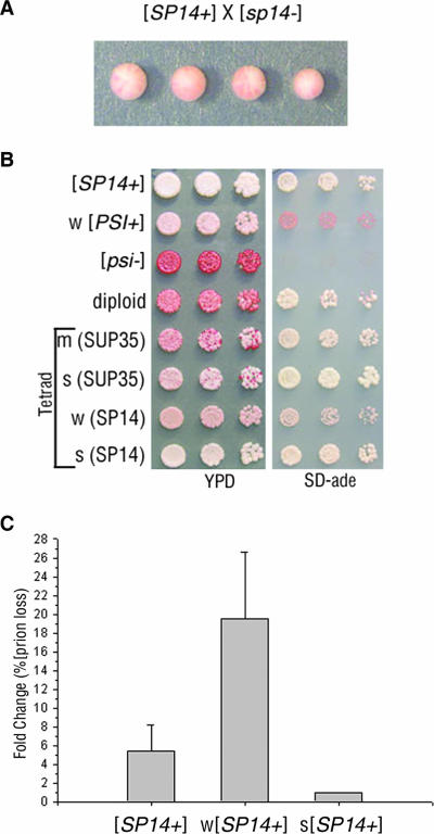
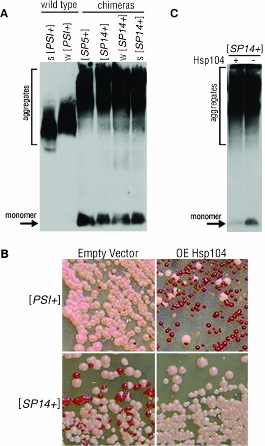
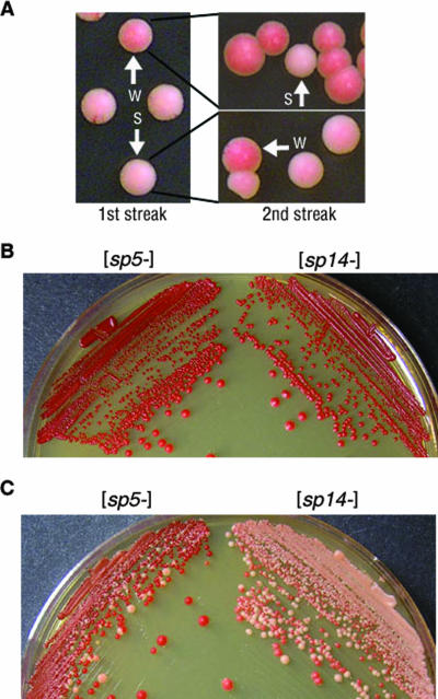
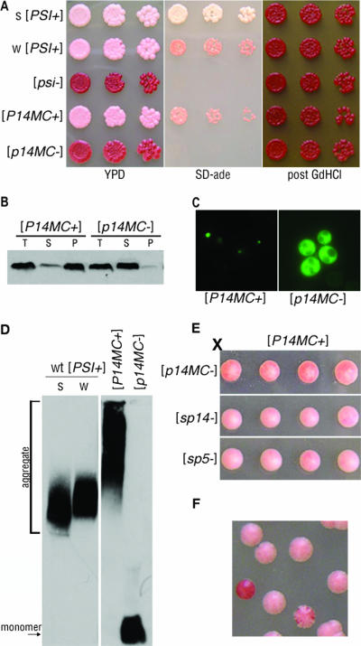
Mammalian prion diseases are fatal neurodegenerative disorders dependent on the prion protein PrP. Expansion of the oligopeptide repeats (ORE) found in PrP is associated with inherited prion diseases. Patients with ORE frequently harbor PrP aggregates, but other factors may contribute to pathology, as they often present with unexplained phenotypic variability. We created chimeric yeast-mammalian prion proteins to examine the influence of the PrP ORE on prion properties in yeast. Remarkably, all chimeric proteins maintained prion characteristics. The largest repeat expansion chimera displayed a higher propensity to maintain a self-propagating aggregated state. Strikingly, the repeat expansion conferred increased conformational flexibility, as observed by enhanced phenotypic variation. Furthermore, the repeat expansion chimera displayed an increased rate of prion conversion, but only in the presence of another aggregate, the [RNQ+] prion. We suggest that the PrP ORE increases the conformational flexibility of the prion protein, thereby enhancing the formation of multiple distinct aggregate structures and allowing more frequent prion conversion. Both of these characteristics may contribute to the phenotypic variability associated with PrP repeat expansion diseases.


Figures

FIG. 1.
FIG. 1.
Chimeric Sup35-PrP proteins demonstrate yeast prion properties. (A) Schematic depiction of Sup35p and PrP highlighting the consensus sequence of each ORD. PrP(PG14) contains nine additional repeats. The chimeric Sup35-PrP protein (SP14) is also shown. Numbers below the diagrams represent Sup35p amino acids, and those above correspond to PrP. (B) Lysates from cells containing the Sup35p, SP5, and SP14 proteins were analyzed by sodium dodecyl sulfate-polyacrylamide gel electrophoresis and Western blotting to determine relative chimeric Sup35 protein levels. The blot was reprobed with antibodies to Pgk1p to confirm equal protein loading. (C) Strong (s) [PSI+] strain variant, weak (w) [PSI+] strain variant, [psi], plasmid-borne SUP35-supported [PSI+] (pSup35[PSI+]), [SP5+], and [SP14+] cells were spotted onto YPD and SD-ade media. GdHCl-cured cells were subsequently spotted onto YPD (post GdHCl). (D) Cell lysates of the chimeras were subjected to ultracentrifugation, and the chimeric proteins in the total (T), supernatant (S), and pellet (P) fractions were analyzed by Western blotting. (E) [SP5+], [sp5], [SP14+], and [sp14] cells expressing the corresponding PFD-GFP constructs were analyzed by fluorescence microscopy. (F) Prion particles isolated from [SP5+] and [SP14+] cells can convert [prion] cells. Protein harvested from [sp5] or [sp14] cells was transformed into [sp5] or [sp14] cells, respectively (top, [prion] → [prion]). Protein harvested from [SP5+] or [SP14+] cells was transformed into [sp5] or [sp14] cells, respectively (bottom three lines, [PRION+] → [prion]). Individual isolates were spotted onto YPD or SD-ade, cured on YPD with 3 mM GdHCl, and subsequently spotted onto YPD (post GdHCl).
FIG. 2.
FIG. 2.
[SP14+] prions display non-Mendelian inheritance and mitotic instability. (A) A representative tetrad from a cross of [SP14+] to [sp14] illustrated epigenetic inheritance of the prion phenotype. (B) [SP14+], weak (w) [PSI+], [psi], an SP14/SUP35 [PRION+] diploid (containing one copy of SP14 and one copy of SUP35), and a tetrad from the SP14/SUP35 [PRION+] diploid were spotted onto YPD and SD-ade media. The relative strength of nonsense suppression in the progeny is described as strong (s), medium (m), or weak (w). The SUP35 allele expressed in the progeny is indicated parenthetically. (C) The original isolate of [SP14+] and phenotypically weak and strong [SP14+] variants were plated on YPD and analyzed for prion loss (red colonies). Prion loss was calculated as the change compared to the most stable [SP14+] variant.
FIG. 3.
FIG. 3.
[SP14+] contains large aggregates which are unaffected by prion stabilization. (A) Protein aggregates and monomeric Sup35p from wild-type [PSI+] (strong [s] and weak [w]) and [SP5+] and [SP14+] (original isolate, weak and strong) strains were separated by SDD-AGE and detected by Western blotting. (B) Wild-type [PSI+] and [SP14+] cells with and without the overexpression (OE) of Hsp104p plated on YPD. (C) Protein aggregates and soluble protein (monomer) from lysates of [SP14+] cells with (+) or without (−) Hsp104p overexpression were separated by SDD-AGE and detected by Western blotting.
FIG. 4.
FIG. 4.
[SP14+] variants show frequent interconversion and higher spontaneous conversion in a [RNQ+]-dependent manner. (A) [SP14+] cells were plated on YPD (1st streak). A single strong [SP14+] variant and a single weak variant were isolated (as indicated by the arrows) and replated on YPD (2nd streak) to analyze variant stability. (B) Red colonies expressing SP5 and SP14 in [rnq] cells remained red on YPD and were stable as [prion]. (C) Red colonies expressing SP5 and SP14 in [RNQ+] cells gave rise to pink colonies and frequently converted to [PRION+].
FIG. 5.
FIG. 5.
PrP repeat expansion maintains prion competence in the absence of the Q/N region of Sup35p. (A) Strong (s) [PSI+] and weak (w) [PSI+], [psi], and P14MC in [P14MC+] and [p14MC] cells were spotted onto YPD and SD-ade media. GdHCl-cured cells were subsequently spotted onto YPD (post GdHCl). (B) Lysates from [P14MC+] and [p14MC] cells were subjected to ultracentrifugation, and the total (T), supernatant (S), and pellet (P) fractions were analyzed by Western blotting. (C) Fluorescence microscopy of SP14-PFD-GFP expressed in [P14MC+] and [p14MC] cells. (D) Cell lysates from wild-type (wt) strong [PSI+] and weak [PSI+] and strains with a P14MC chimera in either the [P14MC+] or the [p14MC] state were analyzed by SDD-AGE and Western blotting. (E) A [P14MC+] strain was crossed to [p14MC], [sp14], and [sp5] strains. Tetrads dissected from the resulting diploids are shown. (F) [P14MC+] cells struck onto YPD generated colonies that maintained stable [P14MC+] (pink), colonies that completely lost the prion (red), and colonies that frequently lost the prion (sectored).

Similar articles

Cited by

References

    1. Bagriantsev, S., and S. W. Liebman. 2004. Specificity of prion assembly in vivo. J. Biol. Chem. 279:51042-51048. - PubMed
    1. Bessen, R. A., D. A. Kocisko, G. J. Raymond, S. Nandan, P. T. Lansbury, and B. Caughey. 1995. Non-genetic propagation of strain-specific properties of scrapie prion protein. Nature 375:698-700. - PubMed
    1. Bessen, R. A., and R. F. Marsh. 1992. Identification of two biologically distinct strains of transmissible mink encephalopathy in hamsters. J. Gen. Virol. 73(Pt. 2):329-334. - PubMed
    1. Borchsenius, A. S., S. Muller, G. P. Newnam, S. G. Inge-Vechtomov, and Y. O. Chernoff. 2006. Prion variant maintained only at high levels of the Hsp104 disaggregase. Curr. Genet. 49:21-29. - PubMed
    1. Borchsenius, A. S., R. D. Wegrzyn, G. P. Newnam, S. G. Inge-Vechtomov, and Y. O. Chernoff. 2001. Yeast prion protein derivative defective in aggregate shearing and production of new ‘seeds’. EMBO J. 20:6683-6691. - PMC - PubMed

Publication types