. 2007 Aug;75(8):4062-70.
doi: 10.1128/IAI.00489-07. Epub 2007 Jun 4.

The secreted pyomelanin pigment of Legionella pneumophila confers ferric reductase activity

Affiliations

The secreted pyomelanin pigment of Legionella pneumophila confers ferric reductase activity

Christa H Chatfield et al. Infect Immun. 2007 Aug.

Abstract

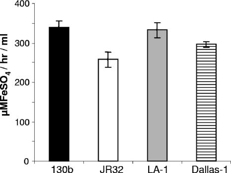
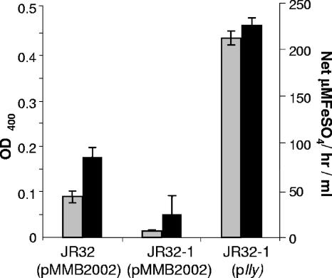
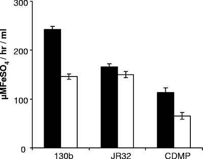
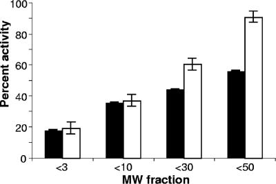
The virulence of Legionella pneumophila is dependent upon its capacity to acquire iron. To identify genes involved in expression of its siderophore, we screened a mutagenized population of L. pneumophila for strains that were no longer able to rescue the growth of a ferrous transport mutant. However, an unusual mutant was obtained that displayed a strong inhibitory effect on the feoB mutant. Due to an insertion in hmgA that encodes homogentisate 1,2-dioxygenase, the mutant secreted increased levels of pyomelanin, the L. pneumophila pigment that is derived from secreted homogentisic acid (HGA). Thus, we hypothesized that L. pneumophila-secreted HGA-melanin has intrinsic ferric reductase activity, converting Fe(3+) to Fe(2+), but that hyperpigmentation results in excessive reduction of iron that can, in the case of the feoB mutant, be inhibitory to growth. In support of this hypothesis, we demonstrated, for the first time, that wild-type L. pneumophila secretes ferric reductase activity. Moreover, whereas the hyperpigmented mutant had increased secreted activity, an lly mutant specifically impaired for pigment production lacked the activity. Compatible with the nature of HGA-melanins, the secreted ferric reductase activity was positively influenced by the amount of tyrosine in the growth medium, resistant to protease, acid precipitable, and heterogeneous in size. Together, these data represent the first demonstration of pyomelanin-mediated ferric reduction by a pathogenic bacterium.


Figures

FIG. 1.
FIG. 1.
Growth inhibition of a feoB mutant by a hyperpigmented mutant of L. pneumophila. Approximately 5 × 10⁵ CFU of either an feoB mutant (A) or wild-type 130b (B) were spread onto BCYE agar lacking its standard ferric iron supplement and then aliquots of BYE medium containing either 5 × 10³ CFU of wild-type 130b (top), hmgA mutant NU326 (left), or complemented NU326 (phmgA) (bottom) or no added bacteria (right) were spotted onto the plates. After 5 days of incubation at 37°C, the growth of the feoB mutant or strain 130b around the spots was recorded. The results are representative of at least three replicate experiments.
FIG. 2.
FIG. 2.
Levels of pigment in culture supernatants of wild-type and hmgA mutant L. pneumophila. Wild-type 130b (black bars), hmgA mutant NU326 (white bars), and complemented mutant NU326 (gray bars) were inoculated into standard BYE (A) or deferrated CDM (B) and then, after the indicated periods of incubation at 37°C, filter-sterilized, culture supernatants were assessed for pigment production as measured by their OD400 over that of medium controls. For ease of visualization, the inset in panel B repeats the data for the wild-type and complemented mutant strains with a smaller scale on the y axis. The data presented are the means and standard deviations from triplicate cultures for each strain. The results presented are representative of at least three replicate experiments in which triplicate cultures for each strain were examined and found to have a level of variation comparable to that presented here. When grown in CDM, the increases in pigment expression by the hmgA mutant NU326 were significant at all time points (P < 0.01). When grown in BYE, pigment expression by the mutant was consistently greater than that of wild type; however, the difference did not achieve the statistical significance seen with CDM cultures.
FIG. 3.
FIG. 3.
Levels of ferric reductase activity in culture supernatants of wild-type and hmgA mutant L. pneumophila. Wild-type 130b (black bars), hmgA mutant NU326 (white bars), and complemented mutant NU326 (gray bars) were inoculated into deferrated CDM and then, after the indicated periods of incubation at 37°C, filter-sterilized, culture supernatants were assessed for the bulk reduction of ferric nitrate to Fe2+. The appearance of Fe2+:ferrozine was monitored at 562 nm and quantified by comparison to a standard curve of ferrous sulfate complexed with ferrozine. The background level of activity exhibited by the CDM is also depicted (hatched bars). The data presented are the means and standard deviations from triplicate cultures. The activities from all strains were significantly above the CDM control, and the activity exhibited by NU326 was significantly greater than that of the wild-type and complemented strain (P < 0.01).
FIG. 4.
FIG. 4.
Effects of substrates, cofactors, and protease treatment on ferric reductase activity. Following growth in CDMP, supernatants from wild-type 130b were tested for their ability to reduce ferric nitrate (FeNit), ferric chloride (FeChl), ferric ammonium citrate (FeCit), or ferric pyrophosphate (FePyr) (black bars). In parallel, the reduction reactions were carried out following either treatment of the supernatants with proteinase K (ProK) (white bars) or the addition of NADH (gray bars) or NADPH (hatched bars). The four horizontal lines depict the level of background activity exhibited by the medium-only negative control. The data presented are the means and standard deviations from triplicate cultures. The asterisk indicates a significant change in activity against ferric citrate following proteinase K treatment (P < 0.001).
FIG. 5.
FIG. 5.
Ferric reductase activity in culture supernatants of different strains of L. pneumophila. Strains 130b (black bar), JR32 (white bar), Los Angeles 1 (gray bar), and Dallas 1 (hatched bar) were grown in CDMP for 24 h and then filter-sterilized culture supernatants were tested for their ability to reduce ferric nitrate as described above. The horizontal line depicts the level of background activity exhibited by the medium control. The data are the means and standard deviations from triplicate cultures.
FIG. 6.
FIG. 6.
Ferric reductase activity of a nonpigmented lly mutant L. pneumophila. Lly+ JR32 (pMMB2002), its Lly-negative derivative JR32-1 (pMMB2002), and a complemented JR32-1 (plly) were cultured in deferrated CDMP, and then filter-sterilized supernatants were assessed for pigmentation (gray bars) and ferric reductase activity (black bars) as described in the text. The background activities exhibited by the medium have been subtracted from the supernatant activities. The data are the means and standard deviations from triplicate cultures. The decreased activities exhibited by the lly mutant relative to both its parent and complemented derivative were significant (P < 0.01). Although the data presented for JR32 and JR32-1 derive from bacteria containing the cloning vector (pMMB2002) that was used to make plly, the same results were obtained when strains devoid of the vector were examined (data not shown).
FIG. 7.
FIG. 7.
Acid precipitation of the ferric reductase activity in culture supernatants of L. pneumophila. After culturing for 48 h in deferrated CDMP, a portion of the cell-free supernatants from wild-type strains 130b and JR32 was subjected to the acid precipitation protocol used to purify melanins. Untreated supernatants (black bars) and precipitated samples (white bars) were tested in the ferrozine assay using ferric nitrate as substrate. Also depicted is an uninoculated medium control that was similarly incubated and acid precipitated. The data presented are the means and standard deviations from triplicate cultures.
FIG. 8.
FIG. 8.
Size fractionation of pigment and ferric reductase activity of L. pneumophila. Aliquots of acid-precipitated material obtained from CDMP cultures of wild-type strain 130b were centrifuged through filter units having a molecular mass cutoff of 3, 10, 30, or 50 kDa, and then the pigmentation and ferric reductase activity in each filtrate were quantified as described in the text. The results are presented as the percentage of pigment (black bars) and ferric reductase activity (white bars) in each of the fractionated filtrates compared to the starting material. The data presented are the means and standard deviations from triplicate cultures.

Similar articles

Cited by

References

    1. Agodi, A., S. Stefani, C. Corsaro, F. Campanile, S. Gribaldo, and G. Sichel. 1996. Study of a melanic pigment of Proteus mirabilis. Res. Microbiol. 147:167-174. - PubMed
    1. Albert-Weissenberger, C., C. Cazalet, and C. Buchrieser. 2007. Legionella pneumophila—a human pathogen that co-evolved with fresh water protozoa. Cell Mol. Life Sci. 64:432-448. - PMC - PubMed
    1. Allard, K. A., V. K. Viswanathan, and N. P. Cianciotto. 2006. lbtA and lbtB are required for production of the Legionella pneumophila siderophore legiobactin. J. Bacteriol. 188:1351-1363. - PMC - PubMed
    1. Arias-Barrau, E., E. R. Olivera, J. M. Luengo, C. Fernandez, B. Galan, J. L. Garcia, E. Diaz, and B. Minambres. 2004. The homogentisate pathway: a central catabolic pathway involved in the degradation of l-phenylalanine, l-tyrosine, and 3-hydroxyphenylacetate in Pseudomonas putida. J. Bacteriol. 186:5062-5077. - PMC - PubMed
    1. Bachman, M. A., and M. S. Swanson. 2004. The LetE protein enhances expression of multiple LetA/LetS-dependent transmission traits by Legionella pneumophila. Infect. Immun. 72:3284-3293. - PMC - PubMed

Publication types

MeSH terms

LinkOut - more resources