. 2007 Jun 4;177(5):843-55.
doi: 10.1083/jcb.200701111.

The Ig cell adhesion molecule Basigin controls compartmentalization and vesicle release at Drosophila melanogaster synapses

Affiliations

The Ig cell adhesion molecule Basigin controls compartmentalization and vesicle release at Drosophila melanogaster synapses

Florence Besse et al. J Cell Biol. .

Abstract

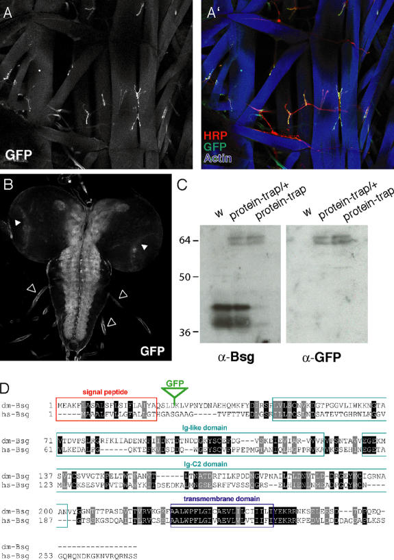
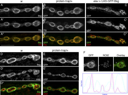
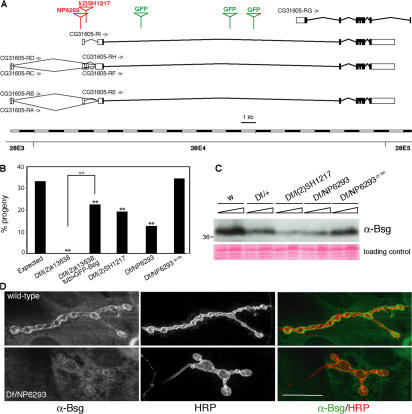
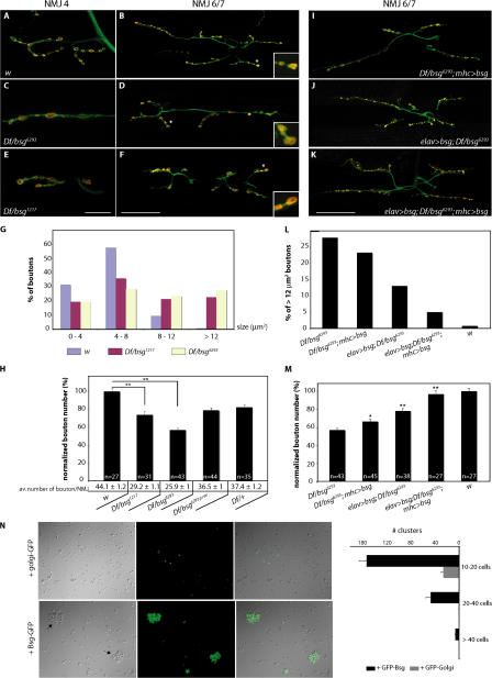
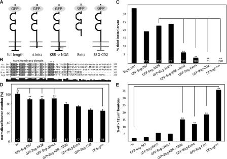
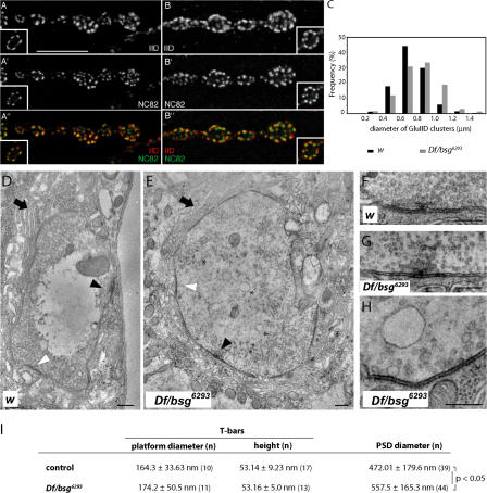
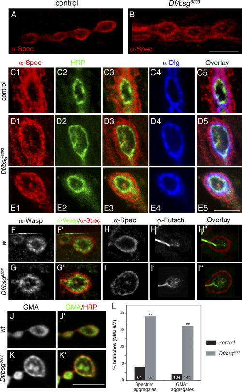
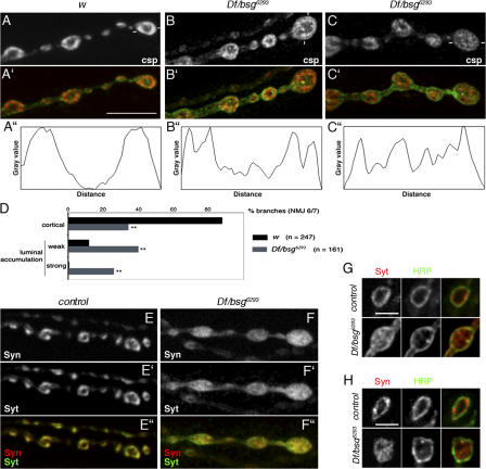
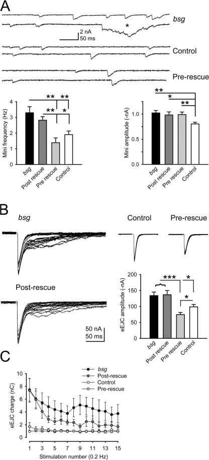
Synapses can undergo rapid changes in size as well as in their vesicle release function during both plasticity processes and development. This fundamental property of neuronal cells requires the coordinated rearrangement of synaptic membranes and their associated cytoskeleton, yet remarkably little is known of how this coupling is achieved. In a GFP exon-trap screen, we identified Drosophila melanogaster Basigin (Bsg) as an immunoglobulin domain-containing transmembrane protein accumulating at periactive zones of neuromuscular junctions. Bsg is required pre- and postsynaptically to restrict synaptic bouton size, its juxtamembrane cytoplasmic residues being important for that function. Bsg controls different aspects of synaptic structure, including distribution of synaptic vesicles and organization of the presynaptic cortical actin cytoskeleton. Strikingly, bsg function is also required specifically within the presynaptic terminal to inhibit nonsynchronized evoked vesicle release. We thus propose that Bsg is part of a transsynaptic complex regulating synaptic compartmentalization and strength, and coordinating plasma membrane and cortical organization.


Figures

Figure 1.
Figure 1.
GFP expression pattern in bsg protein-trap lines. (A and A′) Third instar larva heterozygous for the protein-trap insertion, stained with anti-GFP antibodies (A and A′, green), anti-HRP antibodies (A′, red), and phalloidin (A′, blue). (B) GFP-Bsg accumulation in heterozygous larval brain. (C) Western blot of total extracts from w larvae (left) or larvae heterozygous (protein-trap/+; middle) or homozygous (protein-trap; right) for a protein-trap insertion, probed with anti-Bsg and anti-GFP antibodies. (D) Alignment of D. melanogaster and human Bsg proteins. The green triangle indicates the location of GFP insertion.
Figure 2.
Figure 2.
Bsg accumulation at the NMJ. (A and B) NMJs of w (A) or protein-trap/+ (B) larvae double stained with anti-Bsg (A) or anti-GFP (B) antibodies and anti-Dlg antibodies (A′ and B′). (C) elav-Gal4/+; UAS-GFP-bsg/+ larvae stained with anti-GFP (C) and anti-HRP (C′) antibodies. (D and E) NMJs of w (D) and protein-trap/+ (E) larvae double stained with anti-Bsg (D) or anti-GFP (E) and anti-HRP (D′ and E′) antibodies. A–E correspond to z projections of serial confocal sections. Bar, 10 μm. (F) Tangential confocal section of a protein-trap/+ synaptic bouton stained with anti-GFP and NC82 anti-BRP antibodies and plot of the intensity profiles of GFP (green) and NC82 (pink) stainings across the bouton (section is indicated by white marks on the overlay). A–E are from muscle 6/7 NMJs, and F is from muscle 4 NMJ. Bar, 5 μm.
Figure 3.
Figure 3.
bsg locus and mutants. (A) Genomic organization and intron–exon structure of bsg. Untranslated and coding regions are represented as white and black boxes, respectively. Positions of P element and GFP-containing piggyBac insertions are indicated in red and green, respectively. (B) Percentages of GFP third instar larvae recovered among the progeny of a cross between Df/CyO-GFP females and l(2)k13638/CyO-GFP, l(2)SH1217/CyO-GFP, NP6293/ CyO-GFP, or NP6293pr.ex/CyO-GFP males. 33% of GFP animals are expected in absence of lethality. NP6293pr.ex is the NP6293 chromosome obtained after precise excision of the P element. The third bar corresponds to rescued Df/l(2)k13638; UAS-GFP-Bsg-fl/tub-Gal4 larvae. Note that ∼80% (16/20) of these larvae developed into pharate adults, of which 38% (6/16) hatched. At least 177 larvae were scored per cross. Statistical comparisons to the “expected” control: **, P < 0.001 (χ test). (C) Western blot of wild-type and mutant body wall extracts, probed with anti-Bsg antibodies. Ponceau staining is shown as a loading control. (D) Wild-type (top) and Df/NP6293 (bottom) larvae stained with anti-Bsg and anti-HRP antibodies. It was necessary to use diluted and purified anti-Bsg serum to visualize the difference in expression levels. Both pictures were taken from muscle 4 NMJs using identical confocal settings. Bar, 20 μm.
Figure 4.
Figure 4.
bsg regulates NMJ growth. (A–F) NMJs at muscle 4 (A, C, and E) or muscles 6/7 (B, D, and F) of w (A and B), Df/bsg6293 (C and D), and Df/bsg1217 (E and F) larvae stained with anti-CSP (red) and anti-HRP (green) antibodies. Asterisks in B, D, and F mark the branch terminals chosen for magnification (insets). Bars: (A, C, and E) 20 μm; (B, D, and F) 60 μm. (G) Distribution of synaptic boutons according to their size (in μm2). At least 97 boutons were scored per genotype. (H) Quantification of muscle 6/7 type Ib bouton number. Bouton numbers were normalized to muscle surface area, and the reference was set to 100 for w control larvae. Note that removal of one copy of bsg causes a mild reduction of bouton number, indicating a dosage sensitivity of the phenotype. Because of the smaller muscle size of Df/bsg1217 larvae, their bouton number is increased artificially upon normalization (numerical values below the bars represent the raw data, which are also shown in Table S1, available at http://www.jcb.org/cgi/content/full/jcb.200701111/DC1). Statistical comparisons to the w controls: **, P < 0.001 (t test). n represents the number of junctions scored per genotype. Error bars indicate SEM. (I–K) NMJs at muscle 6/7 of Df/bsg6293; mhc-Gal4/UAS-bsg (I), elav-Gal4/+; Df/bsg6293; UAS-bsg/+ (J), and elav-Gal4/+; Df/bsg6293; mhc-Gal4, UAS-bsg/+ (K) larvae double stained with anti-CSP (red) and anti HRP (green) antibodies. Bar, 60 μm. (L) Percentage of synaptic boutons >12 μm2 in Df/bsg6293 larvae and different rescue contexts. At least 114 boutons were scored per genotype. (M) Quantification of muscle 6/7 type Ib bouton numbers in Df/bsg6293 larvae and different rescue contexts. Bouton numbers were normalized to muscle surface area, and the reference set to 100 for w control larvae. Statistical comparisons to Df/bsg6293 animals: *, P < 0.05; **, P < 0.001 (t test). n represents the numbers of junctions scored per genotype. Error bars indicate SEM. (N) Cell aggregation assays using S2 cells transfected with either a GFP control construct (GFP-Golgi) or GFP-Bsg. (left) Light microscopy pictures; (right) number of clusters containing 10–20 cells, 20–40 cells, or >40 cells (per 5 × 10⁴ cells). Four samples, from two independent transfections, were analyzed per construct.
Figure 5.
Figure 5.
Differential rescue capacity of various mutated Bsg proteins. (A) Scheme of Bsg variants. Δintra corresponds to a form lacking the last 14 amino acids but including the juxtamembrane KRR stretch, Extra to a form lacking both the transmembrane and the cytoplasmic domains, KRR→NGG to a full-length protein where the KRR residues have been substituted to NGG, and Bsg-CD2 to a chimeric protein composed of the two Ig domains of Bsg fused to the transmembrane and cytoplasmic domains of rat CD2. (B) Alignment of Bsg transmembrane and intracellular domains. H.s., Homo sapiens; M.m., Mus musculus; D.r., Danio rerio; Dr. m., D. melanogaster; A.g., Anopheles gambiae. The black box indicates the amino acids deleted in the Δintra construct. (C) Percentages of non–CyO-GFP third instar larvae recovered among the non-TM6 progeny of a cross between Df/CyO-GFP; UAS-GFP-Bsg*/TM6 females and l(2)k13638/CyO-GFP; tub-Gal4/TM6 males. 33% of non–CyO-GFP animals are expected in case of complete rescue (left bar). Numbers correspond to the total numbers of non-TM6 larvae scored in the entire progeny of each cross. Statistical comparison to GFP-Bsg-fl#28 animals: **, P < 0.001 (χ test). (D) Quantification of muscle 6/7 type Ib bouton numbers in Df/bsg6293 larvae and different rescue contexts (driver used for rescue: elav-Gal4). Statistical comparisons to Df/bsg6293 animals: **, P < 0.001 (t test). The number of junctions scored for each genotype is represented in white. Error bars indicate SEM. fl#7 and fl#28 represent two independent insertions of the GFP-tagged full-length transgene. (E) Percentage of synaptic boutons >12 μm2 in Df/bsg6293 larvae and different rescue contexts. Statistical comparison to w animals: **, P < 0.001 (χ test).
Figure 6.
Figure 6.
Pre- and postsynaptic specializations in bsg mutants. (A and B) w (A) and Df/bsg6293 (B) NMJs stained with anti-GluRIID (A and B) and anti-BRP (A′ and B′) antibodies. Insets show cross-sections of single boutons. Bar, 10 μm. (C) Frequency distribution of postsynaptic glutamate receptor cluster sizes in w (black) and Df/bsg6293 (gray) third instar larvae. (D and E) Electron micrographs of w (D) and Df/bsg6293 (E) type Ib boutons. Arrows point to the SSR, whereas black and white arrowheads indicate active zones, with and without T-bars, respectively. Asterisks indicate atypically large vesicles. Although, in E, synaptic vesicles are also localized in the bouton center, an unambiguous identification of ectopically localized synaptic vesicles was more difficult in other samples. Bars, 250 nm. (F–H) Magnification of w (F) and Df/bsg6293 (G) active zones. (H) Example of an abnormally large vesicle in proximity to a Df/bsg6293 active zone (defined by its electron-dense plasma membrane and synaptic vesicle cluster). (I) Transmission EM–based quantification of active zone and PSD parameters.
Figure 7.
Figure 7.
Distribution of actin cytoskeleton markers is altered in bsg larvae. (A and B) Wild-type (A) and Df/bsg6293 (B) muscle 4 NMJs stained with anti–α-Spec antibodies. Bar, 10 μm. (C–E) Heterozygous control (C) and Df/bsg6293 (D and E) boutons stained with anti–α-Spec (C1, D1, and E1), anti-HRP (C2, D2, and E2), and anti-Dlg (C4, D4, and E4) antibodies. Bar, 5 μm. (F and G) w (F) and Df/bsg1217 (G) bouton stained with anti-Wasp (F and G) and anti–α-Spec (F′ and G′, red) antibodies. (H and I) w (H) and Df/bsg6293 (I) boutons stained with anti–α-Spec (H and I) and anti-Fusch (H′ and I′) antibodies. Bar, 5 μm. (J and K) Synaptic boutons of wild-type (J) and Df/bsg6293 (K) larvae expressing a fusion of GFP with the F-actin binding domain of Moesin (GMA), under the control of elav-Gal4. GFP-GMA expression is shown in J and K, and is in green in J′ and K′. HRP staining is shown in red in J′ and K′. Bar, 5 μm. Images A and B correspond to z projections of serial confocal sections throughout entire boutons (step size: 0.3 μm), and images C–K correspond to single optical slices taken through bouton centers. (L) Graph showing the percentage of NMJ 6/7 branches containing presynaptic Spec+ or GMA+ aggregates. **, P < 0.001 (χ test).
Figure 8.
Figure 8.
Synaptic vesicle distribution is altered in bsg larvae. (A–C) w (A) and Df/bsg6293 (B and C) larvae stained with anti-CSP (A–C and A′–C′, red) and anti-HRP antibodies (A′–C′, green). (A″–C″) Fluorescence intensity profiles of CSP staining along a section of the terminal bouton indicated in A–C (white marks). (D) Graph showing the proportion of NMJ 6/7 branches showing either a cortical or luminal accumulation of CSP. **, P < 0.001 (χ test). (E and F) Wild-type (E) and Df/bsg6293 (F) larvae expressing GFP-tagged synaptotagmin and stained with anti-Synapsin (E and F) and anti-GFP (E′ and F′) antibodies. Images in A–F correspond to z projections of serial confocal sections throughout entire boutons (see Fig. S3 A, available at http://www.jcb.org/cgi/content/full/jcb.200701111/DC1, for serial individual pictures corresponding to pictures E and F). Bar, 10 μm. (G and H) Single confocal sections of bsg6293/+ and Df/bsg6293 larvae double stained for Synaptotagmin-GFP (Syt; G) or Synapsin (Syn; H) and HRP. Optical slices all traverse bouton centers (defined by the section with the largest bouton diameter and the ring-like appearance of HRP staining). Bars, 4 μm.
Figure 9.
Figure 9.
Presynaptic loss of Bsg provokes asynchronous vesicle release. (A) Representative traces and quantification of mini frequency and amplitude in bsg mutants and rescued animals. *, P ≤ 0.05; **, P ≤ 0.01. (B) Example traces of eEJCs after 15 stimuli at 0.2 Hz and mean amplitudes. *, P ≤ 0.05; **, P ≤ 0.01;***, P ≤ 0.001. (C) Presynaptic loss of Bsg leads to an increased and atypically delayed release of vesicles, reflected by the larger charge carried by eEJCs of both bsg mutants and postsynaptic rescues. Precise genotypes are as follows: Basigin, Df/bsg6293; control, elav-Gal4/Y; prerescue, elav-Gal4/Y; Df/bsg6293; UAS-bsg/+; postrescue, Df/bsg6293; UAS-bsg/mhc-Gal4.

Similar articles

Cited by

References

    1. Berditchevski, F., S. Chang, J. Bodorova, and M.E. Hemler. 1997. Generation of monoclonal antibodies to integrin-associated proteins. Evidence that alpha3beta1 complexes with EMMPRIN/basigin/OX47/M6. J. Biol. Chem. 272:29174–29180. - PubMed
    1. Beumer, K.J., J. Rohrbough, A. Prokop, and K. Broadie. 1999. A role for PS integrins in morphological growth and synaptic function at the postembryonic neuromuscular junction of Drosophila. Development. 126:5833–5846. - PubMed
    1. Bogdan, S., R. Stephan, C. Lobke, A. Mertens, and C. Klambt. 2005. Abi activates WASP to promote sensory organ development. Nat. Cell Biol. 7:977–984. - PubMed
    1. Bretscher, A., K. Edwards, and R.G. Fehon. 2002. ERM proteins and merlin: integrators at the cell cortex. Nat. Rev. Mol. Cell Biol. 3:586–599. - PubMed
    1. Collins, M.O., H. Husi, L. Yu, J.M. Brandon, C.N. Anderson, W.P. Blackstock, J.S. Choudhary, and S.G. Grant. 2006. Molecular characterization and comparison of the components and multiprotein complexes in the postsynaptic proteome. J. Neurochem. 97(Suppl. 1):16–23. - PubMed

Publication types

MeSH terms