Comparative Study
. 2007 Jun 4;177(5):857-70.
doi: 10.1083/jcb.200612012.

Nodes of Ranvier and axon initial segments are ankyrin G-dependent domains that assemble by distinct mechanisms

Affiliations
Comparative Study

Nodes of Ranvier and axon initial segments are ankyrin G-dependent domains that assemble by distinct mechanisms

Yulia Dzhashiashvili et al. J Cell Biol. .

Abstract

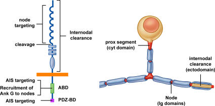
Axon initial segments (AISs) and nodes of Ranvier are sites of action potential generation and propagation, respectively. Both domains are enriched in sodium channels complexed with adhesion molecules (neurofascin [NF] 186 and NrCAM) and cytoskeletal proteins (ankyrin G and betaIV spectrin). We show that the AIS and peripheral nervous system (PNS) nodes both require ankyrin G but assemble by distinct mechanisms. The AIS is intrinsically specified; it forms independent of NF186, which is targeted to this site via intracellular interactions that require ankyrin G. In contrast, NF186 is targeted to the node, and independently cleared from the internode, by interactions of its ectodomain with myelinating Schwann cells. NF186 is critical for and initiates PNS node assembly by recruiting ankyrin G, which is required for the localization of sodium channels and the entire nodal complex. Thus, initial segments assemble from the inside out driven by the intrinsic accumulation of ankyrin G, whereas PNS nodes assemble from the outside in, specified by Schwann cells, which direct the NF186-dependent recruitment of ankyrin G.


Figures

Figure 1.
Figure 1.
NF186 is targeted to initial/proximal segments and nodes of Ranvier. (A) Schematic organization of Hc pyramidal and myelinated DRG neurons (top) and corresponding micrographs (bottom) showing the AIS of Hc neurons and PS and nodes of DRG neurons stained for ankyrin G (red; indicated by arrows). Hc neurons are also stained for MAP2 (blue); myelinated neurites are stained for Caspr (green) and P0 (blue). Bars: (a) 10 μm; (b) 40 μm; (c) 5 μm. (B) Schematic diagram of epitope-tagged NF186 and ICAM1 constructs. (C) HA-NF186 expression. (top) HA-NF186 (green) is targeted to the AIS (arrows) of HC neurons. (middle) HA-NF186-GFP is targeted to the PS (arrows) of DRG neurons, where it colocalizes with ankyrin G (rhodamine). (bottom) HA-NF186-GFP has a diffuse pattern of expression in neurites (without Schwann cells) but concentrates at nodes of myelinated fibers (arrowheads). Bars: (a and b) 20 μm; (c) 10 μm. (D) ICAM1 expression. (top) ICAM1 (green) has a predominantly somatodendritic distribution in Hc neurons; the AIS is shown (arrows). (middle) ICAM1 (green) is diffuse, the PS is enriched in ankyrin G (red), indicated by arrows. (bottom) ICAM1 is diffuse in both neurites and myelinated fibers; a node is indicated by the arrowheads. Bars: (a and b) 20 μm; (c) 10 μm. (E) Quantitative analysis of NF186 and ICAM1 targeting in Hc neurons showing the percentage of expression restricted to the AIS, enriched in the AIS but also present elsewhere in the neuron (AIS+), and expressed in the soma, dendrites, and distal axon (S + D + A). (F) Quantitative analysis of NF186 and ICAM1 targeting in myelinated co-cultures. The percentage of expression of each construct in nodes and heminodes is shown.
Figure 2.
Figure 2.
The cytoplasmic domain of NF186 is necessary and sufficient for AIS targeting. Targeting constructs (A) and photomicrographs of their expression in Hc neurons (B) are shown. In the schematics, domains contributed by NF186 are shown in blue; those from ICAM1 are in red. NFΔABD lacks the FIGQY sequence required for binding to ankyrin G. Transfected proteins were stained with an antibody to ICAM1 (top) and HA (middle and bottom). Ankyrin G immunoreactivity (red) demarcates initial segments (arrows). ICAM1/NF-CD (top) was targeted to the AIS; the NF-ED/ICAM1 (middle) and NFΔABD (bottom) constructs remained diffuse. Bar, 20 μm.
Figure 3.
Figure 3.
NF186 is targeted to nodes and cleared from the internode via extracellular sequences. (A) Schematic diagram of targeting constructs. NF186 sequences are in blue, and ICAM1 sequences are in red. (B) Expression of corresponding constructs in myelinating co-cultures analyzed for targeting to nodes (flanked on both sides by myelin segments; left) and heminodes (associated with a single myelin segment; right); both are indicated by arrowheads. Cultures were stained for the GFP tag (green) and the myelin protein, P0 (blue). Bars, 5 μm. (C) Quantitation of a representative experiment analyzing the targeting of constructs to nodes and heminodes as a percentage of total sites counted. Myelinated axons were analyzed for the presence of GFP immunoreactivity at nodes or heminodes, each identified by staining for ankyrin G. Staining was scored as either strongly positive (++) or increased above background (+). See Fig. S5 (available at http://www.jcb.org/cgi/content/full/jcb.200612012/DC1) for representative images.
Figure 4.
Figure 4.
Clearance of NF186 from the internode is independent of its targeting to the node. (A) Schematic diagram of constructs. NF186 sequences are in blue, and ICAM1 sequences are in red. (B) Expression of the corresponding constructs in myelinating co-cultures, stained for GFP (green), ankyrin G (red), and the myelin protein P0 (blue). NF186 and ICAM1/NFΔIg are both cleared from internodes, indicated by white brackets in each field. The position of the corresponding nodes is indicated by arrows and asterisks; those indicated by asterisks are shown at high power in the insets. ICAM1/NFΔIg remained diffusely expressed in the nonmyelinated regions adjacent to nodes. Bar, 20 μm.
Figure 5.
Figure 5.
NF186 expression is stabilized by interactions with ankyrin G. DRG neurons were nucleofected with either NF186-GFP or NF186ΔABD-GFP; half were maintained as neuron-only cultures (A), and the other half were seeded with Schwann cells after 1 wk and maintained in myelinating conditions (B). Detergent lysates were prepared at weekly intervals; fractionated by SDS-PAGE; blotted; probed for GFP, peripherin (as a loading control), and the myelin protein P0 (co-cultures); incubated with 125I protein A; and analyzed via a phosphorimager. Quantitation is shown in Fig. S3 A (available at http://www.jcb.org/cgi/content/full/jcb.200612012/DC1). Full-length NF186-GFP is indicated (arrowhead); a proteolytic fragment of ∼150 kD (asterisk) is faintly visible in the neuron-only blots and prominently at initial co-culture time points. Addition of Schwann cells accelerates the turnover of NF186-GFP and, in particular, NF186ΔABD-GFP (compare corresponding time points in A and B). (C) DRG neurons expressing either NF186-GFP or NF186ΔABD-GFP under lentiviral control were fixed (control) or extracted with Triton X-100 and then fixed, followed by staining for GFP, P0, and ankyrin G. With extraction, NF186 staining is removed from nonmyelinated fibers but persists at most nodes in contrast to NF186ΔABD, which is removed from all nodes as well as nonmyelinated fibers. Insets show nodes and heminodes, indicated by arrowheads, at higher power. Bar, 20 μm.
Figure 6.
Figure 6.
NF186 is essential for PNS node but not AIS formation. (A) DRG neurons infected with the pLL3.7 vector alone (control) or vector encoding shRNA to NF186 (shNF186) were co-cultured with Schwann cells, fixed, and stained. (left) Co-cultures were stained for P0 (blue) and with an antibody that recognizes both the neuronal and glial isoforms of NF (red). Expression of NF was abolished at nodes (arrowheads) of shRNA-treated but not control co-cultures; staining persists in the flanking glial paranodes. (right) Cultures stained with the pan-NF antibody (red) and for Caspr (green), a marker of the paranodes, demonstrate that shRNA treatment abolished nodal but not paranodal expression of NF. Bars, 5 μm. (B) shRNA to NF186, but not vector alone, abolishes expression of ankyrin G, sodium channels (NaCh), and NrCAM at nodes (arrowheads), shown at higher power in the insets. P0 is stained in blue. Gliomedin (GLDN) expression at nodes was also significantly diminished and was minimally detected at many nodes (arrowhead; white inset); others were only partially affected (asterisk; yellow inset). Bar, 20 μm. (C) Hc neurons infected with control or shRNA-expressing vector were stained for ankyrin G, sodium channel, and MAP2. shRNA treatment had no effect on localization of ankyrin G and sodium channel at the AIS (indicated by arrows); at higher power in the insets. Bars, 20 μm. (D) Western blots show that shRNA treatment substantially reduced NF186 expression; actin is a loading control.
Figure 7.
Figure 7.
Recruitment of ankyrin G by NF186 is critical for PNS node assembly. (A) DRG neurons expressing NF186-GFP or NF186ΔABD-GFP were cultured with Schwann cells under myelinating conditions, fixed, and stained for GFP, P0, and ankyrin G (left) or sodium channels (NaCh; right). Robust levels of NF186ΔABD at nodes were associated with reduced coexpression of ankyrin G and sodium channels, as evident by comparison of nontransfected nodes (asterisks) to transfected nodes (arrowheads). Bars, 10 μm. (B) Schematic diagram and sequence of NF constructs used for rescue experiments. The modified sequence within the NF mucin-like domain is shown with substituted codons marked in red. (C) Western blot of NF186 knockdown and rescue. DRG neurons were infected with pLL3.7 vector alone (sh Con) or encoding shRNA to endogenous NF186. shRNA-treated neurons were either not nucleofected (sh NF) or were nucleofected with the codon-modified NF186-GFP (+NF) or NF186ΔABD-GFP constructs (+NFΔABD). Lysates were blotted with an NF antibody that recognizes a cytoplasmic epitope present on NF186 but not on NF186ΔABD, and antibodies to GFP, NrCAM, and peripherin. The position of endogenous NF186 (bottom arrow) and of the exogenous NF186-GFP protein (top arrow) is shown on the NF blot. Endogenous NF186 is robustly expressed in the first lane but is essentially absent in all neurons treated with shRNA. NF186ΔABD-GFP, which is not recognized by the pan-NF antibody, is strongly detected when lysates are blotted for GFP. NrCAM levels are unchanged with shRNA treatment; peripherin is a loading control. (D) Both full-length NF186 and NF186ΔABD constructs were targeted to nodes (white arrowheads; shown at higher power in insets). Expression of full-length NF186, but not NF186ΔABD, rescued ankyrin G and sodium channel expression at transfected nodes (identified by staining for GFP). A nontransfected node in the NF186 nucleofected cultures (orange arrowhead), which lack sodium channels, is shown. Myelin segments were visualized by staining for P0 (blue). Bars, 20 μm. (E) Quantitation of NF186 knockdown and rescue by modified NF186 constructs. Ankyrin G and sodium channel staining at nodes were scored as robustly (++) or modestly positive (+); absence of staining was also scored but is not shown, to simplify the figure. Representative images from each category are shown in Fig. S5 (available at http://www.jcb.org/cgi/content/full/jcb.200612012/DC1). Results are graphed as the percentages of the total node sites positive for nodal proteins in cultures treated with the control lentivirus (sh Con) or in cultures treated with shRNA and nucleofected with NF186-GFP (shNF + NF) or NF186ΔABD-GFP (shNF + NFΔABD). GFP-negative nodes (N; nontransfected) and GFP-positive nodes (T; transfected) were scored separately in each set of cultures. Expression of ankyrin G and sodium channels was dramatically reduced at nontransfected nodes in both sets of shRNA-treated neurons compared with neurons treated with vector alone (sh Con). Expression of the full-length, codon-modified NF186 at nodes substantially rescued ankyrin G and sodium channel expression in the shRNA-treated co-cultures, whereas expression of NF186ΔABD did not.
Figure 8.
Figure 8.
Ankyrin G knockdown blocks PS and node assembly. (A) DRG neurons were infected with a lentiviral vector alone (sh Con) or expressing shRNA to ankyrin G (sh ankG). Vectors retained the GFP marker in these studies to identify infected cells (not depicted); somas of infected neurons are marked with asterisks. 10 d after infection, expression of ankyrin G and sodium channel is prominent in the PSs (arrows) of control neurons but absent from shRNA-treated neurons. Bar, 40 μm. (B) Lysates prepared from control (sh Con) and shRNA-treated (sh ankG) DRG neurons were blotted for ankyrin G, ankyrin B, pan–sodium channel, pan-NF, and NrCAM. Expression of ankyrin G was markedly reduced in shRNA-treated cultures, whereas the expression of ankyrin B was unaffected; expression of the NF186 isoform was slightly reduced, whereas sodium channels and NrCAM were unchanged. (C) Effects of ankyrin G knockdown on nodes were analyzed in vector alone (sh Con) and shRNA-treated (sh ankG) DRG neurons co-cultured with Schwann cells under myelinating conditions. Ankyrin G and sodium channel were expressed at nodes (arrowheads) of control but not shRNA-treated co-cultures. Bar, 20 μm. (D) Quantitation of ankyrin G and sodium channel expression in control and shRNA-treated cultures shown as a percentage of total nodes counted. (E) Expression of NF186, NrCAM, and βIV spectrin at nodes of control and shRNA–ankyrin G–treated co-cultures is shown. Nodes are indicated by arrowheads and shown at higher power in the insets. Bar, 20 μm.
Figure 9.
Figure 9.
Summary of NF186 localization mechanisms. Schematic figures showing the structural features of NF186 (left) that mediate its targeting to different axonal domains (red) of a pseudounipolar DRG neuron (right). Targeting of NF186 to nodes is mediated by its Ig domains; restricted expression at the node is enhanced by clearance from the internode mediated by multiple regions of the ectodomain. In contrast, targeting to the initial/proximal segments requires interactions of the ABD in the cytoplasmic segment with ankyrin G; AIS targeting also involves the C-terminal PDZ binding sequence. The ABD is critical for recruitment of ankyrin G to the node, which is required for localization of sodium channels and other nodal components. A likely cleavage site between the Ig and FNIII domains is shown.

References

    1. Alessandri-Haber, N., G. Alcaraz, C. Deleuze, F. Jullien, C. Manrique, F. Couraud, M. Crest, and P. Giraud. 2002. Molecular determinants of emerging excitability in rat embryonic motoneurons. J. Physiol. 541:25–39. - PMC - PubMed
    1. Ango, F., G. di Cristo, H. Higashiyama, V. Bennett, P. Wu, and Z.J. Huang. 2004. Ankyrin-based subcellular gradient of neurofascin, an immunoglobulin family protein, directs GABAergic innervation at purkinje axon initial segment. Cell. 119:257–272. - PubMed
    1. Bennett, V., and S. Lambert. 1999. Physiological roles of axonal ankyrins in survival of premyelinated axons and localization of voltage-gated sodium channels. J. Neurocytol. 28:303–318. - PubMed
    1. Berghs, S., D. Aggujaro, R. Dirkx, E. Maksimova, P. Stabach, J.M. Hermel, J.P. Zhang, W. Philbrick, V. Slepnev, T. Ort, and M. Solimena. 2000. βIV spectrin, a new spectrin localized at axon initial segments and nodes of Ranvier in the central and peripheral nervous system. J. Cell Biol. 151:985–1002. - PMC - PubMed
    1. Boiko, T., and B. Winckler. 2003. Picket and other fences in biological membranes. Dev. Cell. 5:191–192. - PubMed

Publication types