. 2007 Jun 4;177(5):871-80.
doi: 10.1083/jcb.200702080.

Alpha4beta1 integrin and erythropoietin mediate temporally distinct steps in erythropoiesis: integrins in red cell development

Affiliations

Alpha4beta1 integrin and erythropoietin mediate temporally distinct steps in erythropoiesis: integrins in red cell development

Shawdee Eshghi et al. J Cell Biol. .

Erratum in

  • J Cell Biol. 2008 Apr 21;181(2):395

Abstract

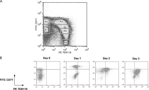
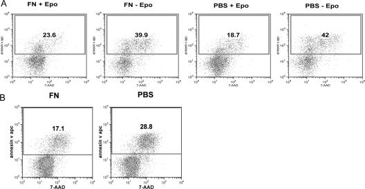
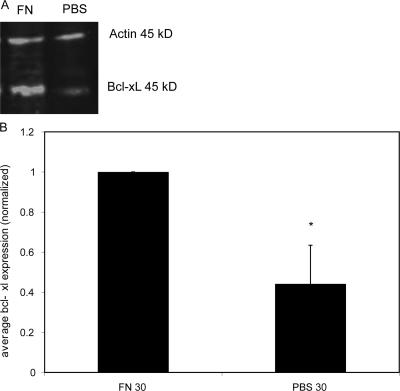
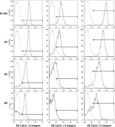
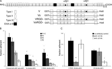
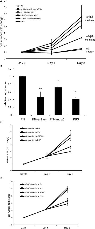
Erythropoietin (Epo) is essential for the terminal proliferation and differentiation of erythroid progenitor cells. Fibronectin is an important part of the erythroid niche, but its precise role in erythropoiesis is unknown. By culturing fetal liver erythroid progenitors, we show that fibronectin and Epo regulate erythroid proliferation in temporally distinct steps: an early Epo-dependent phase is followed by a fibronectin-dependent phase. In each phase, Epo and fibronectin promote expansion by preventing apoptosis partly through bcl-xL. We show that alpha(4), alpha(5), and beta(1) are the principal integrins expressed on erythroid progenitors; their down-regulation during erythropoiesis parallels the loss of cell adhesion to fibronectin. Culturing erythroid progenitors on recombinant fibronectin fragments revealed that only substrates that engage alpha(4)beta(1)-integrin support normal proliferation. Collectively, these data suggest a two-phase model for growth factor and extracellular matrix regulation of erythropoiesis, with an early Epo-dependent, integrin-independent phase followed by an Epo-independent, alpha(4)beta(1)-integrin-dependent phase.


Figures

Figure 1.
Figure 1.
CD71 and TER119 can be used as markers of erythroid differentiation in vivo and in vitro. (A) Five populations of sequentially differentiated erythroid cells can be isolated from day 14.5 fetal liver based on the expression of CD71 and TER119 as previously described (Zhang et al., 2003). (B) CD71 and TER119 can be used as markers of erythroid differentiation in vitro. Day 14.5 TER119 fetal liver cells were cultured on fibronectin-coated plates in Epo-containing media for 1 d and in Epo-free media for an additional 2 d. On each day, cells were dissociated and stained with antibodies to CD71 and TER119 for FACS analysis.
Figure 2.
Figure 2.
Fibronectin supports the expansion of erythroid progenitors. TER119 fetal liver erythroid progenitor cells were cultured on fibronectin or control (uncoated) substrates with Epo-containing media for 1 d and then in Epo-free media for a second day. At days 1 and 2, the number of live cells was counted. Progenitor cells cultured on fibronectin undergo dramatic expansion in cell number, but cells cultured on a control substrate do not. Each point is the mean of three to five independent experiments. Error bars represent the SD.
Figure 3.
Figure 3.
Fibronectin is only required on the second day of culture. (A) TER119 erythroid progenitors were cultured on either fibronectin or uncoated surfaces for 1 d in Epo-containing media and were dissociated and transferred to fresh fibronectin surfaces in Epo-free media. A dramatic expansion in cell number was observed regardless of which substrate was present on the first day of culture. (B) TER119+ cells were cultured on fibronectin or uncoated surfaces in the absence of Epo. Only those cells cultured on fibronectin proliferate; the addition of Epo to the culture media (dashed line) had no effect on cell number. Each point is the mean of three to five independent experiments. Error bars represent the SD.
Figure 4.
Figure 4.
Epo and fibronectin protect against apoptosis. (A) TER119 progenitor cells were cultured overnight on fibronectin or uncoated substrates in the presence or absence of Epo. Annexin v binding (shown on the y axis) was assayed via flow cytometry, and the percentage of positive cells is indicated in each plot. The absence of Epo during the first day of culture leads to an increase in the percentage of apoptotic cells, but the absence of fibronectin does not have an effect. (B) Fibronectin protects from apoptosis in the second stage of proliferation. TER119 progenitor cells were cultured overnight on uncoated surfaces in the presence of Epo and were dissociated and transferred to fresh fibronectin or control surfaces in Epo-free media. Annexin v binding was assayed at 4 h. The absence of fibronectin in the second phase of culture led to an increased percentage of annexin v–positive cells. These data are representative of three to five independent experiments.
Figure 5.
Figure 5.
Fibronectin up-regulates bcl-xL in the second stage. TER119 progenitor cells were cultured overnight on uncoated surfaces in the presence of Epo and were dissociated and serum starved for 1 h before being transferred to fresh fibronectin or control surfaces in Epo-free media. bcl-xL expression was assayed at 30 min via Western blotting. (A) Representative Western blot indicating the up-regulation of bcl-xL on fibronectin and the equivalent expression of actin as a loading control. The anti-actin and anti–bcl-xL antibodies were raised in different hosts, thereby allowing the visualization of both proteins on the same membrane. (B) Quantification of five identical experiments. Integrated fluorescence intensity on the control substrate was normalized to that on fibronectin, indicating a statistically significant decrease in the absence of fibronectin. *, P ≤ 0.01. Error bars represent the SD.
Figure 6.
Figure 6.
Erythroid progenitors express fibronectin receptors. α4, α5, and β1 integrins are highly expressed on early erythroid progenitors, and expression is down-regulated as cells differentiate. Fetal livers were stained with antibodies against CD71, TER119, and each of α4, α5, and β1 integrins. Cells were gated into regions as in Fig. 1 A, and integrin expression within each population was determined. The light trace is the background staining of the secondary antibody alone. These data are representative of at least three experiments. The horizontal bars represent the fraction of the population with integrin expression above the level of the background control.
Figure 7.
Figure 7.
Adhesion to fibronectin is correlated with integrin receptor expression. (A) Adhesion of R1 + R2, R3, R4, and R5 cells to either 10 (left) or 3 μg/ml (right) human plasma fibronectin at an acceleration of ∼1,000 g. Cells from each population were sorted and allowed to adhere to fibronectin-coated plates for 30 min before the adhesion assay. Adhesion to fibronectin decreases as cells differentiate. (B) Adhesion of R1 + R2 and R5 cells to 10 μg/ml fibronectin at a range of centrifugal accelerations. (A and B) In all cases, each bar represents the mean fraction adherent from three wells, and error bars represent the SD. *, P ≤ 0.05; **, P ≤ 0.01 when compared with R1 + R2 cells.
Figure 8.
Figure 8.
Role of integrins in the adhesion of erythroid progenitors. (A) Schematic diagram of recombinant fibronectin fusion proteins. The V fragment contains both the α4β1- and α5β1-binding sites, Vo contains only the α5β1-binding site, VRGD contains only the α4β1-binding site, and VoRGD contains neither integrin-binding site. (B) Characterization of the adhesion of sorted R1 + R2, R3, R4, and R5 cells to 10 μg/ml V, Vo, or VRGD substrates at an acceleration of ∼1,000 g. (C) α4β1 and α5β1 integrins mediate adhesion to different fibronectin domains. Adhesion of sorted R1 + R2 cells to 10 μg/ml recombinant fibronectin fragments Vo or VRGD with and without pretreatment with function-blocking antibodies to α4 and α5 integrins. Each bar represents the mean fraction of cells adherent in each of three replicate wells. In all cases, error bars are the SD. *, P ≤ 0.05; **, P ≤ 0.01.
Figure 9.
Figure 9.
α4 integrin supports terminal proliferation. (A) α4β1-Mediated adhesion is required for the proliferation of differentiating fetal liver erythroid cells. TER119 fetal liver erythroid progenitor cells were cultured on V, Vo, VRGD, or VoRGD substrates with Epo-containing media for 1 d and then in Epo-free media for a second day. At days 1 and 2, the number of live cells was counted. Only α4β1-binding substrates V and VRGD support an expansion in cell number similar to that seen on intact fibronectin. α5β1-Binding substrate Vo supports a moderate level of expansion, and VoRGD, which does not engage either integrin, does not support any cell expansion. (B) Blocking α4 integrin with an antibody blocks proliferation. TER119+ cells were isolated and incubated with function-blocking antibodies to either α4 or α5 integrin and were cultured on fibronectin-coated or control substrates for 1 d. The mean normalized increase in cell number from three experiments is shown. *, P ≤ 0.05; **, P ≤ 0.01. (C and D) Expansion of erythroid progenitors during the second day occurs only in conditions in which α4β1 is engaged. TER119 cells were cultured on Vo (C) or VRGD (D) substrates in Epo-containing media for the first day and were dissociated and transferred to wells coated with each of the various integrin-binding substrates in Epo-free media for a second day. Error bars represent the SD.
Figure 10.
Figure 10.
α4 integrin provides protection from apoptosis. (A) α4 integrin engagement provides protection from apoptosis in the second phase. TER119 fetal liver cells were purified and cultured overnight on uncoated substrates in Epo-containing media. Cells were then dissociated, incubated with or without antibodies to α4 or α5 integrins, and cultured in Epo-free media on fibronectin or control substrates. 4 h after Epo removal, the cells were dissociated and stained with annexin V and 7-AAD to assay for apoptosis. Annexin V binding is shown on the y axis. Data are representative of three independent experiments. The percentage of positive cells is indicated in each plot. (B) Summary of apoptosis data. Error bars represent the SD. *, P ≤ 0.05; **, P ≤ 0.01.

References

    1. Arroyo, A.G., J.T. Yang, H. Rayburn, and R.O. Hynes. 1999. Alpha4 integrins regulate the proliferation/differentiation balance of multilineage hematopoietic progenitors in vivo. Immunity. 11:555–566. - PubMed
    1. Avraham, H., S.Y. Park, K. Schinkmann, and S. Avraham. 2000. RAFTK/Pyk2-mediated cellular signalling. Cell. Signal. 12:123–133. - PubMed
    1. Campos, L.S. 2005. Beta1 integrins and neural stem cells: making sense of the extracellular environment. Bioessays. 27:698–707. - PubMed
    1. Fawcett, D.W., and R.P. Jensh. 1997. Bloom & Fawcett: Concise Histology. Chapman and Hall, New York. 84–93 pp.
    1. Gregory, C.J., and A.C. Eaves. 1977. Human marrow cells capable of erythropoietic differentiation in vitro: definition of three erythroid colony responses. Blood. 49:855–864. - PubMed

Publication types