. 2007 Jun 4;177(5):893-903.
doi: 10.1083/jcb.200703010.

Contact-dependent inhibition of EGFR signaling by Nf2/Merlin

Affiliations

Contact-dependent inhibition of EGFR signaling by Nf2/Merlin

Marcello Curto et al. J Cell Biol. .

Abstract

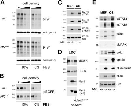
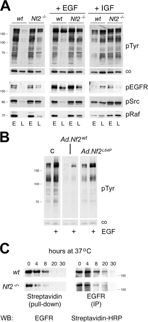
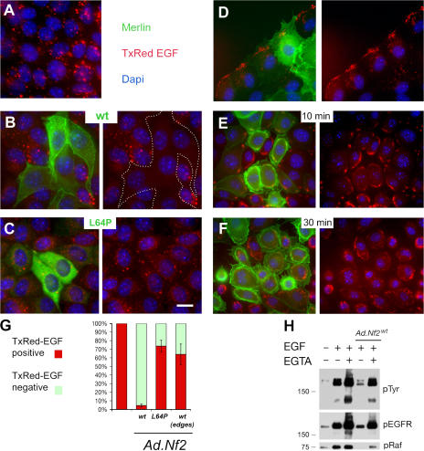
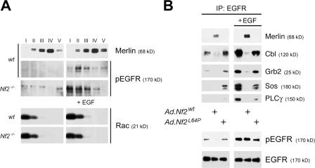
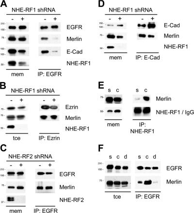
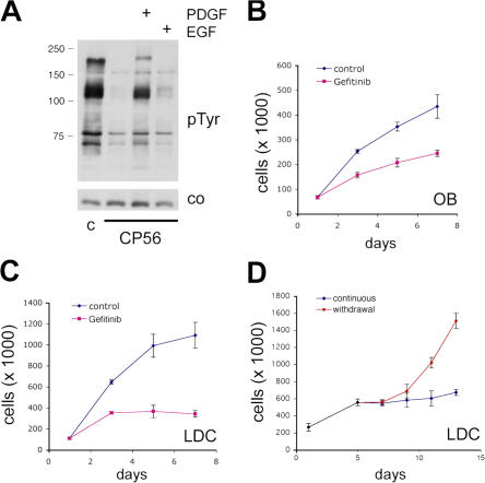
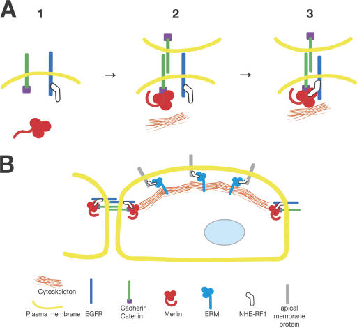
The neurofibromatosis type 2 (NF2) tumor suppressor, Merlin, is a membrane/cytoskeleton-associated protein that mediates contact-dependent inhibition of proliferation. Here we show that upon cell-cell contact Merlin coordinates the processes of adherens junction stabilization and negative regulation of epidermal growth factor receptor (EGFR) signaling by restraining the EGFR into a membrane compartment from which it can neither signal nor be internalized. In confluent Nf2(-/-) cells, EGFR activation persists, driving continued proliferation that is halted by specific EGFR inhibitors. These studies define a new mechanism of tumor suppression, provide mechanistic insight into the poorly understood phenomenon of contact-dependent inhibition of proliferation, and suggest a therapeutic strategy for NF2-mutant tumors.


Figures

Figure 1.
Figure 1.
Persistent EGFR signaling in multiple types of confluent Nf2−/− cells. (A) Tyrosine-phosphorylated proteins in total membrane preparations from MEFs at increasing stages of confluence. At early confluence (*) in the presence of serum, wild-type (top) and Nf2 −/− (bottom) cells display a similar pattern of pTyr; (see Fig. S2 for the definition of confluence). Progression to late confluence is accompanied by marked down-regulation of tyrosine kinase activity in wild-type, but not Nf2 −/− cells (left). In contrast, both wild-type and Nf2 −/− cells can down-regulate membrane pTyr after serum starvation (right). Actin = loading control. (B) EGFR activation, detected with an antibody against phosphorylated Y845, parallels the pTyr content observed in response to increasing confluence and serum starvation. In wild-type MEFs, pEGFR levels decrease as confluence progresses, but remain high in Nf2 −/− cells in a serum-dependent manner. (C) Phosphorylation of multiple EGFR tyrosine residues is down-regulated in wild-type late-confluent primary MEFs and OBs grown in serum, but persist in the absence of Merlin. (D) Membranes of confluent Nf2 −/− LDCs display high pEGFR levels that are markedly reduced upon adenoviral reintroduction of Nf2wt, but not mutant Nf2L64P. Nf2L64P is underrepresented in the membrane fraction, but comparable to that of Nf2wt in the total cell extract (tce). (E) Persistent activation of multiple EGFR targets is evident in membrane extracts from confluent Nf2 −/− cells compared with wild-type. Phosphorylated active forms are indicated by the letter “p” preceding the name of the protein. All experiments were performed at least three times.
Figure 2.
Figure 2.
Merlin prevents EGFR signaling in confluent cells. (A) In late-confluent (L) wild-type MEFs, EGF-induced pTyr is limited to the EGFR without concomitant activation of EGFR downstream effectors such as Src and Raf. Increased membrane pTyr is already present in late-confluent Nf2 −/− MEFs. The response to IGF-I is not prevented by Merlin. (EGF: 40 ng/ml, 30 min; IGF-I: 100 ng/ml, 30 min) (E = early confluence; co = loading controls). (B) Reintroduction of Nf2wt but not Nf2L64P abrogates persistent membrane pTyr in late-confluent Nf2 −/− MEFs; as in wild-type MEFs, EGF stimulation of Nf2wt-expressing MEFs affects pTyr of only the EGFR itself. (C) Surface EGFR turnover in late-confluent OBs. Confluent wild-type and Nf2 −/− OBs were surface biotinylated at 4°C for 1 h and then shifted to 37°C in the presence of serum. At the indicated time points, the amount of biotinylated EGFR remaining in the cell was evaluated in both the total biotinylated fraction and in EGFR immunoprecipitates. Compared with wild-type OBs (top panels), Nf2 −/− OBs (bottom panels) show accelerated clearing of surface-biotinylated EGFR. All experiments were performed at least three times.
Figure 3.
Figure 3.
Control of EGFR internalization and signaling by Merlin is contact dependent. (A–F) Internalization of fluorescent Tr-EGF (2 μg/ml, 30 min) in confluent LDCs. (green = Merlin; red = Tr-EGF; blue = DAPI). (A) Tr-EGF containing vesicles are found within virtually every Nf2 −/− LDC cell. (B) Internalized Tr-EGF is rarely observed in cells expressing wild-type Merlin. Dotted lines demarcate non-internalizing Nf2wt-expressing cells. (C) Tr-EGF internalization is not prevented in cells expressing Nf2L64P or, (D) in cells that express Nf2wt but are situated at the free edge of a scrape wound. In these cells, Tr-EGF internalization occurs along the free edge itself. (E and F) Disruption of intercellular adhesion by Ca2+ depletion promotes Tr-EGF internalization in confluent Nf2wt-expressing LDCs. E = 10 min; F = 30 min after Tr-EGF addition. Bar, 10 μm. (G) Average percentage distribution of ligand-internalizing cells in relation to Merlin expression and cell density. 200 Nf2wt or Nf2L64P-expressing cells enclosed within a confluent monolayer, or Nf2wt cells situated at the free edges of nonconfluent cultures, were scored in each of four separate experiments. Cells with internalized Tr-EGF were designated as positive (error bars, ± SD). (H) Disruption of intercellular adhesion by Ca2+-depletion restores EGFR signaling in Nf2wt-expressing LDCs. EGF (40 ng/ml, 30 min) was added to starved LDCs after 30 min preincubation in EGTA/Ca2+-free medium.
Figure 4.
Figure 4.
Merlin controls EGFR membrane distribution and access to downstream effectors. (A) Optiprep gradient fractionation of Triton-insoluble membranes from late-confluent wild-type and Nf2 −/− MEFs reveals altered physical distribution of pEGFR in Nf2 −/− cells after EGF stimulation (40 ng/ml, 30 min). In wild-type cells, pEGFR appears predominantly in fractions II and III, where Merlin is also present; both are excluded from the lowest density fraction I. In the absence of Merlin a unique pool of pEGFR appears in fraction I. In contrast, distribution of the small GTPase Rac is independent of Merlin status. (B) After reintroduction into Nf2 −/− LDCs, Nf2wt but not Nf2L64P complexes with EGFR and prevents its interaction with multiple signaling effectors in response to EGF (40 ng/ml, 10 min). Experimental conditions are as in Fig. 1 D. All experiments were performed at least three times.
Figure 5.
Figure 5.
NHE-RF1–mediated, contact-dependent association of Merlin and EGFR. (A) Immunoprecipitation of EGFR from total membrane extracts of Nf2wt-expressing LDCs with and without lentiviral NHE-RF1 shRNA expression revealed that the association of both Nf2wt and E-cadherin with the EGFR are nearly eliminated by NHE-RF1 knockdown. (B) Immunoprecipitation of Ezrin reveals that the association of Nf2wt with Ezrin occurs in a NHE-RF1–independent manner. (C) NHE-RF2 knockdown does not affect EGFR-Merlin association. (D) The association between Nf2wt and E-cadherin is not affected by NHE-RF1 down-regulation. (E) Immunoprecipitation of NHE-RF1 from total membrane extracts of subconfluent (s) and confluent (c) Nf2wt-expressing LDCs revealed that the association between Nf2wt and NHE-RF1 occurs only in confluent conditions. Total IgG is shown as a control for the NHE-RF1 immunoprecipitations. (F) Immunoprecipitation of EGFR from total membrane extracts of Nf2wt-expressing LDCs when sparse (s), confluent (c), and after cell–cell contact disruption by incubation in Ca2+-depleted medium for 45 min (d). Complexing of Merlin and EGFR markedly increases from sparse to confluent cells and is abrogated by acute loss of intercellular contacts. All experiments were performed at least three times.
Figure 6.
Figure 6.
Pharmacological inhibitors of EGFR arrest proliferation of Nf2−/− cells. (A) Tyrosine phosphorylation of membrane proteins in late-confluent Nf2 −/− MEFs is eliminated by the EGFR inhibitor Compound 56. In the presence of the inhibitor (added 2 h before EGF addition), pTyr induction is blocked after stimulation with EGF (40 ng/ml) but not PDGF (35 ng/ml) for 30 min. Similar specificity was observed for Gefitinib (not depicted). Proliferation of Nf2 −/− OBs (B) and LDCs (C) at high cell density is inhibited by Gefinitib. (D) Inhibition of EGFR by Gefitinib does not permanently restore contact inhibition of growth in LDCs. All experiments were performed at least three times. (Error bars, ± SD).
Figure 7.
Figure 7.
Molecular and spatial attributes of Merlin function. (A) Model depicting the mechanism of Merlin-mediated coordination of AJ stabilization and EGFR down-regulation. In non-contacting cells (1), Merlin does not associate with cadherin or EGFR; however, upon cell–cell contact (2), Merlin is recruited to nascent junctions and activated, stabilizing the interaction between junctional proteins and the cortical cytoskeleton. Activated Merlin then “captures” NHE-RF1-associated EGFR (3), preventing it from internalizing or signaling. Active Merlin is likely hypophosphorylated and could be in an “open” or “closed” (self-associated) conformation; indeed, more than two conformational states of Merlin may exist. Although Merlin lacks the C-terminal actin-binding domain present in the ERM proteins, some studies suggest that Merlin can interact directly with filamentous actin via the FERM domain. Despite an abundance of evidence linking Merlin physically and functionally to the actin cytoskeleton, the precise mechanism whereby Merlin associates with the actin cytoskeleton remains unclear. (B) Although Merlin and the ERM proteins can all interact with NHE-RF1 and NHE-RF1 can, in turn, interact with several membrane receptors, Merlin may be functionally dedicated to the junctional domain and the ERM proteins to the apical domain. In this way, spatial and temporal regulation of NHE-RF1–associated receptors may be achieved.

Similar articles

Cited by

References

    1. Adams, C.L., W.J. Nelson, and S.J. Smith. 1996. Quantitative analysis of cadherin-catenin-actin reorganization during development of cell-cell adhesion. J. Cell Biol. 135:1899–1911. - PMC - PubMed
    1. Bailey, S.N., S.M. Ali, A.E. Carpenter, C.O. Higgins, and D.M. Sabatini. 2006. Microarrays of lentiviruses for gene function screens in immortalized and primary cells. Nat. Methods. 3:117–122. - PubMed
    1. Baser, M.E., J.M. Friedman, A.J. Wallace, R.T. Ramsden, H. Joe, and D.G. Evans. 2002. Evaluation of clinical diagnostic criteria for neurofibromatosis 2. Neurology. 59:1759–1765. - PubMed
    1. Bennett, F.C., and K.F. Harvey. 2006. Fat cadherin modulates organ size in Drosophila via the Salvador/Warts/Hippo signaling pathway. Curr. Biol. 16:2101–2110. - PubMed
    1. Betson, M., E. Lozano, J. Zhang, and V.M. Braga. 2002. Rac activation upon cell-cell contact formation is dependent on signaling from the epidermal growth factor receptor. J. Biol. Chem. 277:36962–36969. - PubMed

Publication types