Comparative Study
. 2007 Jun 11;204(6):1335-47.
doi: 10.1084/jem.20070081. Epub 2007 Jun 4.

Altering the distribution of Foxp3(+) regulatory T cells results in tissue-specific inflammatory disease

Affiliations
Comparative Study

Altering the distribution of Foxp3(+) regulatory T cells results in tissue-specific inflammatory disease

Blythe D Sather et al. J Exp Med. .

Abstract

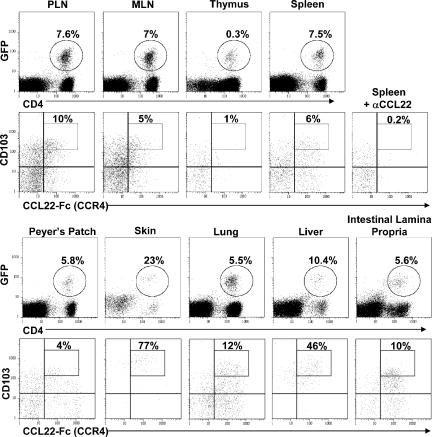
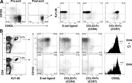
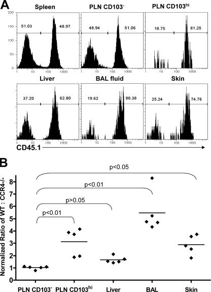
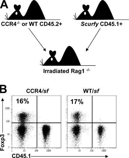
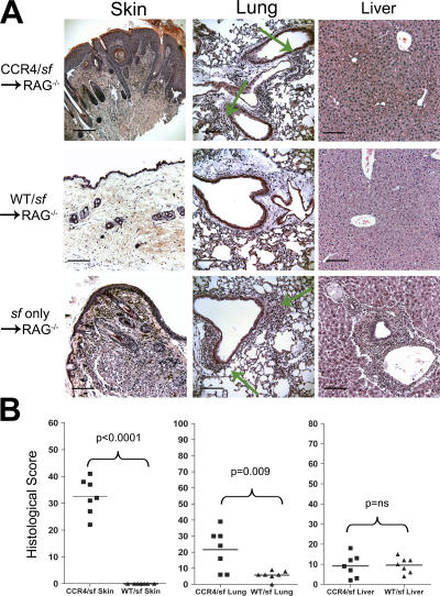
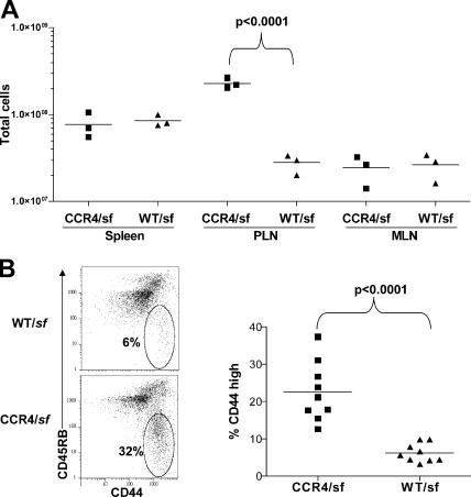
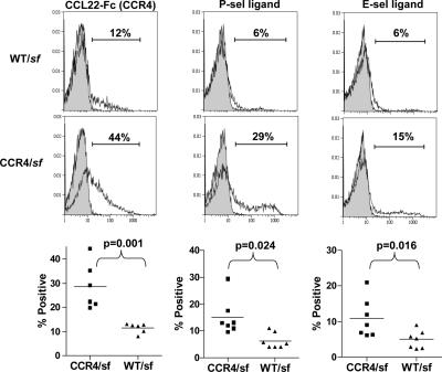
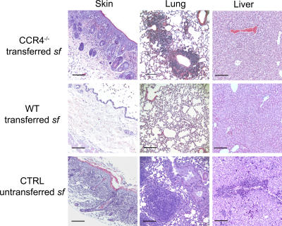
CD4(+)Foxp3(+) regulatory T cells (T reg) are essential for maintaining self-tolerance, but their functional mechanisms and sites of action in vivo are poorly defined. We examined the homing receptor expression and tissue distribution of T reg cells in the steady state and determined whether altering their distribution by removal of a single chemokine receptor impairs their ability to maintain tissue-specific peripheral tolerance. We found that T reg cells are distributed throughout all nonlymphoid tissues tested, and are particularly prevalent in the skin, where they express a unique CCR4(+)CD103(hi) phenotype. T reg cell expression of CCR4 and CD103 is induced by antigen-driven activation within subcutaneous lymph nodes, and accumulation of T reg cells in the skin and lung airways is impaired in the absence of CCR4 expression. Mice with a complete loss of CCR4 in the T reg cell compartment develop lymphocytic infiltration and severe inflammatory disease in the skin and lungs, accompanied by peripheral lymphadenopathy and increased differentiation of skin-tropic CD4(+)Foxp3(+) T cells. Thus, selectively altering T reg cell distribution in vivo leads to the development of tissue-specific inflammatory disease.


Figures

Figure 1.
Figure 1.
CD4+Foxp3+ T cells are resident in both lymphoid and nonlymphoid tissues. (first and third rows) Flow cytometry analysis of CD4 and GFP expression by lymphocytes isolated from the spleen, skin-draining PLNs, MLNs, thymus, Peyer's patches, skin, lung parenchyma, liver, and intestinal lamina propria of a 12-wk-old Foxp3gfp mouse. Percentage indicates the fraction of CD4+ cells expressing GFP. (second and fourth rows) Expression of CD103 and CCR4 by gated CD4+GFP+cells from each tissue. Percentage indicates the fraction of T reg cells that are CD103highCCR4+ as defined by the rectangular gate in the upper right quadrant. For the CCR4-negative control, spleen cells were stained with CCL22-Fc fusion protein that was preincubated for 5 min with a neutralizing anti-CCL22 monoclonal antibody. Results are representative of greater than six mice analyzed in this fashion
Figure 2.
Figure 2.
T reg cells alter their tissue tropism after antigen stimulation. (A) Flow cytometry analysis of CD62L and CD103 expression by gated CD4+CD25+ cells from the pooled spleen and PLN of a DO11. 10xRIP-mOVA mouse before and after sorting of CD62L+CD103 cells (left). (right) Expression of the DO11.10 clonotypic TCR (KJ1-26), E-selectin ligand, CCR4, and CCR7 by the sorted CD62L+CD103 T reg cells. (B) Flow cytometry analysis of lymphocytes from the draining inguinal LNs of recipient mice 5 d after immunization with OVA+CT (top) or OVA alone (bottom). (left) Gates used to define the OVA-specific T reg cells. Percentages indicate the frequency of KJ1-26+ cells among total CD4+ cells. (right) Expression of CD103, E-selectin ligand, CCR4, and CCR7 by gated CD4+KJ1-26+ cells.
Figure 3.
Figure 3.
Impaired accumulation of CCR4−/− T reg cells in the skin and lung airways. (A) Representative flow cytometry analysis of CD45.1 expression by gated CD4+Foxp3+ cells from the indicated tissues of a WT+CCR4−/− mixed BM chimera. WT cells are CD45.1+. (B) The normalized ratio of WT to CCR4−/− T reg cells in the indicated tissues/compartments was derived by dividing the ratio of WT/CCR4−/− T reg cells in each by the ratio of WT/CCR4−/− T reg cells in the spleen. Each data point represents the normalized ratio from one individual chimera. Horizontal lines indicate the average normalized ratio (n = 5) in each tissue/compartment. Statistical analysis was performed using a one-way repeated measures ANOVA (P < 0.0001). P-values for the indicated pairwise comparisons were then computed using Dunnett's Multiple Comparison Test.
Figure 4.
Figure 4.
Schematic of mixed BM chimera experimental setup and Foxp3+ T cell development. (A) Schematic representation of mixed BM chimera experimental set-up. (B) Representative flow cytometry analysis of CD45.1 and Foxp3 expression by gated CD4+ T cells from the PLN of one pair of CCR4/sf and WT/sf chimeras killed 153 d after BM transplant.
Figure 5.
Figure 5.
Inflammatory disease in the skin and lungs of CCR4/sf chimeras. (A) Photomicrographs (20×) of hematoxylin and eosin–stained sections of the skin, lung, and liver from CCR4/sf, WT/sf (killed 114 d after BM transplant), or sf only chimeras (killed 38 d after BM transplant). Green arrows indicate the location of inflammatory infiltrates in the lungs of CCR4/sf and sf chimeras. (B) Blinded analysis of tissue sections from seven pairs of CCR4/sf and WT/sf chimeras (all killed in pairs 114–250 d after BM transplant). Each section was scored based on the severity and extent of inflammation. Statistical analysis was performed using two-tailed, paired Student's t test. ns, not significantly different (P > 0.05). Bars, 100 μm.
Figure 6.
Figure 6.
Peripheral lymphadenopathy and enhanced CD4+ T cell activation in CCR4/sf chimeras. (a) Lymphocytes were isolated and counted from the spleen, subcutaneous PLNs, and MLNs of a matched group of three CCR4/sf chimeras (squares) and three WT/sf chimeras (triangles) killed 140 d after BM transplant. Data are representative of six experiments. Statistical analysis was performed using a two-tailed, unpaired Student's t test. (b) Representative flow cytometry analysis of CD44 and CD45RB expression by gated CD4+CD45.1+ sf-derived T cells from the PLN of one pair of WT/sf and CCR4/sf chimeras killed 140 d after BM transplant. (right) The frequency of CD44hi cells amongst gated CD4+CD45.1+ PLN cells from nine matched pairs of WT/sf- and CCR4/sf chimeras (killed 114–250 d after BM transplant). Statistical analysis was performed using a two-tailed, paired Student's t test.
Figure 7.
Figure 7.
CCR4/sf chimeras have an elevated frequency of skin-tropic CD4+ T cells. (top) Representative flow cytometry analysis of CCR4, P-selectin ligand, and E-selectin ligand expression by gated CD4+CD45.1+ sf-derived T cells from the PLN of WT/sf and CCR4/sf chimeras (open area) killed 140 d after BM transplant. Shaded areas of the graph indicate background staining in the presence of anti-CCL22 (left) or 10 mM EDTA (middle and right). (bottom) Graphs indicating the frequency of gated CD4+CD45.1+ cells expressing the indicated receptor in the PLNs of five matched pairs of WT/sf- and CCR4/sf chimeras (all killed 114–250 d after BM transplant). Statistical analysis was performed using a two-tailed, paired Student's t test.
Figure 8.
Figure 8.
CCR4−/− T reg cells fail to prevent cutaneous and pulmonary inflammation after transfer into neonatal sf mice. Photomicrographs of hematoxylin and eosin–stained sections of skin, lung, and liver from either an unmanipulated sf mouse (bottom) or from sf mice given purified WT or CCR4−/− CD4+CD25+ T reg cells (top and middle) shortly after birth. Data are representative of more than eight mice analyzed in each group. Bars, 100 μm

References

    1. Sakaguchi, S. 2004. Naturally arising CD4+ regulatory t cells for immunologic self-tolerance and negative control of immune responses. Annu. Rev. Immunol. 22:531–562. - PubMed
    1. Szanya, V., J. Ermann, C. Taylor, C. Holness, and C.G. Fathman. 2002. The subpopulation of CD4+CD25+ splenocytes that delays adoptive transfer of diabetes expresses L-selectin and high levels of CCR7. J. Immunol. 169:2461–2465. - PubMed
    1. Salomon, B., D.J. Lenschow, L. Rhee, N. Ashourian, B. Singh, A. Sharpe, and J.A. Bluestone. 2000. B7/CD28 costimulation is essential for the homeostasis of the CD4+CD25+ immunoregulatory T cells that control autoimmune diabetes. Immunity. 12:431–440. - PubMed
    1. Hori, S., M. Haury, A. Coutinho, and J. Demengeot. 2002. Specificity requirements for selection and effector functions of CD25+4+ regulatory T cells in anti-myelin basic protein T cell receptor transgenic mice. Proc. Natl. Acad. Sci. USA. 99:8213–8218. - PMC - PubMed
    1. Kohm, A.P., P.A. Carpentier, H.A. Anger, and S.D. Miller. 2002. Cutting edge: CD4+CD25+ regulatory T cells suppress antigen-specific autoreactive immune responses and central nervous system inflammation during active experimental autoimmune encephalomyelitis. J. Immunol. 169:4712–4716. - PubMed

Publication types

MeSH terms