. 2007 Jun 15;178(12):7598-606.
doi: 10.4049/jimmunol.178.12.7598.

Impaired accumulation of antigen-specific CD8 lymphocytes in chemokine CCL25-deficient intestinal epithelium and lamina propria

Affiliations

Impaired accumulation of antigen-specific CD8 lymphocytes in chemokine CCL25-deficient intestinal epithelium and lamina propria

Marc-André Wurbel et al. J Immunol. .

Abstract

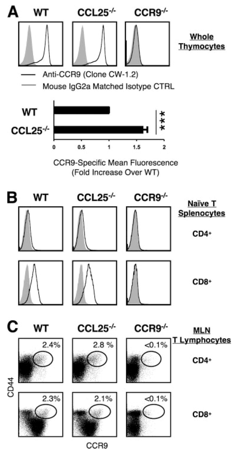
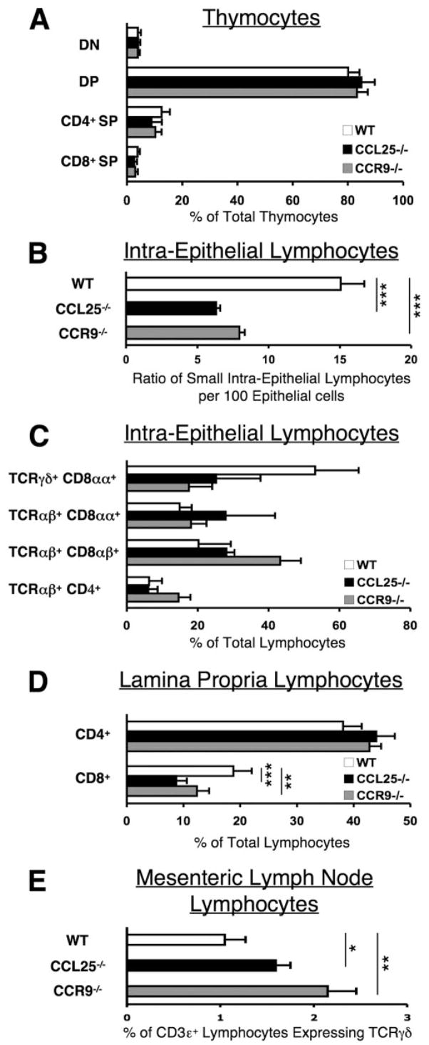
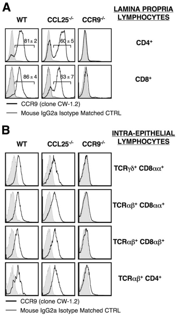
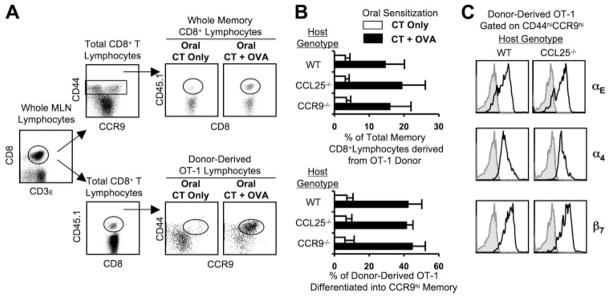
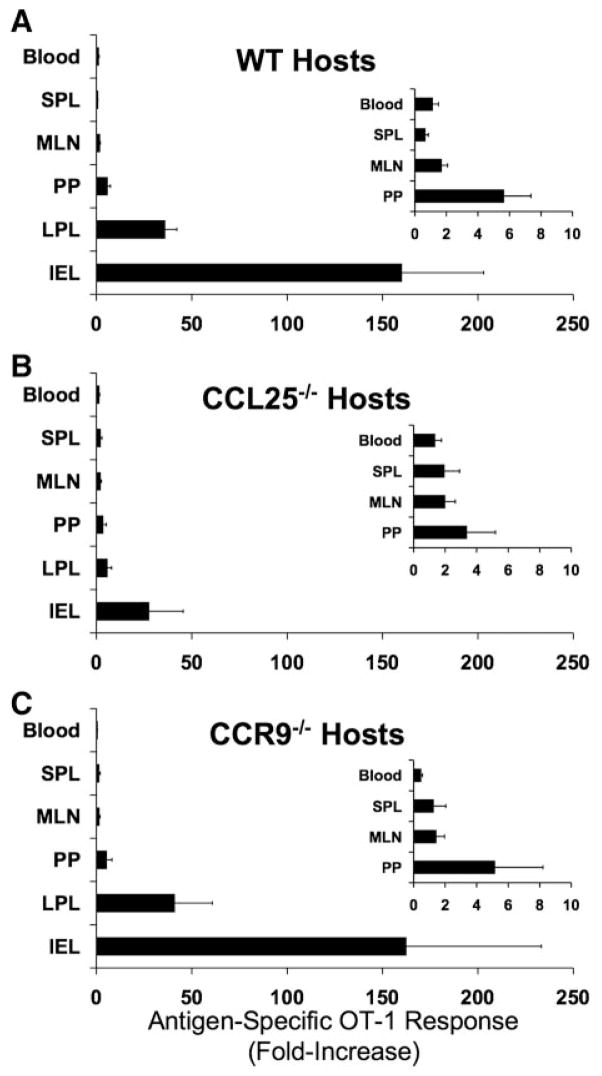
CCL25 and CCR9 constitute a chemokine/receptor pair involved in T cell development and in gut-associated immune responses. In this study, we generated CCL25(-/-) mice to answer questions that could not be addressed with existing CCR9(-/-) mice. Similar phenotypes were observed for both CCL25(-/-) and CCR9(-/-) mice, consistent with the notion that CCL25 and CCR9 interact with each other exclusively. We assessed the requirement for CCL25 in generating CCR9(high) CD8 intestinal memory-phenotype T cells and the subsequent accumulation of these cells within effector sites. TCR-transgenic naive CD8 T cells were transferred into wild-type or CCL25-deficient hosts. Oral sensitization with Ag allowed these naive donor cells to efficiently differentiate into CCR9(high) memory-phenotype cells within the mesenteric lymph nodes of wild-type hosts. This differentiation event occurred with equal efficiency in the MLN of CCL25-deficient hosts, demonstrating that CCL25 is not required to induce the CCR9(high) memory phenotype in vivo. However, we found that CCL25 deficiency severely impaired the Ag-dependent accumulation of donor-derived CD8 T cells within both lamina propria and epithelium of the small intestine. Thus, although CCL25 is not necessary for generating memory-phenotype CD8 T cells with "gut-homing" properties, this chemokine is indispensable for their trafficking to the small intestine.


Conflict of interest statement

Disclosures

The authors have no financial conflict of interest.

Figures

Figure 1
Figure 1
Generation and identification of CCL25-deficient mice. A, 1) Partial restriction map of the WT CCL25 gene. Exons 1– 6 are shown as gray boxes. The restriction sites are B (BglII), E (EcoR V), H (HindIII), and X (XbaI) (2). Targeting vector used for the deletion of the 3′ end of exon 2 (starting at the BglII restriction site and including ATG), exon 3 and exon 4 (containing the four cysteine residues crucial for the folding of CCL25). One diagnostic XbaI restriction site was introduced at the 5′ end of the loxP-flanked neomycin resistance gene (neo), whereas a diagnostic BglII restriction site was lost in exon 2. □, Correspond to the thymidine kinase expression cassette (tk), to the loxP-flanked neomycin gene, and to the pBluescript IIKS+ vector (pBS) (3). Structure of the targeted allele following homologous recombination (4). Final structure of the targeted allele after removal of the neomycin-resistance gene via Cre-mediated recombination with Cre Deleter mice. ■, The 5′ and 3′ single-copy probes used to verify the targeting events; the position of the primers used to monitor the germline transmission of the intended mutation are indicated by arrows. B, Southern blot analysis of the rES cell clone IIID7 that gave germline transmission before deletion of the neomycin-resistance gene. DNA was digested with BglII and hybridized with the 5′ single copy probe. C, DNA-PCR analysis of wild-type (+/+), neomycin-deleted heterozygous (+/−), and neomycin-deleted homozygous (−/−) littermates using the pair of primers shown in A. Targeted CCL25 allele gave an amplified PCR product of 510 bp, whereas the wild-type allele gave an amplified PCR product of 280 bp. Amplified products were run on an agarose gel and stained with ethidium bromide.
Figure 2
Figure 2
CCR9 expression by thymocytes and mature lymphocytes from WT and CCL25-deficient mice. A and B, Cells from thymus and SPL of WT, CCL25−/−, and CCR9−/− mice were stained and analyzed by flow cytometry. CCR9 cell surface expression was assessed by mAb CW-1.2 (black lines) and overlaid on isotype-matched mIgG2a control (gray filled curves). A, Upper panels, CCR9 expression on unfractionated thymocytes (gated only by scatter). Lower panel, CCL25-deficient thymocytes expressed CCR9 >1.5-fold more brightly (by mean fluorescence) than WT thymocytes (***, p < 0.05 in a Wilcoxon-signed rank, n = 5). B, Upper panels, CCR9 expression by CD4+ naive T splenocytes (gated as CD4+/B220/CD44low/CD45RBhigh lymphocytes). No significant CCR9 expression was observed for any of the three genotypes. Bottom row, CCR9 expression by CD8+ naive T splenocytes (gated as CD3ε+/CD8+/CD44low/PNAlow lymphocytes). CCR9 expression was not significantly different between WT and CCL25−/− mice. C, CD44 vs CCR9 expression on CD4+ and CD8+ T cells from MLN of WT, CCL25−/−, and CCR9−/− mice. Top row, Staining of CD4+ T cells (gated as CD4+/B220 lymphocytes). There was no significant difference in the representation of CD44high/CCR9high memory cells between WT and CCL25−/− MLN within the CD4+ population. Bottom row, Staining of CD8+ T cells (gated as CD3ε+/CD8+ lymphocytes). There was no significant difference in the representation of CD44high/CCR9high memory cells between WT and CCL25−/− MLN within the CD8+ population. It should also be noted that this staining protocol also highlights the fact that CD44low naive CD8 T cells are clearly CCR9low in the WT and CCL25−/− MLN, but are CCR9neg in the CCR9−/− MLN. All flow cytometry panels in this figure depict individual experiments and represent more than five repeats.
Figure 3
Figure 3
Analysis of subset composition within thymus, small intestine compartments and MLN of WT and CCL25-deficient mice. A, The percentage of DN, DP, CD4+ SP, and CD8+ SP thymocytes are shown for thymocytes from WT, CCL25−/−, and CCR9−/− mice, as indicated (gated by scatter and CD4 vs CD8 expression). Mean and SEM are shown for 10 experiments. No significant differences were detected among the three different genotypes. B, Ratio of small IEL per 100 EC was assessed on duodenal tissue sections from WT, CCL25−/−, and CCR9−/− intestines, as indicated. Mean and SEM are shown for six experiments. Values of p for divergence from WT ratios were calculated by ANOVA with Bonferroni correction as indicated by asterisks (***, p < 0.001). C, The percentage of major IEL subsets are shown from WT, CCL25−/−, and CCR9−/− small intestine, as indicated. Mean and SEM are shown for 12 experiments. D, The percentage of LPL T cell subsets (as percent of total lymphocytes) is shown from WT, CCL25−/−, and CCR9−/− small intestines, as indicated. LPL were stained with CD3ε, CD4, CD8, and B220 mAbs, and analyzed by flow cytometry. Data are presented as mean ± SEM for 12 experiments. Values of p for divergence from WT ratios were calculated by an ANOVA with Bonferroni correction as indicated by asterisks (***, p < 0.001; **, p < 0.01). E, The percentage of TCRγδ+ T cell subset (as percent of total CD3ε+ T lymphocytes) in MLN is depicted for WT, CCL25−/−, and CCR9−/− mice. Mean and SEM are shown for six experiments. Values of p for divergence from WT ratios were calculated by an ANOVA with Bonferroni correction as indicated by asterisks (*, p < 0.01; **, p < 0.001).
Figure 4
Figure 4
CCR9 cell surface expression on LPL and IEL. A, Whole LPL from WT, CCL25-deficient, and CCR9-deficient mice were obtained by collagenase/DNase digestion of WT, CCL25-deficient, and CCR9-deficient small intestines after elimination of all IEL with EGTA. CD4+ and CD8+ LPL were stained with anti-CCR9 mAb (clone CW-1.2, in black) overlaid on mouse IgG2a-matched isotype control (in gray) and analyzed by flow cytometry. Data are from one representative experiment of six with similar results. B, Flow cytometry analysis of CCR9 cell surface expression (clone CW-1.2, in black) on major small intestinal IEL subsets from WT, CCL25-deficient, and CCR9-deficient mice (TCRγδ+ CD8αα+, TCRαβ+ CD8αα+, TCRαβ+ CD8αβ+, and TCRαβ+ CD4+) overlaid on mouse IgG2a matched isotype control. Data are from a single representative experiment of six repeats.
Figure 5
Figure 5
Ag-specific CCR9high memory MLN lymphocytes are generated in the absence of CCL25 chemokine expression. Splenocytes were isolated from CD45.1+ OT-1 TCR-Tg mice and adoptively transferred into CD45.2+ WT hosts. The hosts were then orally sensitized with OVA + cholera toxin (CT) or with CT alone. Four days after oral sensitization, MLN lymphocytes were harvested and analyzed by flow cytometry. A, Two different cytometry gating schemes are shown for analyzing OT-1 cells in adoptive hosts after oral immunization with OVA + CT or CT only. The gating scheme on top displays the percentage of total memory CD8 T cells within the host MLN that were derived from the OT-1 donor after oral immunization, and was used to calculate the data shown in part B, upper panel. The gating scheme on the bottom displays the percentage of donor OT-1 CD8 T cells that have differentiated into CD44high/CCR9high memory cells after oral immunization, and was used to calculate the data shown in part B, lower panel. B, Generation of CCR9high memory-phenotype OT-1 cells in WT, CCL25−/−, and CCR9−/− MLN using gating criteria described in A. Percentages of donor-derived OT-1 cells that differentiated into CCR9high memory-phenotype (lower panel) and percentage of total memory CD8+ lymphocytes derived from OT-1 donor (upper panel) are shown. Data are presented as mean and SEM of eight experiments. No significant differences were detected among the three different genotypes in ANOVA with Bonferroni correction. C, Expression of αE, α4, and β7 integrins by donor-derived OT-1 cells that differentiated into CCR9high memory. CCR9high differentiated donor-derived OT-1 memory lymphocytes in WT and CCL25-deficient hosts depicted in A and B (lower panel) were staining with anti-αE (clone M290), anti-α4 (clone 9C10), and anti-β7 (clone FIB-504) mAbs (black lines) and overlaid on isotype-matched rIgG2a controls (gray filled curves). Data are from a single representative experiment of three repeats.
Figure 6
Figure 6
Response to oral immunization, but not i.p. immunization, results in differentiation of intestinal memory-phenotype cells that are largely restricted to intestine and GALT. Naive CD45.1+ OT-1 TCR-Tg lymphocytes were isolated and adoptively transferred into CD45.2+ WT and CD45.2+ CCL25-deficient recipient mice. The hosts were then treated either orally or i.p. with OVA + CT. Four days after sensitization, PLN and MLN lymphocytes were harvested and analyzed by flow cytometry as depicted in Fig. 5 (A and B, lower panel). The gating scheme displays the percentage of donor OT-1 CD8 T cells that have differentiated into CD44high/CCR9high memory cells after i.p. (left) and oral (right) immunization obtained in PLN (upper panel) and in MLN (lower panel) in WT and CCL25-deficient host mice. Dot plots are from a single representative experiment of four repeats.
Figure 7
Figure 7
Ag-specific lymphocyte homing to small intestine LP and epithelium is dramatically impaired in CCL25-deficient mice. A, Lamina propria CD8+ T lymphocytes (LPL) and (B) intraepithelial CD8+ lymphocytes (IEL) were harvested, stained, and analyzed by flow cytometry. The percentage of total CD8+ T cells derived from the OT-1 donor at day 4 after oral gavage is shown. Data are presented as the mean and SEM of four or six mice as indicated by N. Values of p as calculated by ANOVA with Bonferroni correction as indicated by asterisks (***, p < 0.05; ns: not significant).
Figure 8
Figure 8
Ag-specific enrichment of OT-1 lymphocytes after oral gavage is specific to intestine-associated tissues. Ag-specific fold-increase in OT-1 representation was calculated for the total CD8+ T cell populations between pairs of mice treated with CT only or OVA + CT in various organs at day 4 after oral gavage. The ratios were calculated for blood, SPL, MLN, PP, LPL, and IEL. This was calculated for (A) WT hosts, (B) CCL25−/− hosts, and (C) CCR9−/− hosts. Mean and SEM are shown for four pairs of mice for each genotype. The differences between WT and CCL25−/− hosts were highly significant for LPL (p < 0.05) and IEL (p < 0.001) by ANOVA test with Bonferroni correction, but were not significant between WT and CCR9−/− hosts. Insets for each genotype (A–C) show the identical data for only lymphoid organs on a smaller scale.

References

    1. Vicari AP, Figueroa DJ, Hedrick JA, Foster JS, Singh KP, Menon S, Copeland NG, Gilbert DJ, Jenkins NA, Bacon KB, Zlotnik A. TECK: a novel CC chemokine specifically expressed by thymic dendritic cells and potentially involved in T cell development. Immunity. 1997;7:291–301. - PubMed
    1. Wurbel MA, Philippe JM, Nguyen C, Victorero G, Freeman T, Wooding P, Miazek A, Mattei MG, Malissen M, Jordan BR, et al. The chemokine TECK is expressed by thymic and intestinal epithelial cells and attracts double- and single-positive thymocytes expressing the TECK receptor CCR9. Eur J Immunol. 2000;30:262–271. - PubMed
    1. Zabel BA, Agace WW, Campbell JJ, Heath HM, Parent D, Roberts AI, Ebert EC, Kassam N, Qin S, Zovko M, et al. Human G protein-coupled receptor GPR-9 – 6/CC chemokine receptor 9 is selectively expressed on intestinal homing T lymphocytes, mucosal lymphocytes, and thymocytes and is required for thymus-expressed chemokine-mediated chemotaxis. J Exp Med. 1999;190:1241–1256. - PMC - PubMed
    1. Benz C, Heinzel K, Bleul CC. Homing of immature thymocytes to the subcapsular microenvironment within the thymus is not an absolute requirement for T cell development. Eur J Immunol. 2004;34:3652–3663. - PubMed
    1. Uehara S, Hayes SM, Li L, El-Khoury D, Canelles M, Fowlkes BJ, Love PE. Premature expression of chemokine receptor CCR9 impairs T cell development. J Immunol. 2006;176:75– 84. - PubMed

Publication types

MeSH terms