. 2007 Jun 15;178(12):7814-21.
doi: 10.4049/jimmunol.178.12.7814.

Protein kinase Cdelta regulates antigen receptor-induced lytic granule polarization in mouse CD8+ CTL

Affiliations

Protein kinase Cdelta regulates antigen receptor-induced lytic granule polarization in mouse CD8+ CTL

Jennifer S Y Ma et al. J Immunol. .

Abstract

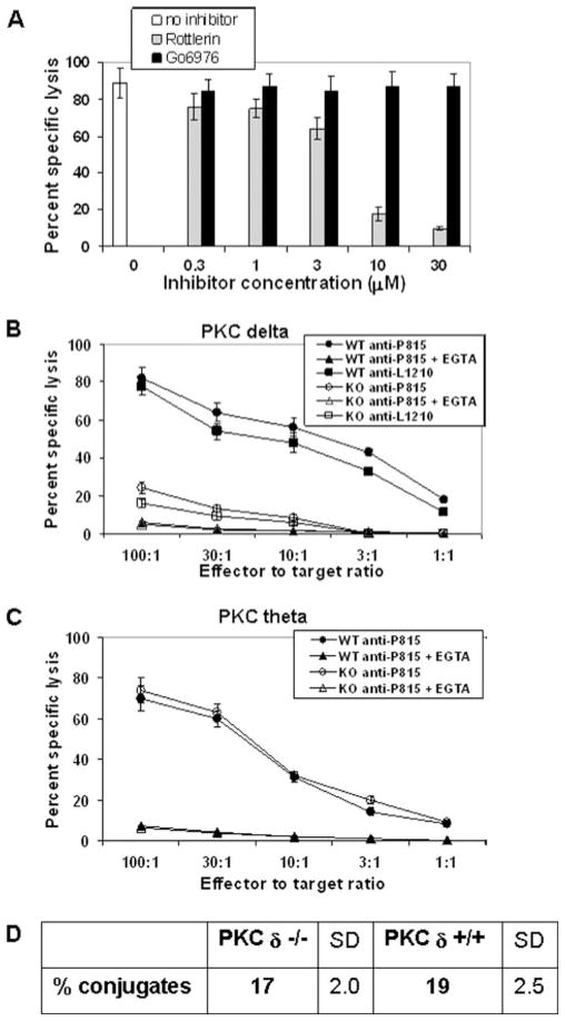
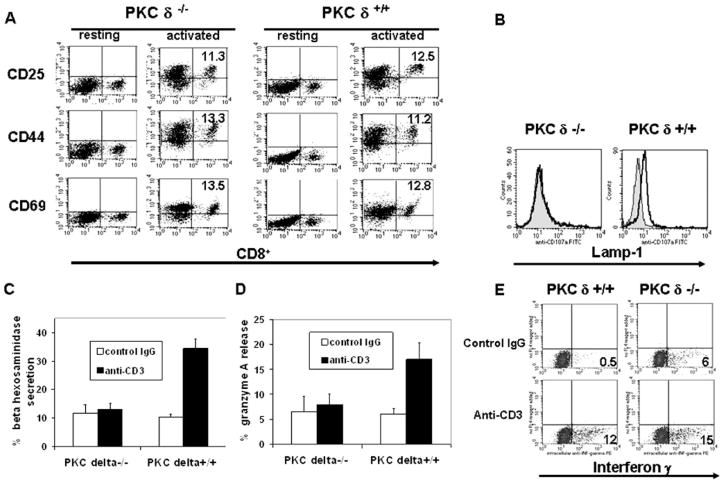
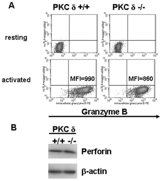
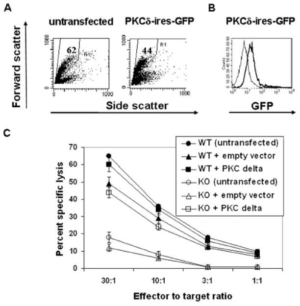
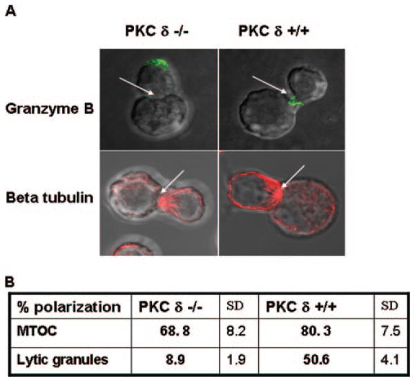
Lytic granule exocytosis is the major pathway used by CD8+ CTL to kill virally infected and tumor cells. Despite the obvious importance of this pathway in adaptive T cell immunity, the molecular identity of enzymes involved in the regulation of this process is poorly characterized. One signal known to be critical for the regulation of granule exocytosis-mediated cytotoxicity in CD8+ T cells is Ag receptor-induced activation of protein kinase C (PKC). However, it is not known which step of the process is regulated by PKC. In addition, it has not been determined to date which of the PKC family members is required for the regulation of lytic granule exocytosis. By combination of pharmacological inhibitors and use of mice with targeted gene deletions, we show that PKCdelta is required for granule exocytosis-mediated lytic function in mouse CD8+ T cells. Our studies demonstrate that PKCdelta is required for lytic granule exocytosis, but is dispensable for activation, cytokine production, and expression of cytolytic molecules in response to TCR stimulation. Importantly, defective lytic function in PKCdelta-deficient cytotoxic lymphocytes is reversed by ectopic expression of PKCdelta. Finally, we show that PKCdelta is not involved in target cell-induced reorientation of the microtubule-organizing center, but is required for the subsequent exocytosis step, i.e., lytic granule polarization. Thus, our studies identify PKCdelta as a novel and selective regulator of Ag receptor-induced lytic granule polarization in mouse CD8+ T cells.


Conflict of interest statement

Disclosures

The authors have no financial conflict of interest.

Figures

FIGURE 1
FIGURE 1
PKCδ is required for granule exocytosis-mediated cytotoxicity of mouse CD8+ CTL. A, C57BL/6 spleen cells (H-2b) were stimulated in vitro with irradiated BALB/c splenocytes (H-2d) for 5 days. CD8+ T cells (effectors) were isolated from the responder population by magnetic immunobead-ing mixed with 51Cr-labeled target P815 cells (H-2d) at different E:T ratios and assessed for the ability to lyse the targets after 4 h of coculture in the presence or absence of increasing concentrations of indicated inhibitors. Each E:T ratio was assayed in quadruplicate samples. For simplicity of the data interpretation, only the E:T of 10:1 is shown. This is a representative of four independent experiment yielding similar results. Error bars, SD. B and C, Cytolytic activity of in vitro-generated alloantigen-specific WT (H-2b) and (B) PKCδ-deficient (H-2b) or (C) PKCθ-deficient CTL (H-2b) against P815 cells, in the presence or absence of 1 mM EGTA, was tested in a 4-h chromium release assay at the indicated E:T ratios. Each E:T ratio was done in quadruplicate samples. B, In addition to P815 cells, Fas-resistant L1210 cells (H-2d) were used as targets. Data shown in B and C are representatives of five independent experiments yielding similar results. Error bars, SD. D, Quantification of conjugate formation of in vitro-generated alloantigen-specific WT (H-2b) and PKCδ-deficient (H-2b) CTL with P815 cells, as described in Materials and Methods. The mean values of three independent experiments are shown. KO, Knockout.
FIGURE 2
FIGURE 2
TCR-induced degranulation is defective in PKCδ-deficient CD8+ T cells. Total splenocytes from WT or PKCδ-deficient mice were cultured in the presence of anti-CD3 Ab. A, After 24 h, cell surface activation marker expression on CD8+ T cells was assessed by flow cytometry. B, Purified 2-day in vitro-activated CD8+ T cells were stimulated for 4 h with plate-bound anti-CD3 (tracing) or isotype-matched control Ab (filled histogram) in the presence of 10 μM monensin and 5 μg/ml FITC-conjugated anti-Lamp-1 Ab, followed by flow cytometry analysis of Lamp-1 expression. Staining of the cells with the Lamp-1 isotype-matched control Ab resulted in the fluorescence intensity equivalent to the one observed in the unstained cells. This experiment was repeated four times, giving similar results. C and D, After 2 days of activation, CD8+ T cells were purified by magnetic immunobeading, stimulated for 4 h with the plate-bound anti-CD3- or isotype-matched control Ab and were assayed for β-hexosaminidase (C) or granzyme A (D) release in quadruplicate samples. Error bars, SD. C and D, A representative of three independent experiments is shown. In all degranulation experiments, no significant death of CD8+ T cells upon degranulation was observed, as determined by trypan blue exclusion and propidium iodide staining followed by flow cytometry. E, After 2 days of activation, CD8+ T cells were purified by magnetic immunobeading and stimulated for 4 h with plate-bound anti-CD3- or isotype-matched control Ab in the presence of brefeldin A, followed by intracellular staining for IFN-γ and flow cytometry analyses. Staining of the cells with the isotype-matched control Ab for IFN-γ resulted in the fluorescence intensity equivalent to the one observed in the unstained cells. The numbers in the flow cytometry dot plots refer to the percentage of cells in a given quadrant. A representative of three independent experiments giving similar results is shown.
FIGURE 3
FIGURE 3
PKCδ-deficient CD8+ T cells express cytolytic molecules in response to TCR stimulation. A, Purified resting or 2-day in vitro-activated WT or PKCδ-deficient CD8+ T cells were stained intracellularly for granzyme B followed by flow cytometry. Staining of the cells with the granzyme B isotype-matched control Ab resulted in a fluorescence intensity equivalent to the one observed in the unstained cells. MFI, Mean fluorescent intensity. A representative of three independent experiments giving similar results is shown. B, Purified 2-day in vitro-activated WT or PKCδ-deficient CD8+ T cells were lysed and subjected to SDS-PAGE, followed by immunoblotting using Abs specific for mouse perforin or β-actin (loading control). A representative of three independent experiments giving similar results is shown.
FIGURE 4
FIGURE 4
Ectopic expression of PKCδ restores cytolytic activity in PKCδ-deficient CTL. Total resting splenocytes from PKCδ-deficient mice were stimulated in the presence of anti-CD3 Ab for 36 h. A, The cells were either left untreated (untransfected), or nucleofected with the PKCδ-IRES-GFP expression vector. Sixteen to 24 h posttransfection, the cells were analyzed by flow cytometry. Numbers in the gates in forward vs side scatter plots indicate the percentage of total cells. B, The cells were nucleo-fected with control, non-GFP coding, pcDNA3.1 plasmid vector (thin lines) or with PKCδ-IRES-GFP expression vector (thick lines). Sixteen to 24 h posttransfection, the cells were analyzed by flow cytometry. The gated cells (A) were analyzed for GFP expression. Identical results were obtained if splenocytes from WT C57BL/6 mice were nucleofected. Representative of four independent experiments giving similar results is shown. C, WT or PKCδ-deficient splenocytes were stimulated with anti-CD3 for 36 h and then either left untreated (untransfected) or nucleofected with pIRES2-Ac-GFP1 vector (empty vector) or with PKCδ-IRES-GFP vector (PKCδ). After 16 h, CD8+ T cells were purified by magnetic immunobeading followed by a redirected 4-h chromium release assay against P815 cells. Each E:T ratio was done in triplicate samples. A representative of three independent experiments giving similar results is shown. Error bars, SD. KO, Knockout.
FIGURE 5
FIGURE 5
PKCδ regulates lytic granule polarization in CTL responding to target cell recognition. A, CD8+ T cells purified by magnetic im-munobeading from WT or PKCδ-deficient mouse splenocytes activated in vitro with anti-CD3 Ab for 2 days were allowed to form conjugates with P815 cells for 15 min at 37°C in the presence of anti-CD3 Ab. The cells were transferred to coverslips, stained intracellularly for β- tubulin or gran-zyme B as described in Materials and Methods, and analyzed by confocal microscopy. Arrowheads, CTL/target cells synapse. A representative from three independent experiments is shown. B, Quantification of MTOC and lytic granule polarization frequencies in WT or PKCδ-deficient CTL responding to target cell recognition. Percent polarization refers to the ratio of the number of CTL that polarize MTOC/lytic granules to the total number of scored conjugates, multiplied by 100%. The mean values of four independent experiments are shown. In each experiment, 40 or more CTL target cell conjugates were scored for the polarization of MTOC or lytic granules toward the contact sites.

Similar articles

Cited by

References

    1. Russell JH, Ley TJ. Lymphocyte-mediated cytotoxicity. Annu Rev Immunol. 2002;20:323–370. - PubMed
    1. Kagi D, Ledermann B, Burki K, Zinkernagel RM, Hengartner H. Molecular mechanisms of lymphocyte-mediated cytotoxicity and their role in immunological protection and pathogenesis in vivo. Annu Rev Immunol. 1996;14:207–302. - PubMed
    1. Berry M, Bleackley R. Cytotoxic lymphocytes: all roads lead to death. Nat Rev Immunol. 2002;2:401–409. - PubMed
    1. Blott EJ, Griffiths GM. Secretory lysosomes. Nat Rev Mol Cell Biol. 2002;3:122–131. - PubMed
    1. Lancki DW, Weiss A, Fitch FW. Requirements for triggering of lysis by cytolytic T lymphocyte clones. J Immunol. 1987;138:3646–3653. - PubMed

Publication types

MeSH terms