. 2007 Jun 12;104(24):9970-5.
doi: 10.1073/pnas.0701515104. Epub 2007 Jun 4.

nanos gene control DNA mediates developmentally regulated transposition in the yellow fever mosquito Aedes aegypti

Affiliations

nanos gene control DNA mediates developmentally regulated transposition in the yellow fever mosquito Aedes aegypti

Zach N Adelman et al. Proc Natl Acad Sci U S A. .

Abstract

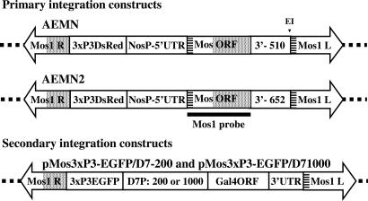
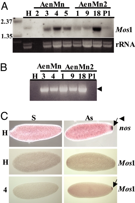
Transposable elements (TEs) are proposed as a basis for developing drive systems to spread pathogen resistance genes through vector mosquito populations. The use of transcriptional and translational control DNA elements from genes expressed specifically in the insect germ line to mediate transposition offers possibilities for mitigating some of the concerns about transgene behavior in the target vector species and eliminating effects on nontarget organisms. Here, we describe the successful use of the promoter and untranslated regions from the nanos (nos) orthologous gene of the yellow fever mosquito, Aedes aegypti, to control sex- and tissue-specific expression of exogenously derived mariner MosI transposase-encoding DNA. Transgenic mosquitoes expressed transposase mRNA in abundance near or equal to the endogenous nos transcript and exclusively in the female germ cells. In addition, MosI mRNA was deposited in developing oocytes and localized and maintained at the posterior pole during early embryonic development. Importantly, four of five transgenic lines examined were capable of mobilizing a second MosI transgene into the mosquito genome, indicating that functional transposase was being produced. Thus, the nos control sequences show promise as part of a TE-based gene drive system.


Conflict of interest statement

The authors declare no conflict of interest.

Figures

Fig. 1.
Fig. 1.
Schematic representations of the primary and secondary transformation constructs. All constructs were flanked by the mariner MosI right- (MosI R) and left-hand (MosI L) inverted repeat sequences. The dotted lines represent A. aegypti genomic DNA into which the constructs are transposed. Primary integration constructs carry the marker gene (3xP3DsRed), which confers red eye-specific fluorescence, and the MosI transposase ORF (MosORF) flanked by the nos promoter and 5′- and 3′-UTRs (NosP + 5′-UTR). AEMN has a 510-bp 3′-UTR (3′- 510) and an internal EcoRI site (E1). AEMN2 has a 652-bp 3′-UTR (3′- 652) and lacks the restriction endonuclease site. The striped and dotted boxes indicate regions of identity derived from the MosI left- and right-hand repeat regions, respectively. The thick line represents the extent of probes (MosI probe) used to verify the presence of the MosI ORF-encoding DNA or mRNA in Southern and Northern blot analyses, respectively, and in hybridizations in situ. Secondary integration constructs carry the marker gene (3xP3EGFP), which confers green eye-specific fluorescence, one of two versions of the D7 gene promoter (42) 200 or 1,000 nucleotides in length (D7P: 200 or 1,000), and 3′-UTR flanking the Gal4 ORF (Gal4ORF).
Fig. 2.
Fig. 2.
Northern blot analyses of adult AenMn and AenMn2 transgenic mosquitoes. (A) Northern blot analysis conducted by using 10 μg of total RNA isolated from fully developed ovaries from untransformed Higgs strain mosquitoes (H), AenMn transgenic line 3 or AenMn2 transgenic lines 1 and 18. Duplicate membranes were hybridized with a probe specific for either the MosI ORF (MosI) or the A. aegypti nanos ORF (nos). The RNA size marker lane is indicated (M). Ethidium bromide-stained rRNA loading control of samples is shown below. (B) Northern blot analysis conducted by using 10 μg of total RNA isolated from fully developed ovaries (O), dissected carcasses (C), or whole males (♂) from AenMn and AenMn2 transgenic lines or from untransformed Higgs strain mosquitoes (H). Membranes were hybridized with a radiolabeled probe specific for the MosI ORF (MosI). rRNA loading control of samples is shown below. RNA size markers (M) are shown on the left.
Fig. 3.
Fig. 3.
RNA analysis of AenMn and AenMn2 transgenic embryos. (A) Northern blot analysis was conducted by using 10 μg of total RNA isolated from 0- to 5-h-old embryos collected from untransformed Higgs strain mosquitoes (H), AenMn lines 2, 3, 4, and 5, and AenMn2 lines 1, 9, 18, and P1. Membranes were hybridized with a radiolabeled probe specific for the MosI ORF (MosI), with rRNA loading control of samples shown below. The RNA size marker lane is indicated (M). (B) One-Step RT-PCR of 0- to 5-h-old embryos RNA collected from untransformed (H) or transgenic mosquitoes (1, 3, 4, 9, 18, and P1), using the MosI-specific forward primer MLF2 and the A. aegypti nos-specific reverse primer nosUTRR2. The arrow indicates the expected transcript size. (C) In situ hybridization of MosI in AenMn transgenic embryos. Control (H) and transgenic line (4) embryos hybridized with sense (S) or antisense (AS) nos and MosI probes. The arrow points to specific localization of the mRNA to the posterior pole.
Fig. 4.
Fig. 4.
Characterization of secondary integrations in transgenic mosquitoes. (A) Marker gene expression in eyes of a mosquito containing a primary insertion with the DsRed reporter gene (DsRed) and a secondary insertion with the EGFP reporter gene (EGFP). The image on the right shows the same head under visible light (visible). (B) Southern blot analyses of secondary lines with or without primary integrations. Hybridization patterns of the EGFP-specific probe are shown with both primary and secondary insertions (5.1, 5.3, 2.4, 3.12, and 3.22), and lines derived from these with only the secondary insertions (5.3.1, 2.4.1, 3.22.1, and 18.13.1). Control Higgs (H) also is shown. Numbers on the left refer to the relative positions of molecular-weight makers. (C) Transposon-chromosome junction fragments from a secondary insertion. The nucleotide sequence of genomic DNA adjacent to the right- and left-hand terminal inverted repeats of the 2.4.1 secondary insertion. The bolded GATC and CATG are the endogenous Sau3AI and CviAII restriction endonuclease cleavage sites, respectively, used to recover the junction fragments. The double-headed block arrow represents the location of the secondary insertion DNA. The TA dinucleotides in bold are the duplications of the endogenous target sites. Sequence polymorphisms between the transformed line and the strain from which the genomic sequence was derived are shown in bold italics.

References

    1. Calisher CH. Emerg Infect Dis. 2005;11:738–739. - PMC - PubMed
    1. Gubler DJ. Comp Immunol Microbiol Infect Dis. 2004;27:319–330. - PubMed
    1. Gubler DJ. Nat Med. 2004;10:129–130. - PubMed
    1. Greenwood B, Mutabingwa T. Nature. 2002;415:670–672. - PubMed
    1. Curtis CF, Graves PM. J Trop Med Hyg. 1988;91:43–48. - PubMed

Publication types

MeSH terms

LinkOut - more resources