. 2007 Jun 12;104(24):10057-62.
doi: 10.1073/pnas.0700169104. Epub 2007 Jun 4.

Fragile X mental retardation protein FMRP and the RNA export factor NXF2 associate with and destabilize Nxf1 mRNA in neuronal cells

Affiliations

Fragile X mental retardation protein FMRP and the RNA export factor NXF2 associate with and destabilize Nxf1 mRNA in neuronal cells

Meiqin Zhang et al. Proc Natl Acad Sci U S A. .

Abstract

Fragile X syndrome is caused by the inactivation of the X-linked FMR1 gene, leading to the loss of its encoded protein FMRP. Although macroorchidism and defects in neuronal architecture and function have been associated with lack of FMRP, the exact molecular mechanism underlying this disease remains unclear. We have reported previously that in the brain and testis of mice, FMRP specifically interacts with a distinct mRNA nuclear export factor NXF2 but not with its close relative NXF1, a ubiquitously expressed essential mRNA nuclear export factor. This interaction marked NXF2 as a putative functional partner of FMRP. Here, we demonstrate by immunoprecipitation and quantitative real-time RT-PCR that, in cultured mouse neuronal cells, both FMRP and NXF2 are present in Nxf1 mRNA-containing ribonucleoprotein particles. Further, we show that expression of NXF2 leads to the destabilization of Nxf1 mRNA and that this effect is abolished when Fmr1 expression is reduced by siRNA, arguing that both proteins collaborate to exert this effect. Importantly, these findings correlate well with our observations that in both mouse hippocampal neurons and male germ cells where the expression of FMRP and NXF2 is most prominent, the expression of NXF1 is relatively poorly expressed. Our studies thus identify Nxf1 mRNA as a likely biologically relevant in vivo target of both FMRP and NXF2 and implicate FMRP, in conjunction with NXF2, as a posttranscriptional regulator of a major mRNA export factor. Such regulation may prove important in the normal development and function of neurons as well as of male germ cells.


Conflict of interest statement

The authors declare no conflict of interest.

Figures

Fig. 1.
Fig. 1.
Immunofluorescence of adult mouse testicular cryostatic sections by using antibodies specific for FMRP (A and D), NXF2 (B and E), and NXF1 (C and F). (A–C) High-power images of the selected areas (white boxes in D–F). Each panel is a merged image of DAPI nuclear staining (blue) and the indicated antibody staining (green for FMRP and red for NXF2 and NXF1). The spermatogonia (arrowheads) are the cells residing at the periphery of the seminiferous tubules of the testis. The more mature germ cells including spermatocytes (large arrows) and spermatids (small arrows) are located closer to the lumen of the tubules. (Scale bars, 20 μm.)
Fig. 2.
Fig. 2.
Immunohistochemistry of adult mouse brain paraffin sections with antibodies specific for FMRP (A and D), NXF2 (B and E), and NXF1 (C and F). (A–C) High-power images of the selected areas (white boxes in D–F). Arrowheads indicate the cell bodies of hippocampal neurons. Note that the NXF1 staining of the hippocampal neurons is relatively weaker than that of cells located away from the hippocampal area (white arrows). (Scale bars, 20 μm.)
Fig. 3.
Fig. 3.
FMRP and NXF2 preferentially associate with Nxf1 mRNA-containing RNPs. FLAG-NXF2 was transfected into N2a cells, and IP was carried out 48 h after transfection. (A) Proteins from purified IP complexes or from 3% of supernatants were resolved on SDS/10% PAGE. The presence of the FMRP and NXF2 proteins in the IP complexes was confirmed by Western blot analyses. (Upper) IP with anti-FMRP (αFMRP) using mouse IgG as a negative control, followed by Western blotting using anti-FMRP. FMRP was in the anti-FMRP IP complexes (lane 4) but not in the IgG IP complexes (lane 3). (Lower) IP with anti-NXF2 (αNXF2) using preimmune serum as a negative control followed by Western blotting using anti-NXF2. NXF2 was in the anti-NXF2 IP complexes (lane 4) but not in the preimmune IP complexes (lane 3). Lanes 1 and 2 indicate that FMRP and NXF2 were present in the cell lysates. (B and C) RNAs were extracted from purified IP complexes followed by qRT-PCR. mRNA levels associated with FMRP (white bars) or NXF2 (gray bars) RNPs are indicated relative to those with negative control IP samples, which were arbitrarily set as 1. (B) A polyclonal anti-NXF2 antibody was used. (C) A monoclonal anti-FLAG antibody was used to immunoprecipitate the transfected FLAG-NXF2. Each bar represents mean ± SEM (B, n = 4; C, n = 2). (D) mRNA expression levels. RNAs were extracted from 10% of IP supernatants, and qRT-PCR was carried out using primers specific for each mRNA. Levels are plotted relative to Gapdh mRNA levels, which were arbitrarily set a value of 100. Each bar represents mean ± SEM (n = 4).
Fig. 4.
Fig. 4.
NXF2 expression results in the destabilization of Nxf1 mRNA. (A) (Upper) Representative Western blots showing NXF2 expression levels in empty vector-transfected (lane 1) or FLAG-NXF2-transfected (lane 2) N2a cells or in the mouse testis (lane 3). (Lower) β-Actin was used as a loading control. (B) N2a cells were transfected with FLAG-NXF2 or empty vector, together with a YFP expression vector. Forty-eight hours after the transfection, cells were harvested and subjected to FACS. YFP-positive cells were collected followed by RNA extraction and qRT-PCR using primers specific for the indicated mRNAs. The levels of mRNAs from FLAG-NXF2-transfected cells are shown relative to those from empty vector-transfected cells, which were arbitrarily set as 100%. Each bar represents mean ± SEM (n = 3≈5). (C) N2a cells were transfected with FLAG-NXF2 or empty vector. Twenty-four hours after transfection, actinomycin D was added to inhibit transcription, and Nxf1 mRNA levels at 0 h, 3 h, and 6 h time points, respectively, were measured by qRT-PCR. The mRNA levels at the 0 h time point were arbitrarily set as 100%. Each time point represents mean ± SEM (n = 4).
Fig. 5.
Fig. 5.
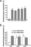
FMRP contributes to Nxf1 mRNA destabilization induced by NXF2. (A) The mouse Fmr1 gene is specifically down-regulated by siRNA. An siRNA specific for the Fmr1 gene or a control siRNA was transfected into N2a cells. Twenty-four hours after transfection, RNAs were isolated and analyzed by qRT-PCR using primers specific for the indicated mRNAs. mRNA levels from Fmr1-specific siRNA-treated cells are shown relative to those treated with control siRNA. Each bar indicates mean ± SEM (n = 3≈6). (B) Inhibition of FMRP expression abolishes the Nxf1 mRNA destabilization effect induced by NXF2. Fmr1-specific siRNA or control siRNA, together with FLAG-NXF2 or empty vector, was transfected into N2a cells. Seventy-two hours after transfection, RNAs were extracted, and levels were measured by qRT-PCR. The levels of mRNAs from FLAG-NXF2-transfected cells are shown relative to those from empty vector-transfected cells, which were arbitrarily set as 100%. Each bar represents mean ± SEM (n = 3≈4).

Similar articles

Cited by

References

    1. Bardoni B, Davidovic L, Bensaid M, Khandjian EW. Expert Rev Mol Med. 2006;8:1–16. - PubMed
    1. Koukoui SD, Chaudhuri A. Brain Res Rev. 2006;53:27–38. - PubMed
    1. Devys D, Lutz Y, Rouyer N, Bellocq JP, Mandel JL. Nat Genet. 1993;4:335–340. - PubMed
    1. Bakker CE, Otero Y, Bontekoe C, Raghoe P, Luteijn T, Hoogeveen AT, Oostra BA, Willemsen R. Exp Cell Res. 2000;258:162–170. - PubMed
    1. Willemsen R, Oostra BA, Bassell GJ, Dictenberg J. Ment Retard Dev Disabil. 2004;10:60–67. - PubMed

Publication types

Substances