. 2007 Jun;117(6):1595-604.
doi: 10.1172/JCI31450.

Sustained hippocampal IL-1 beta overexpression mediates chronic neuroinflammation and ameliorates Alzheimer plaque pathology

Affiliations

Sustained hippocampal IL-1 beta overexpression mediates chronic neuroinflammation and ameliorates Alzheimer plaque pathology

Solomon S Shaftel et al. J Clin Invest. 2007 Jun.

Abstract

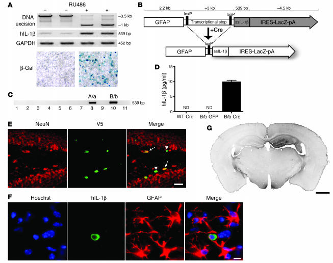
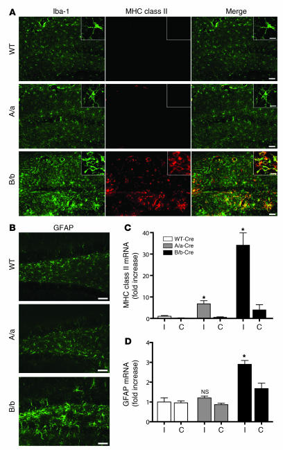
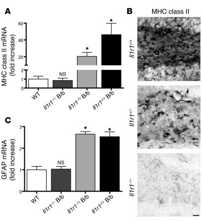
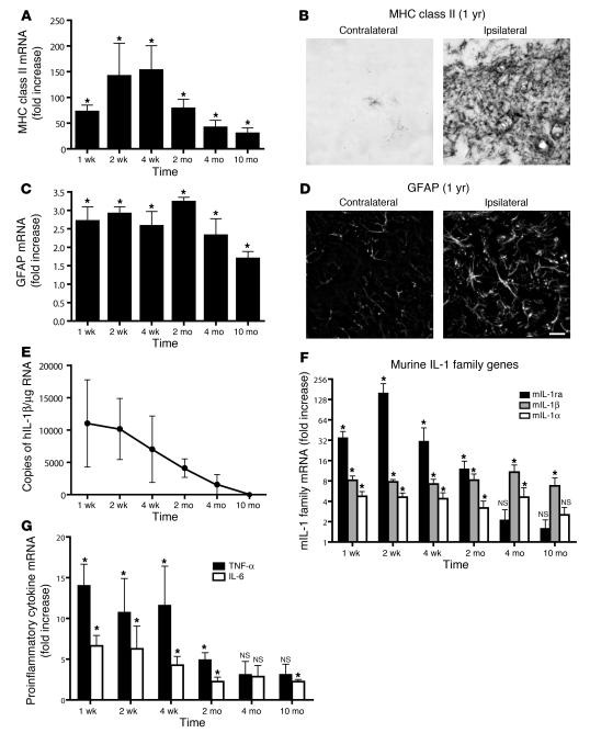
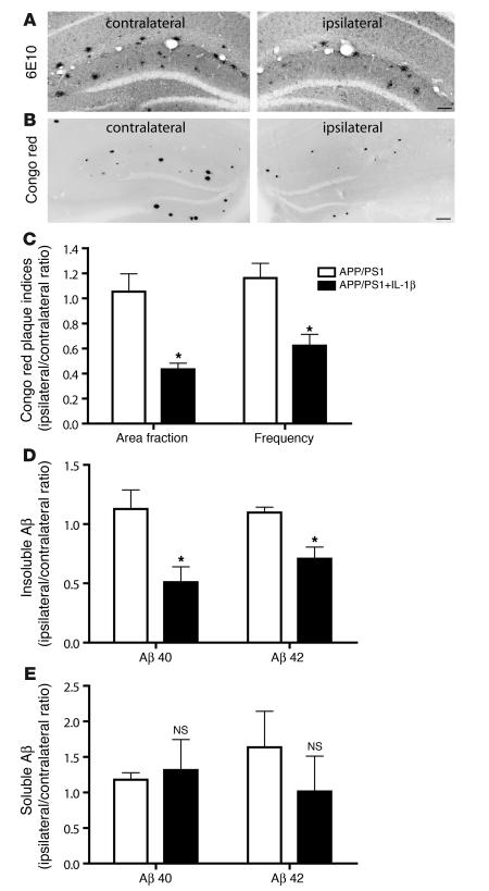
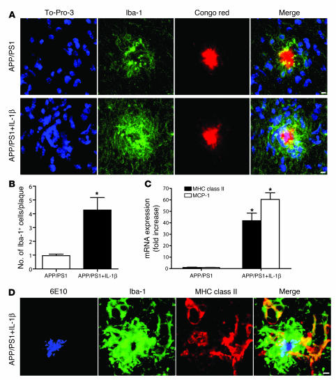
Neuroinflammation is a conspicuous feature of Alzheimer disease (AD) pathology and is thought to contribute to the ultimate neurodegeneration that ensues. IL-1 beta has emerged as a prime candidate underlying this response. Here we describe a transgenic mouse model of sustained IL-1 beta overexpression that was capable of driving robust neuroinflammation lasting months after transgene activation. This response was characterized by astrocytic and microglial activation in addition to induction of proinflammatory cytokines. Surprisingly, when triggered in the hippocampus of the APPswe/PS1dE9 mouse model of AD, 4 weeks of IL-1 beta overexpression led to a reduction in amyloid pathology. Congophilic plaque area fraction and frequency as well as insoluble amyloid beta 40 (A beta 40) and A beta 42 decreased significantly. These results demonstrate a possible adaptive role for IL-1 beta-driven neuroinflammation in AD and may help explain recent failures of antiinflammatory therapeutics for this disease.


Figures

Figure 1
Figure 1. Engineering and testing of the IL-1βXAT mouse.
(A) The 293GLVP/CrePr stable cell line was transiently transfected with CMV/IL-1βXAT and cultured with or without RU486. RU486 caused DNA excision and expression of hIL-1β and β-galactosidase by RT-PCR and X-gal histochemistry, respectively. (B) The linear IL-1βXAT construct (~10 kb) consisted of a murine GFAP promoter; a transcriptional stop element flanked by loxP sequences; downstream ssIL-1β cDNA (11); and an internal ribosome entry site (IRES) followed by the β-gal coding sequence (LacZ) and bovine growth hormone polyadenylation signal (pA). Exposure to Cre recombinase caused excision of the transcriptional stop and subsequent transcriptional activation of ssIL-1β. (C) PCR screening of the 11 live-born IL-1βXAT pups using ssIL-1β–specific primers. Transgenic founder lines A/a and B/b are shown. (D) ELISA quantification (mean ± SEM) of hIL-1β protein supernatant concentration in individual primary astrocyte (n = 4) cultures from B/b and WT astrocytes transduced with FIV-Cre or FIV-GFP. ND, not detected (i.e., below detection limits). (E) The epicenter of viral transduction, the dentate gyrus, was bounded by NeuN-positive neurons (red stain). Colocalization of the epitope tag V5 (green stain) expressed by FIV-Cre demonstrated transduction of both neuronal (arrows) and non-neuronal cells (arrowheads). (F) Demonstration of hIL-1β expression (green stain) by astrocytes (red stain) in the dentate gyrus. Hoechst (blue stain) labeled cell nuclei. (G) MHC class II–stained coronal section from a B/b animal 1 week after intrahippocampal FIV-Cre injection (right hemisphere). Scale bars: 25 μm (E); 10 μm (F); 1 μm (G).
Figure 2
Figure 2. Induction of hIL-1β mediates a robust neuroinflammatory response in the mouse hippocampus.
IL-1βXAT A/a and B/b mice and WT controls were injected unilaterally in the dentate gyrus with approximately 1.5 × 10⁴ infectious units of FIV-Cre. Inflammatory indices were assayed 2 weeks later. (A) Microglial activation was demonstrated by increased staining intensity of Iba-1 (green) and MHC class II (red) in B/b and A/a relative to WT mice, with colocalization appearing yellow. Insets show higher-magnification morphologic changes among Iba-1–positive cells residing in the dentate gyrus. (B) Astrocyte activation, as evidenced by increased GFAP expression, was demonstrated in the dentate gyrus of B/b animals only. Scale bars: 50 μm (A and B); 10 μm (insets in A). (C and D) qRT-PCR analysis compared relative abundance of gene transcripts in ipsilateral (I) and contralateral (C) hippocampi. Analysis revealed significant upregulation of MHC class II in A/a and B/b mice (C), but significant upregulation of GFAP in B/b mice only (D), compared with WT controls. n = 3–4 per group. Data are mean ± SEM. *P < 0.05 versus respective WT.
Figure 3
Figure 3. The neuroinflammatory response requires IL-1R1.
IL-1βXAT B/b animals that were lacking (Il1r1–/–), heterozygous for (Il1r1+/–), or with 2 copies (Il1r1+/+) of the gene encoding IL-1R1 were examined 2 weeks following FIV-Cre hippocampal injections. qRT-PCR analysis of ipsilateral hippocampi was performed relative to WT controls. (A) MHC class II induction was absent in Il1r1–/– B/b animals, whereas Il1r1+/– B/b mice exhibited a significant, intermediate phenotype compared with WT mice. (B) Histochemical analysis of MHC class II expression within the dentate gyrus of B/b mice revealed a pattern of expression mirroring the results in A. Scale bar: 25 μm. (C) GFAP induction was absent in Il1r1–/– B/b mice, whereas Il1r1+/– and Il1r1+/+ B/b mice displayed significant GFAP upregulation. n = 3–5 animals per group. Data are mean ± SEM. *P < 0.05 versus WT.
Figure 4
Figure 4. Transgene activation in the IL-1βXAT mouse elicits a chronic neuroinflammatory response.
IL-1βXAT B/b and WT control mice received intrahippocampal injections of FIV-Cre and were analyzed over a prolonged time course for neuroinflammatory indices. (A, C, F, and G) qRT-PCR generated a ratio of gene expression in the ipsilateral hippocampi of B/b compared with WT mice at the same time point, except for MHC class II analysis, in which the 4-week time point was used for all comparisons. (A) MHC class II expression was significantly upregulated at all time points assayed. (B) MHC class II staining in the dentate gyrus of a B/b mouse 1 year after FIV-Cre injection. (C) GFAP expression was also significantly upregulated at all time points examined. (D) GFAP expression in the dentate gyrus at 1 year. Scale bar: 20 μm. (E) GFAP and MHC class II upregulation coincided with prolonged expression of ssIL-1β. (F and G) In addition to glial activation markers, hIL-1β expression caused significant increases in qRT-PCR gene transcripts coding for all members of the mIL-1 family (F) and for proinflammatory cytokines IL-6 and TNF-α (G). n = 4–5 animals per group. Data are mean ± SEM. *P < 0.05 versus WT as described.
Figure 5
Figure 5. IL-1β overexpression ameliorates plaque pathology in a mouse model of AD.
IL-1βXAT B/b animals were crossed with APP/PS1 mice, generating APP/PS1+IL-1β animals heterozygous for all 3 transgenes. Intrahippocampal FIV-Cre injections were performed at 6 months of age in APP/PS1 and APP/PS1+IL-1β animals to control both for the injection and for viral transduction. After 4 weeks, (7 months of age) the ratio of pathologic indices between the ipsilateral (FIV-Cre injected) and contralateral (uninjected) hemispheres within individual animals was determined. (A and B) Histochemical analysis using the 6E10 antibody (A) and Congo red (B; shown inverted) revealed a reduction in amyloid deposition in the injected ipsilateral hippocampi of APP/PS1+IL-1β mice compared with that of the uninjected contralateral hemispheres. Scale bars: 100 μm (A); 200 μm (B). (C) hIL-1β induction caused significant reductions in Congo red plaque area fraction and frequency. (D and E) Furthermore, hIL-1β overexpression mediated significant reductions in both insoluble Aβ40 and Aβ42 peptides (D), but did not significantly alter levels of their soluble forms (E), as assessed by ELISA. For additional data from CE, see Supplemental Table 1. n = 6–7 per group. Graphs represent mean ± SEM. *P < 0.05.
Figure 6
Figure 6. IL-1β driven microglial activation likely underlies reductions in plaque pathology.
Histological analysis was performed in 7-month-old APP/PS1 mice and APP/PS1+IL-1β mice 4 weeks following viral injections. (A) Confocal microscopy revealed an increase in Iba-1–positive (green) microglial cells (nuclei stained blue) directly in contact with Congophilic plaques (red) in APP/PS1+IL-1β mice compared with their APP/PS1 counterparts. (B) Quantitative analysis of mice in A revealed a greater than 4-fold increase in the number of Iba-1–positive cell nuclei overlapping Congophilic plaques. (C) qRT-PCR analysis showed robust upregulation of MHC class II and MCP-1 in APP/PS1+IL-1β mice compared with APP/PS1 mice. (D) 6E10 amyloid staining (blue) in conjunction with Iba-1 (green) and MHC class II (red) staining revealed activation of microglia in contact with amyloid plaque in an APP/PS1+IL-1β mouse. Scale bars: 5 μm. n = 6–7 per group. Data are mean ± SEM. *P < 0.05.

Comment in

References

    1. Basu A., Krady J.K., Levison S.W. Interleukin-1: a master regulator of neuroinflammation. J. Neurosci. Res. 2004;78:151–156. - PubMed
    1. Allan S.M., Tyrrell P.J., Rothwell N.J. Interleukin-1 and neuronal injury. Nat. Rev. Immunol. 2005;5:629–640. - PubMed
    1. Emsley H.C., et al. A randomised phase II study of interleukin-1 receptor antagonist in acute stroke patients. J. Neurol. Neurosurg. Psychiatr. 2005;76:1366–1372. - PMC - PubMed
    1. Griffin W.S., et al. Brain interleukin 1 and S-100 immunoreactivity are elevated in Down syndrome and Alzheimer disease. Proc. Natl. Acad. Sci. U. S. A. 1989;86:7611–7615. - PMC - PubMed
    1. Van Everbroeck B., et al. The role of cytokines, astrocytes, microglia and apoptosis in Creutzfeldt-Jakob disease. Neurobiol. Aging. 2002;23:59–64. - PubMed

Publication types

MeSH terms