. 2007 Jun;117(6):1616-26.
doi: 10.1172/JCI31581.

The hypoxia-inducible factor alpha pathway couples angiogenesis to osteogenesis during skeletal development

Affiliations

The hypoxia-inducible factor alpha pathway couples angiogenesis to osteogenesis during skeletal development

Ying Wang et al. J Clin Invest. 2007 Jun.

Abstract

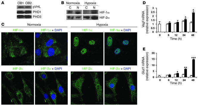
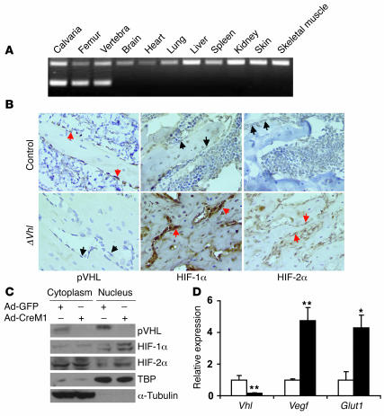
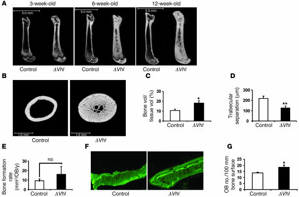
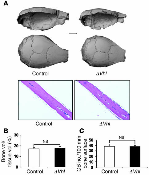
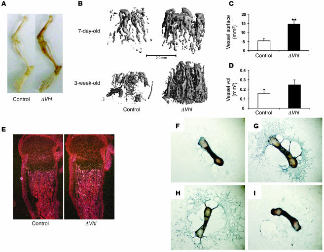
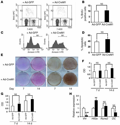
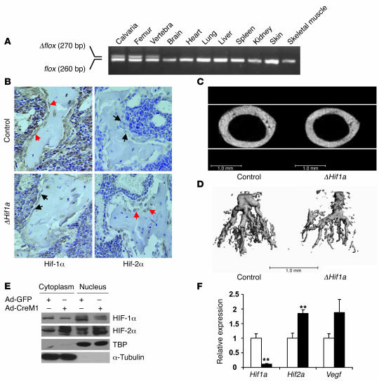
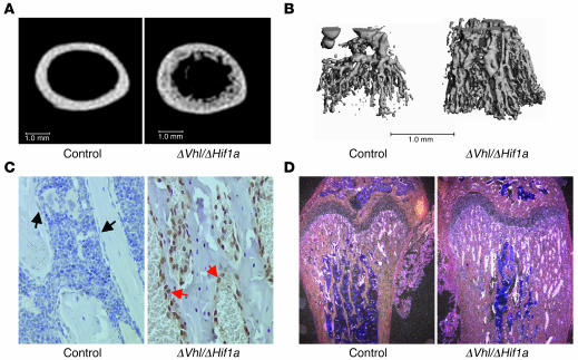
Skeletal development and turnover occur in close spatial and temporal association with angiogenesis. Osteoblasts are ideally situated in bone to sense oxygen tension and respond to hypoxia by activating the hypoxia-inducible factor alpha (HIF alpha) pathway. Here we provide evidence that HIF alpha promotes angiogenesis and osteogenesis by elevating VEGF levels in osteoblasts. Mice overexpressing HIF alpha in osteoblasts through selective deletion of the von Hippel-Lindau gene (Vhl) expressed high levels of Vegf and developed extremely dense, heavily vascularized long bones. By contrast, mice lacking Hif1a in osteoblasts had the reverse skeletal phenotype of that of the Vhl mutants: long bones were significantly thinner and less vascularized than those of controls. Loss of Vhl in osteoblasts increased endothelial sprouting from the embryonic metatarsals in vitro but had little effect on osteoblast function in the absence of blood vessels. Mice lacking both Vhl and Hif1a had a bone phenotype intermediate between those of the single mutants, suggesting overlapping functions of HIFs in bone. These studies suggest that activation of the HIF alpha pathway in developing bone increases bone modeling events through cell-nonautonomous mechanisms to coordinate the timing, direction, and degree of new blood vessel formation in bone.


Figures

Figure 1
Figure 1. Primary mouse osteoblasts express components of the HIFα pathway.
(A) Calvarial primary osteoblasts were obtained from wild-type mice and cultured in α-MEM until confluent. Whole-cell lysate was analyzed by immunoblotting using antibodies against pVHL (top), PHD1 (middle), and PHD3 (bottom) in experiments performed in duplicate. OB, osteoblast. (B and C) Confluent cell monolayers were exposed to 21% (normoxia) or 2% (hypoxia) O2 for 24 hours. (B) Proteins from cytoplasm (C) and nucleus (N) were extracted separately and analyzed by immunoblotting with antibodies against HIF-1α (top row) and HIF-2α (bottom row). (C) Nuclear translocation of HIF-1α (top row) and HIF-2α (bottom row) was assessed by confocal microscopy as described in Methods. DAPI was used to stain the nuclei. Original magnification, ×100. (D and E) Quantitative real-time PCR analysis was performed to determine Vegf and Glut1 mRNA expression after cell monolayers were exposed to normoxia (white bars) or hypoxia (black bars) for the indicated times. *P < 0.05; **P < 0.01; ***P < 0.001.
Figure 2
Figure 2. Osteoblast-specific, Cre-mediated deletion of Vhl.
(A) PCR analysis of Cre-mediated recombination in selected tissues from a ΔVhl mouse. The recombined allele (Δflox) was present exclusively in bone tissue. (B) Representative histological sections of distal femurs from 6-week-old control and ΔVhl mice after staining with antibodies against pVHL (left), HIF-1α (middle), or HIF-2α (right) as described in Methods. Sections were counterstained with hematoxylin. Red arrows indicate positive staining and black arrows negative staining in osteoblasts. Original magnification, ×400. (C and D) Confluent monolayers of Vhl floxed primary osteoblasts were infected with either Ad-GFP or Ad-CreM1 (100 MOI) for 48 hours. (C) Proteins in the cytoplasm and nucleus were extracted separately and analyzed by immunoblotting with antibodies against pVHL, HIF-1α, and HIF-2α. Immunoblots for TBP and α-tubulin were used as loading controls for nuclear and cytoplasmic proteins, respectively. TBP, TATA box–binding protein. (D) Total mRNA was extracted from confluent monolayers of osteoblasts 48 hours after adenoviral infection, and gene expression for Vhl, Vegf, and Glut1 was determined by quantitative real-time PCR. White bars represent Ad-GFP infection; black bars represent Ad-CreM1 infection. *P < 0.05; **P < 0.01.
Figure 3
Figure 3. Disruption of Vhl in osteoblasts increases long bone volume.
(A) Representative μCT images of the femurs from ΔVhl and control mice at the age of 3, 6, and 12 weeks. Scale bars: 5.0 mm. (B) Representative femoral cross sections from 6-week-old ΔVhl and control mice. Scale bars: 1.0 mm. (CE) Histomorphometric analyses were performed on femoral sections from ΔVhl mice and controls at 3 weeks of age as described in Methods. Comparisons of trabecular bone volume (C), trabecular separation (D), and bone formation rate per osteoblast (E) in control (white bars; n = 6) and ΔVhl mice (black bars; n = 7) are shown. Data represent mean ± SEM. *P < 0.05; **P < 0.01. (F) Seven-day-old mice were labeled with sequential doses of calcein before sacrifice. Representative calcein-labeled sections of distal femur from control and ΔVhl mice are shown. Original magnification, ×400. (G) Quantitative histomorphometric measurement of osteoblast number was performed at the distal femur of 7-day-old ΔVhl (black bar; n = 3) and control mice (white bar; n = 3). Data represent mean ± SEM. *P < 0.05.
Figure 4
Figure 4. Loss of Vhl in osteoblasts does not alter calvarial bone.
(A) Representative μCT images of calvaria from 12-week-old ΔVhl and control mice (top). Scale bar: 2.0 mm. The bottom panels show H&E-stained sections of calvaria from 6-week-old control and ΔVhl mice. Original magnification, ×100. (B and C) Quantitative histomorphometric measurement of bone volume and osteoblast number was performed on calvarial sections from 6-week-old ΔVhl (black bars; n = 3) and control mice (white bars; n = 3). Data represent mean ± SEM.
Figure 5
Figure 5. Increased angiogenesis in long bones of ΔVhl mice.
(A) Photograph of hind limbs from ΔVhl and control mice. (B) Representative μCT images of vasculature in Microfil-perfused femurs from 7-day-old and 3-week-old ΔVhl and control mice. Scale bar: 1.0 mm. (C and D) Morphological analysis of vessel surface and volume within femurs from Microfil-perfused ΔVhl (black bars; n = 3) and control (white bars; n = 3) mice. Data represent mean ± SEM. **P < 0.01. (E) In situ hybridization analysis with Vegf mRNA on histological sections from 3-day-old control and Vhl mutant femurs. Original magnification, ×40. (FI) In vitro angiogenesis assay. Metatarsals were dissected from control and ΔVhl E17.5 fetuses and cultured in α-MEM for 14 days. The assay was performed using anti-CD31 antibody as described in Methods. Representative images are shown. Original magnification, ×25. (F) Little detectable endothelial sprouting from control metatarsal. (G) Massive endothelial sprouting in control metatarsal treated with recombinant VEGF (10 ng/ml). (H) Extensive endothelial sprouting from ΔVhl metatarsal remained intact after treatment with a mouse control IgG (100 ng/ml). (I) Specific inhibition of endothelial sprouting in ΔVhl metatarsal using a VEGF-neutralizing antibody (100 ng/ml). Data are representative of 3 independent experiments.
Figure 6
Figure 6. Deletion of Vhl in primary osteoblasts in vitro does not affect osteoblast proliferation and apoptosis.
Confluent Vhl floxed primary osteoblast monolayers were infected with either adeno-GFP or adeno-CreM1 (100 MOI). Vhl mRNA expression in infected osteoblasts was determined by real-time PCR 48 hours after infection to assess deletion efficiency. Cell proliferation, apoptosis, and differentiation assays were performed as described in Methods. (A and B) Cell proliferation was assessed by flow cytometry using BrdU incorporation. (C and D) Cell apoptosis was assessed by flow cytometry using annexin V–PE staining. (E) Mineralized nodule formation was determined by ALP (left) and von Kossa staining (right) 7 and 14 days after cells were cultured in osteogenic medium. (F and G) Densitometric analysis of ALP and von Kossa staining observed in E using NIH ImageJ 1.36b. Data represent mean ± SEM. (H) Measurement of Vhl, Hif1a, runt-related transcription factor 2 (Runx2), and OC mRNA expression by quantitative real-time PCR at day 14 of osteogenic induction. **P < 0.01; ***P < 0.001.
Figure 7
Figure 7. Mice lacking Hif1a in osteoblasts have narrow, poorly vascularized long bones.
(A) PCR analysis of Cre-mediated recombination in selected tissues from a ΔHif1a mouse. The recombined allele (Δflox) was present exclusively in bone tissue. (B) Representative histological sections of distal femurs from 6-week-old control and ΔHif1a mice after staining with antibodies against HIF-1α (left) or HIF-2α (right) as described in Methods. Sections were counterstained with hematoxylin. Red arrows indicate positive and black arrows negative staining in osteoblasts. Original magnification, ×400. (C) Representative images of femoral cross sections from control and ΔHif1a mice. Scale bars: 1.0 mm. (D) Representative μCT images of vasculature in Microfil-perfused femurs from 3-week-old ΔHif1a and control mice. Scale bar: 1.0 mm. (E and F) Confluent monolayers of Hif1a floxed primary osteoblasts were infected with either Ad-GFP or Ad-CreM1 (100 MOI). (E) Proteins in the cytoplasm and nucleus were extracted separately 48 hours after infection. Immunoblotting analysis was performed with antibodies against HIF-1α and HIF-2α. Immunoblots for TBP and α-tubulin were used as loading controls for nuclear and cytoplasmic proteins, respectively. (F) Total mRNA was extracted from confluent monolayers of osteoblasts 48 hours after infection. Hif1a, Hif2a, and Vegf mRNA expression was determined by quantitative real-time PCR. **P < 0.01.
Figure 8
Figure 8. Bone and vascular phenotype in Vhl and Hif1a double-knockout mice.
(A) Representative μCT images of femoral cross sections from 6-week-old ΔVhl/ΔHif1a double mutant mice and wild-type controls. Scale bars: 1.0 mm. (B) Representative images of Microfil-perfused femurs from 3-week-old ΔVhl/ΔHif1a double-mutant mice and control littermates. Scale bar: 1.0 mm. (C) Immunohistochemical analysis of HIF-2α level in femoral sections from 6-week-old ΔVhl/ΔHif1a double-mutant mice and control littermates. Sections were counterstained with hematoxylin. Red arrows indicate positive and black arrows negative staining in osteoblasts. Original magnification, ×400. (D) In situ hybridization analysis with Vegf mRNA on histological sections from 6-week-old control and double-mutant femurs. Original magnification, ×100.

Comment in

Similar articles

Cited by

References

    1. Steinbrech D.S., et al. VEGF expression in an osteoblast-like cell line is regulated by a hypoxia response mechanism. Am. J. Physiol. Cell Physiol. 2000;278:C853–C860. - PubMed
    1. Semenza G.L. Regulation of mammalian O2 homeostasis by hypoxia-inducible factor 1. . Annu. Rev. Cell Dev. Biol. 1999;15:551–578. - PubMed
    1. Semenza G.L. Hypoxia-inducible factor 1: oxygen homeostasis and disease pathophysiology. Trends Mol. Med. 2001;7:345–350. - PubMed
    1. Huang L.E., Gu J., Schau M., Bunn H.F. Regulation of hypoxia-inducible factor 1alpha is mediated by an O2-dependent degradation domain via the ubiquitin-proteasome pathway. . Proc. Natl. Acad. Sci. U. S. A. 1998;95:7987–7992. - PMC - PubMed
    1. Kaelin W.G., Jr. Molecular basis of the VHL hereditary cancer syndrome. Nat. Rev. Cancer. 2002;2:673–682. - PubMed

Publication types

MeSH terms

Substances