. 2007 Jul;31(1):5-17.

RNA interference-mediated targeting of urokinase plasminogen activator receptor and matrix metalloproteinase-9 gene expression in the IOMM-lee malignant meningioma cell line inhibits tumor growth, tumor cell invasion and angiogenesis

Affiliations

RNA interference-mediated targeting of urokinase plasminogen activator receptor and matrix metalloproteinase-9 gene expression in the IOMM-lee malignant meningioma cell line inhibits tumor growth, tumor cell invasion and angiogenesis

Padmaja Tummalapalli et al. Int J Oncol. 2007 Jul.

Abstract

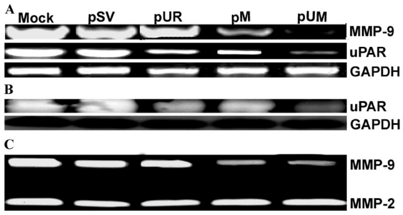
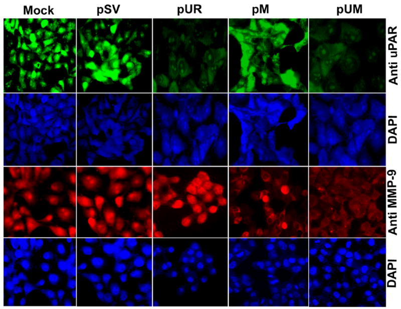
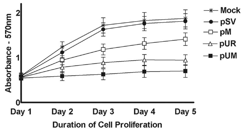
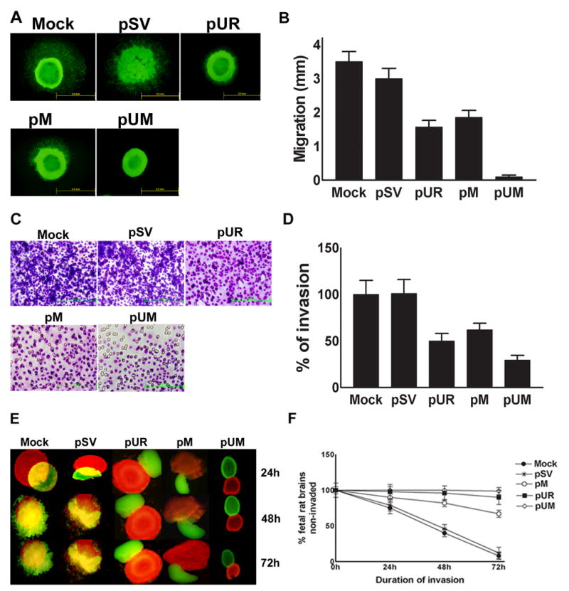
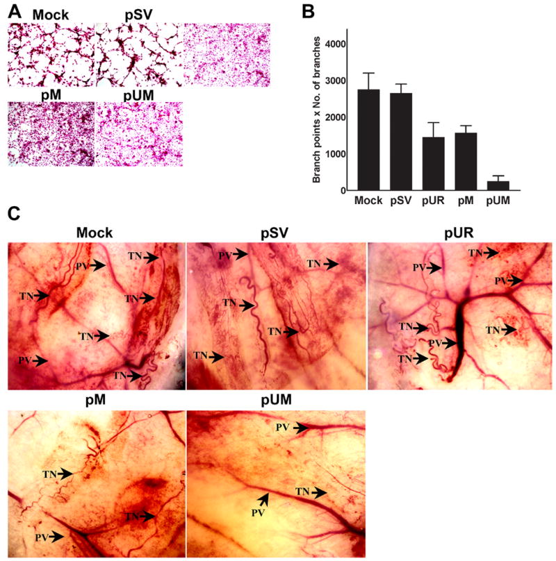
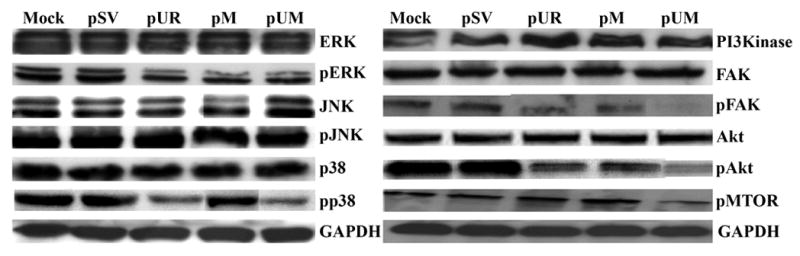
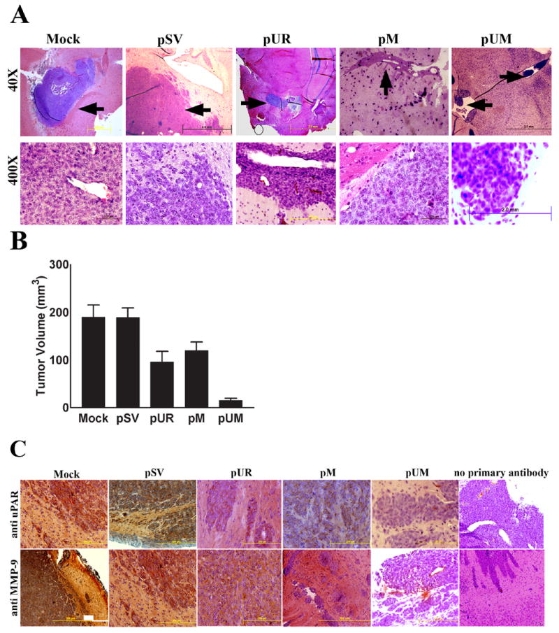
Meningiomas are the most commonly occurring tumors of the central nervous system including the brain and spinal cord. Malignant meningiomas are highly aggressive and frequently recur after surgical resection of the tumor. Our previous studies have reported that urokinase plasminogen activator receptor (uPAR) and matrix metalloproteinase-9 (MMP-9) play important roles in tumor progression. In the present study, we have attempted to evaluate the roles of these molecules in the malignant meningioma tumor microenvironment and to determine the effectiveness of using single or bicistronic small interfering RNA constructs for uPAR and MMP-9 on tumor cell proliferation, migration, invasion, angiogenesis and regression of pre-established orthotopic tumors. Transfection of single or bicistronic constructs downregulated uPAR and MMP-9 in meningioma cells compared to controls. A significant reduction in tumor invasion was determined with matrigel gel and spheroid invasion assays in meningioma cells after transfection of these plasmids. Furthermore, downregulation of uPAR and MMP-9 reduced migration of tumor spheroids on vitronectin-coated plates. uPAR and MMP-9 downregulation suppressed capillary network formation, in both in vitro and in vivo models. Also, it is well known that tumor cells manipulate intracellular signaling pathways to aid in various processes involved in tumor progression. Our study revealed that downregulation of uPAR and MMP-9 leads to a decrease in the activation of some of the important enzymes participating in the MAPK and PI3 kinase pathways, which in turn, might decrease cell survival and proliferation. In addition, we analyzed the efficiency of RNAi-mediated targeting of uPAR and MMP-9 in pre-established tumor growth in vivo. We observed a significant regression of pre-established orthotopic tumors upon RNAi-mediated targeting of uPAR and MMP-9. In addition, the present study indicated that targeting both the proteins simultaneously augmented the therapeutic treatment of human meningiomas.


Figures

Figure 1
Figure 1. RNAi-mediated knockdown of uPAR and MMP-9 expression in IOMM-Lee cells
(A) Semi-quantitative RT-PCR of RNA extracted from IOMM-Lee parental cells (mock) and cells transfected with shRNA plasmids pSV, pUR, pM and pUM. RT-PCR reaction was also performed for GAPDH and served as a loading control. (B) Following transfection with cell lysates from pSV, pUR, pM and pUM cells and parental cells (mock) were probed for uPAR using a specific antibody for uPAR by western blotting analysis. GAPDH was analyzed as a loading control. (C) Enzymatic activity of MMP-9 was analyzed in the conditioned media collected from cells transfected with pSV, pUR, pM, pUM and parental cells. 30 μg of protein was loaded onto 10% SDS-PAGE containing gelatin.
Figure 2
Figure 2. Immunofluorescence detection for in situ expression of uPAR and MMP-9
5 × 10⁴ IOMM-Lee cells were cultured in 8-well chamber slides and transfected with pSV, pUR, pM, and pUM. Parental cells (mock) were simultaneously maintained. 72 h following transfection, the cells were fixed in formaldehyde and processed to detect expression of uPAR and MMP-9. The cells were mounted with 4′, 6-diamidino-2-phenylindole (DAPI) to visualize the nucleus.
Figure 3
Figure 3. IOMM-Lee cell proliferation decreases following RNAi-mediated reduction of uPAR and MMP-9 gene expression
IOMM-Lee cells were maintained in triplicate in 96-well plates at a concentration of 5 × 10³ and transfected with pSV, pUR, pM and pUM as described in Materials & Methods. 48 h after transfection, viable cell mass was measured in both parental and treated cells at different time intervals (1 to 5 days). A570 was plotted against the respective time intervals. Mean ± S.D. values from 3 different experiments are shown (p<0.001).
Figure 4
Figure 4. Downregulation of uPAR and MMP-9 expression through RNAi treatment abate spheroid migration on vitronectin-coated substrate
(A) 7 × 10⁴ IOMM-Lee-GFP cells were cultured in 96-well low attachment plates and spheroids were allowed to grow for 3 to 4 dayswith shaking at 40–60 rpm at 37°C. The spheroids were then transfected with shRNA plasmids pSV, pUR, pM and pUM. Untreated spheroids were also maintained to serve as the control (mock). 48 h after transfection, the spheroids were transferred to vitronectin-coated (50 mg/mL) 8-well chamber slides and maintained for another 72 h in serum-free media. Migration of spheroids was analyzed after taking pictures under a fluorescent microscope. (B) Migration was quantified as the distance spheroid cells moved away from spheroids on vitronectin-coated substrate. Values are mean ± S.D. from three different experiments (p<0.001). (C) IOMM-Lee cells were transfected with pSV, pUR, pM and pUM. Untransfected cells (mock) were also maintained to serve as a control. 48 h later, the cells were trypsinized, counted and 1×10⁵ cells were cultured in the upper chamber of a Transwell insert coated with matrigel (1 mg/mL) and processed per manufacturer’s instructions. (D) Number of cells was counted in three different fields for each sample and the percentage invasion of cells treated with shRNA plasmids was analyzed in comparison with the untreated (mock) cells. The graph represents the percentage invasion shown by the cells transfected with pSV, pUR, pM and pUM in comparison with untreated cells (mock). Values are mean ± S.D. from three different experiments (p<0.001). (E) 7 × 10⁴ IOMM-Lee cells were cultured in 96-well low attachment plates and allowed to grow for 3 to 4 days with shaking at 40–60 rpm at 37°C. The spheroids were later transfected with pSV, pUR, pM and pUM. 48 h after transfection, the cells were labeled with Dil (red fluorescent dye). Untreated spheroids (mock) were maintained as control under similar conditions. Simultaneously fetal rat brain aggregates (FRBA) were grown from 16 to 17 day old fetal rat brain cells and labeled with DiO (green fluorescent dye). Later, both the IOMM-Lee spheroids and spheroids from fetal rat brain cells were co-cultured and maintained in serum-free media. Invasion of fetal rat brain spheroids by tumor cell spheroids was recorded at 24, 48 and 72 h using a fluorescent microscope and quantified. (F) The percentage invasion of the untransfected spheroids and spheroids treated with shRNA plasmids was quantified using image analysis software and plotted as the fetal rat brain aggregates remaining uninvaded against different time intervals. Values shown are the mean ± S.D. from three different experiments (p<0.001).
Figure 5
Figure 5. Targeting uPAR and MMP-9 through RNAi treatment reduced angiogenesis initiated by IOMM-Lee cells
(A) 4 × 10⁴ IOMM-Lee cells were cultured in 8-well chamber slides and transfected with pSV, pUR, pM and pUM and grown for 24 h. At the same time, untreated cells (mock) were maintained for the control. After 24 h, the media was replaced with serum-free media and maintained for another 24 h. Simultaneously, HMEC cells (3 × 10⁴) were maintained in 8-well chamber slides. 24 h following addition of serum-free media to transfected IOMM-Lee cells, the conditioned media was collected and added to the HMEC cells. The HMEC cells were grown in conditioned media for another 72 h, stained with H&E and capillary network formation was analyzed with light microscopy. (B) The ability of capillary network formation was analyzed as number of branch points and number of branches per branch point and plotted against the respective cells. Values represent mean ± S.D. from three different experiments. (C) Dorsal skin fold chamber model revealed inhibition of in vivo angiogenesis as a result of RNAi-mediated abrogation of uPAR and MMP-9. Diffusion chambers holding 2 × 10⁶ IOMM-lee parental cells and cells transfected with pSV, pUR, pM and pUM were introduced into dorsal air sacs of athymic nude mice as described in Materials & Methods. Ten days after introduction of the diffusion chambers, the animals were sacrificed. The skin around the chamber was carefully removed and observed under light microscope. Delicate zigzag-shaped microvessels showing irregular arrangement when compared to more organized pre-existing vasculature (PV) were recorded as tumor-induced neovasculature (TN).
Figure 6
Figure 6. RNAi-mediated downregulation of uPAR and MMP-9 modulates downstream signaling events
IOMM-Lee cells were transfected with pSV, pUR, pM and pUM. Simultaneously, untreated cells (mock) were maintained to serve as the control. The cells were lysed after 48 h and the cell lysates were analyzed for various proteins involved in the MAPK and PI3 kinase intracellular signaling pathways. Phosphorylated and total FAK was also analyzed as described in Materials and Methods.
Figure 7
Figure 7. Regression of pre-established orthotopic tumors upon RNAi-mediated downregulation of uPAR and MMP-9
(A) Orthotopic intracranial tumors were established in nude mice and treated with shRNA plasmids as described in Materials & Methods. Following extraction, the brains were embedded in paraffin, sectioned and stained with H&E. Photomicrographs of tumor sections revealing total tumor (40X) and rapidly dividing tumor cells (400X) are shown in the figure. (B) Semi-quantification of tumor volume was performed as described in Materials & Methods. Values represent mean ± S.D. from five different animals. (C) Immunohistochemical detection for uPAR and MMP-9 expression in pre-established orthotopic tumors following treatment with shRNA plasmids: Paraffin-embedded tumor sections were subjected to immunohistochemical detection for uPAR and MMP-9 following standard protocol. Appropriate protein-specific antibodies were used. The slides were stained with hematoxylin to visualize the nucleus, mounted and observed under a light microscope. Fields with brown stain resulting from DAB interaction were scored for protein expression.

Similar articles

Cited by

References

    1. Bianchini F, D’Alessio S, Fibbi G, Del RM, Calorini L. Cytokine-dependent invasiveness in B16 murine melanoma cells: role of uPA system and MMP-9. Oncol Rep. 2006;15:709–714. - PubMed
    1. Kim TD, Song KS, Li G, Choi H, Park HD, Lim K, Hwang BD, Yoon WH. Activity and expression of urokinase-type plasminogen activator and matrix metalloproteinases in human colorectal cancer. BMC Cancer. 2006;6:211. - PMC - PubMed
    1. Puli S, Lai JC, Bhushan A. Inhibition of matrix degrading enzymes and invasion in human glioblastoma (U87MG) Cells by isoflavones. J Neurooncol. 2006;79:135–142. - PubMed
    1. Schwab W, Schulze-Tanzil G, Mobasheri A, Dressler J, Kotzsch M, Shakibaei M. Interleukin-1beta-induced expression of the urokinase-type plasminogen activator receptor and its co-localization with MMPs in human articular chondrocytes. Histol Histopathol. 2004;19:105–112. - PubMed
    1. Tan X, Egami H, Nozawa F, Abe M, Baba H. Analysis of the invasion-metastasis mechanism in pancreatic cancer: involvement of plasmin(ogen) cascade proteins in the invasion of pancreatic cancer cells. Int J Oncol. 2006;28:369–374. - PubMed

Publication types

MeSH terms

Substances