. 2007 Jul;31(1):161-8.

Selected flavonoids potentiate the toxicity of cisplatin in human lung adenocarcinoma cells: a role for glutathione depletion

Affiliations

Selected flavonoids potentiate the toxicity of cisplatin in human lung adenocarcinoma cells: a role for glutathione depletion

Remy Kachadourian et al. Int J Oncol. 2007 Jul.

Abstract

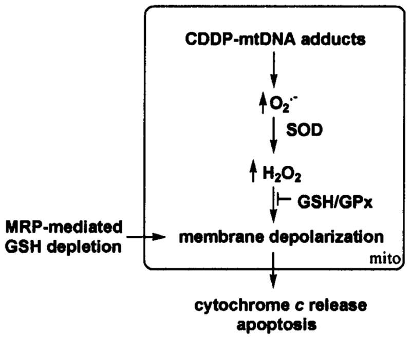
Adjuvant therapies that enhance the anti-tumor effects of cis-diammineplatinum(II) dichloride (cisplatin, CDDP) are actively being pursued. Growing evidence supports the involvement of mitochondrial dysfunction in the anti-cancer effect of cisplatin. We examined the potential of using selective flavonoids that are effective in depleting tumor cells of glutathione (GSH) to potentiate cisplatin-mediated cytotoxicity in human lung adenocarcinoma (A549) cells. We found that cisplatin (40 microM, 48-h treatment) disrupts the steady-state levels of mitochondrial respiratory complex I, which correlates with elevated mitochondrial reactive oxygen species (ROS) production and cytochrome c release. The flavonoids, 2',5'-dihydroxychalcone (2',5'-DHC, 20 microM) and chrysin (20 microM) potentiated the cytotoxicity of cisplatin (20 microM), which could be blocked by supplementation of the media with exogenous GSH (500 microM). Both 2',5'-DHC and chrysin were more effective than the specific inhibitor of GSH synthesis, L-buthionine sulfoximine (BSO, 20 microM), in inducing GSH depletion and potentiating the cytotoxic effect of cisplatin. These data suggest that the flavonoid-induced potentiation of cisplatin's toxicity is due, in part, to synergetic pro-oxidant effects of cisplatin by inducing mitochondrial dysfunction, and the flavonoids by depleting cellular GSH, an important antioxidant defense.


Figures

Figure 1
Figure 1
Molecular structures of chalcone (no hydroxyl group), 2′-hydro-xychalcone (2′-HC), 4′-hydroxychalcone (4′-HC), 2′,5′-dihydroxychalcone (2′,5′-DHC) and chrysin.
Figure 2
Figure 2
Cisplatin (CDDP) disrupted the steady-state level mitochondrial respiratory complex I and increased mitochondrial superoxide (O2•−) formation in A549 cells. (A) Lower levels of the mitochondrial respiratory complex I were detected when compared to complex II as shown by immunoblotting in CDDP-treated A549 cells (40 μM, 48-h treatment) versus control (Co). Each figure is representative of three samples and the experiment repeated twice. (B) CDDP (20 μM, 48-h treatment) induced an increase of O2•− formation in the mitochondria as detected by flow cytometry using the mitochondrial O2•− sensitive dye MitoSOX. (C) Flow cytometry analysis of CDDP-mediated mitochondrial O2•− over time at 24 and 48 h. Bars with different letters are statistically different from one another (n=3, P<0.05).
Figure 3
Figure 3
2′,5′-DHC and chrysin induced GSH depletion and potentiated cisplatin-mediated cytotoxicity in A549 cells. (A) At 6-h treatment, the abilities of 2′,5′-DHC (DHC, 20 μM) and chrysin (Chr, 20 μM) to deplete GSH were more important compared to the inhibitor of γ-glutamylcysteine synthetase BSO (20 μM) (n=3). In the case of 2,5-DHC, however, a rebound of GSH levels was observed at 24 h. (B) 2,5-DHC and chrysin potentiated the toxicity of cisplatin (CDDP, 20 μM) in A549 cells. (C) Exogenous GSH (500 μM) restored cytosolic GSH levels (n=3). (D) Exogenous GSH (500 μM) partially protected the cells towards the toxicity of cisplatin and blocked the 2′,5′-DHC-mediated potentiation of cisplatin’s toxicity. Similar results were obtained with chrysin (data not shown). Bars with different letters are statistically different from one another (n=4, P<0.05).
Figure 4
Figure 4
2′,5′-DHC increased the cisplatin-mediated depolarization of the mitochondrial membrane and cytochrome c release. (A and B) As shown using flow cytometry and JC-1 as dye, cisplatin treatment (CDDP, 20 μM, 48 h) induced a depolarization of the mitochondrial membrane, effect that was amplified by adding 2′,5′-DHC (DHC, 20 μM). The FL2/FL1 ratio was measured using the mean fluorescence in each channel. Bars with different letters are statistically different from one another (n=3, P<0.05). (C) As shown by immunoblotting using GAPDH as internal standard, cisplatin treatment (CDDP, 20 μM, 48 h) induced the release of cytochrome c (Cyt. c), which was increased by adding 2′,5′-DHC (DHC, 20 μM). 2′,5′-DHC alone had no significant effect (not shown). Each figure was representative of three samples and the experiment was repeated at least twice.
Figure 5
Figure 5
Working hypothesis. The formation of cisplatin-mitochondrial DNA (CDDP-mtDNA) adducts disrupts the assembly of the apoproteins of the mitochondrial respiratory chain, which in turn induces the formation of mitochondrial superoxide (O2•−). The latter is converted by superoxide dismutase (SOD) into hydrogen peroxide (H2O2), which mediates the depolarization of the mitochondrial membrane and the release of cytochrome c, thus triggering the cascade of events leading to nuclear DNA fragmentation and apoptosis. Flavonoid-induced GSH depletion adds to the mitochondrial membrane depolarization by mechanisms that remain to be clarified.

Similar articles

Cited by

References

    1. Wang D, Lippard SJ. Cellular processing of platinum anticancer drugs. Nat Rev Drug Discov. 2005;4:307–320. - PubMed
    1. Yen HC, Tang YC, Chen FY, Chen SW, Majima HJ. Enhancement of cisplatin-induced apoptosis and caspase 3 activation by depletion of mitochondrial DNA in a human osteosarcoma cell line. Ann NY Acad Sci. 2005;1042:516–522. - PubMed
    1. Schweyer S, Soruri A, Heintze A, Radzun HJ, Fayyazi A. The role of reactive oxygen species in cisplatin-induced apo-ptosis in human malignant testicular germ cell lines. Int J Oncol. 2004;25:1671–1676. - PubMed
    1. Huang HL, Fang LW, Lu SP, Chu CK, Luh TY, Lai MZ. DNA-damaging reagents induce apoptosis through reactive oxygen species-dependent Fas aggregation. Oncogene. 2003;22:8168–8177. - PubMed
    1. Troyano A, Sancho P, Fernandez C, De Blas E, Bernardi P, Aller P. The selection between apoptosis and necrosis is differentially regulated in hydrogen peroxide-treated and glu-tathione-depleted human promonocytic cells. Cell Death Differ. 2003;10:889–898. - PubMed

Publication types

MeSH terms