. 2007 Nov;16(11):1867-74.
doi: 10.1007/s00586-007-0373-3. Epub 2007 Jun 5.

Kinematics of the lumbar spine in trunk rotation: in vivo three-dimensional analysis using magnetic resonance imaging

Affiliations

Kinematics of the lumbar spine in trunk rotation: in vivo three-dimensional analysis using magnetic resonance imaging

Ryutaro Fujii et al. Eur Spine J. 2007 Nov.

Abstract

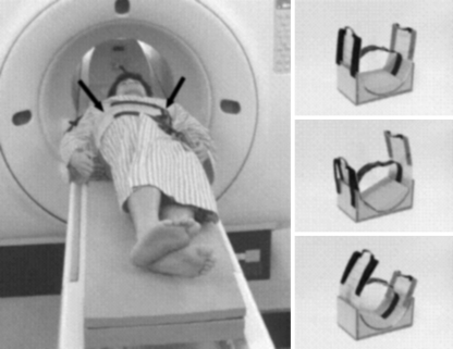
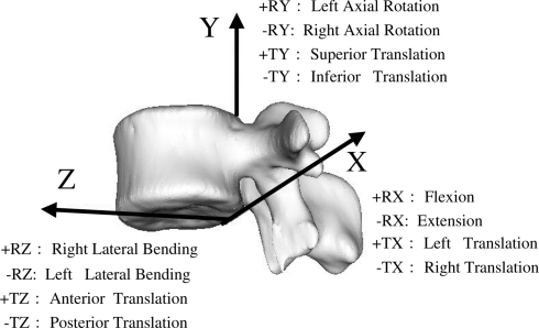
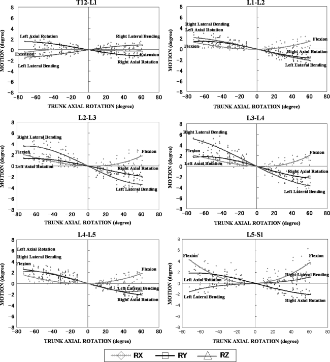
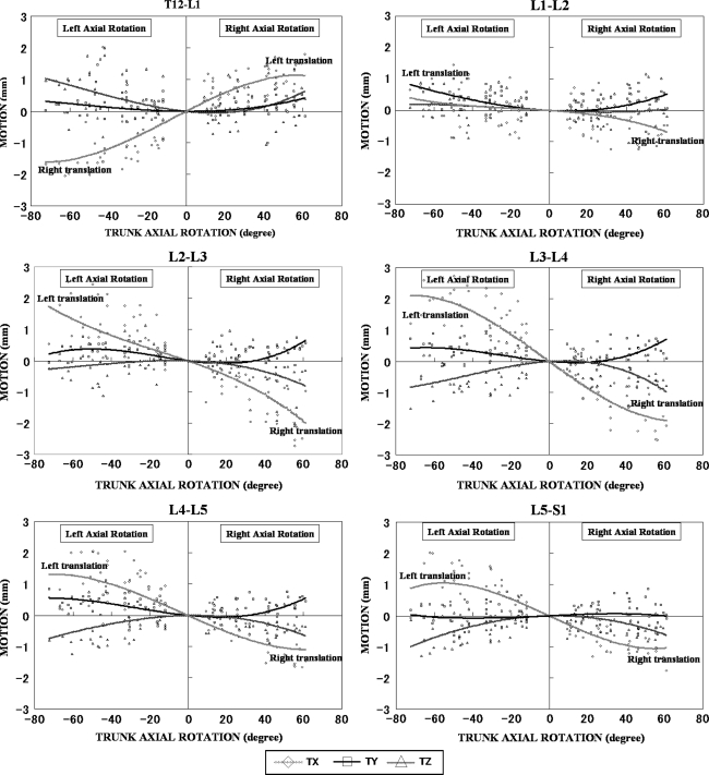
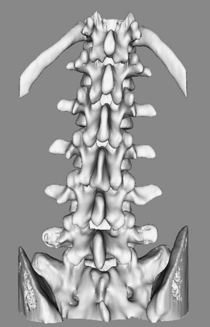
In vivo three-dimensional (3D) kinematics of the lumbar spine has not been well evaluated by the conventional methods because of their methodological limitations, while 3D intervertebral motions have been quantitatively determined by cadaver studies. We thus developed a novel 3D analyzing system for the relative motions of individual vertebrae using 3D magnetic resonance imaging (MRI) and analyzed in vivo 3D intervertebral motions of the lumbar spine during trunk rotation. Ten healthy volunteers underwent 3D MRI of the lumbar spine in nine positions with 15 degrees increments during trunk rotation (0 degrees , 15 degrees , 30 degrees , 45 degrees , and maximum). Relative motions of the lumbar spine were calculated by automatically superimposing a segmented 3D MRI of the vertebra in the neutral position over images of each position using the voxel-based registration method. These 3D motions were represented with 6 degrees of freedom by Euler angles and translations on the coordinate system. The mean axial rotation of ten healthy volunteers of each lumbar spinal segment in 45 degrees trunk rotation to each side ranged from 1.2 degrees to 1.7 degrees . Coupled flexion with axial rotation was observed at the segments from L1/2 to L5/S1. Coupled lateral bending of the segments from L1/2 to L4/5 was in the opposite direction of the trunk rotation, while that of T12/L1 and L5/S1 was in the same direction. The direction of the coupled lateral bending in the present study was different from that in the previous cadaver study only at L4/5. This difference might result from the non-load state of the supine position in the current study and/or the non-physiological state in the cadaver study. Our system has two limitations: (1) the study was conducted with each volunteer in the supine position, and (2) because the rotation device regulated trunk rotation, trunk rotation might not have been physiological. In vivo 3D intervertebral motions of the lumbar spine during trunk rotation were evaluated using our novel motion analysis system. These data may be useful for the optimal orthopaedic management of lumbar spinal disorders.


Figures

Fig. 1
Fig. 1
Acquisition of 3D-MRI. Our original device (arrow) was used to rotate the trunk of the subjects as reproducibly as possible. 3D MRIs were obtained in nine positions with 15° increments of trunk rotation (0°, 15°, 30°, 45°, and maximum). Maximum trunk rotation was defined as the position in which the trunks were rotated as much as possible without the bilateral shoulders pulled away from the MRI table
Fig. 2
Fig. 2
Anatomical orthogonal coordinate system. The origin was located at the most inferior point on the posterior wall of the vertebral body in the mid-sagittal plane. The Z-axis was defined as the line connecting the anterior and posterior points in the inferior plane of the vertebral body (anterior considered to be positive). The Y-axis was defined as perpendicular to the Z-axis, with superior being positive. The positive X-axis was directed to the left. Translation was calculated based on the origin of the coordinate system
Fig. 3
Fig. 3
Intervertebral motions of the lumbar spine during trunk axial rotation. The degree of trunk axial rotation was plotted on the X-axis. The degree of rotational motion along each axis on the anatomical orthogonal coordinate system and the length of translational motion at each level were plotted on the Y-axis. Approximate curves, which were prepared as tertiary approximate curves based on multiple function approximation were drawn. Statistical analysis of right and left rotation data showed no significant difference, thus ruling out asymmetric kinematics
Fig. 3
Fig. 3
Intervertebral motions of the lumbar spine during trunk axial rotation. The degree of trunk axial rotation was plotted on the X-axis. The degree of rotational motion along each axis on the anatomical orthogonal coordinate system and the length of translational motion at each level were plotted on the Y-axis. Approximate curves, which were prepared as tertiary approximate curves based on multiple function approximation were drawn. Statistical analysis of right and left rotation data showed no significant difference, thus ruling out asymmetric kinematics
Fig. 4
Fig. 4
Three-dimensional motions of the lumbar spine during trunk axial rotation. (To access the video illustration, visit the online issue of European Spine Journal)

References

    1. Goto A, Moritomo H, Murase T, Oka K, Sugamoto K, Arimura T, Masumoto J, Tamura S, Yoshikawa H, Ochi T. In vivo three-dimensional wrist motion analysis using magnetic resonance imaging and volume-based registration. J Orthop Res. 2005;23(4):750–756. doi: 10.1016/j.orthres.2004.10.001. - DOI - PubMed
    1. Ishii T, Mukai Y, Hosono N, Sakaura H, Nakajima Y, Sato Y, Sugamoto K, Yoshikawa H. Kinematics of the upper cervical spine in rotation: in vivo three-dimensional analysis. Spine. 2004;29(7):E139–E144. doi: 10.1097/01.BRS.0000116998.55056.3C. - DOI - PubMed
    1. Ishii T, Mukai Y, Hosono N, Sakaura H, Fujii R, Nakajima Y, Tamura S, Sugamoto K, Yoshikawa H. Kinematics of the subaxial cervical spine in rotation in vivo three-dimensional analysis. Spine. 2004;29(24):2826–2831. doi: 10.1097/01.brs.0000147806.31675.6b. - DOI - PubMed
    1. Ishii T, Mukai Y, Hosono N, Sakaura H, Fujii R, Nakajima Y, Tamura S, Iwasaki M, Yoshikawa H, Sugamoto K. Kinematics of the cervical spine in lateral bending: in vivo three-dimensional analysis. Spine. 2006;31(2):155–160. doi: 10.1097/01.brs.0000195173.47334.1f. - DOI - PubMed
    1. Lee RY. Kinematics of rotational mobilisation of the lumbar spine. Clin Biomech. 2001;16(6):481–488. doi: 10.1016/S0268-0033(01)00036-5. - DOI - PubMed