. 2007 Jul;37(7):1924-37.
doi: 10.1002/eji.200737089.

A homodimeric complex of HLA-G on normal trophoblast cells modulates antigen-presenting cells via LILRB1

Affiliations

A homodimeric complex of HLA-G on normal trophoblast cells modulates antigen-presenting cells via LILRB1

Richard Apps et al. Eur J Immunol. 2007 Jul.

Abstract

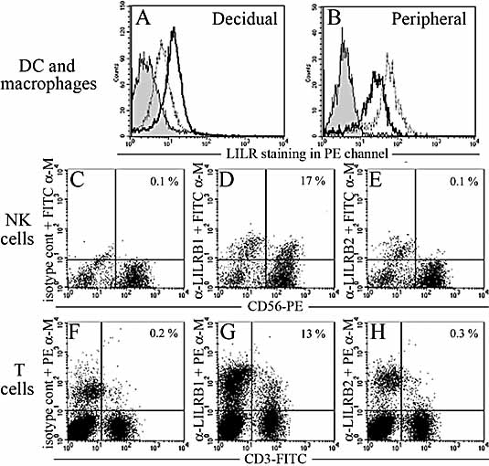
In healthy individuals, the non-classical MHC molecule HLA-G is only expressed on fetal trophoblast cells that invade the decidua during placentation. We show that a significant proportion of HLA-G at the surface of normal human trophoblast cells is present as a disulphide-linked homodimer of the conventional beta(2)m-associated HLA-I complex. HLA-G is a ligand for leukocyte immunoglobulin-like receptors (LILR), which bind much more efficiently to dimeric HLA-G than to conventional HLA-I molecules. We find that a LILRB1-Fc fusion protein preferentially binds the dimeric form of HLA-G on trophoblast cells. We detect LILRB1 expression on decidual myelomonocytic cells; therefore, trophoblast HLA-G may modulate the function of these cells. Co-culture with HLA-G(+) cells does not inhibit monocyte-derived dendritic cell up-regulation of HLA-DR and costimulatory molecules on maturation, but did increase production of IL-6 and IL-10. Furthermore, proliferation of allogeneic lymphocytes was inhibited by HLA-G binding to LILRB1/2 on responding antigen-presenting cells (APC). As HLA-G is the only HLA-I molecule that forms beta(2)m-associated dimers with increased avidity for LILRB1, this interaction could represent a placental-specific signal to decidual APC. We suggest that the placenta is modulating maternal immune responses locally in the uterus through HLA-G, a trophoblast-specific, monomorphic signal present in almost every pregnancy. See accompanying commentary: (http://dx.doi.org/10.1002/eji.200737515).


Figures

Figure 1
Figure 1
HLA-G is present on the surface of normal trophoblast cells as a disulphide-linked homodimer of the conventional β2m-associated class I complex. 721.221 + HLA-G1 transfectants (A, B), JEG-3 cell line (C–E) and trophoblast (F, G) were surface biotinylated, immunoprecipitated with the indicated antibodies and resolved by reducing (A, C, F) or non-reducing (B, D, E, G) PAGE before Western blotting with streptavidin-HRP. All were exposed for less than 1 min, with the exception of (E) which was exposed for 10 min. Complexes immunoprecipitated with G233 from 721.221 HLA-G+ transfectants (H) and trophoblast (J) were also analysed by 2D-PAGE. These gels are representative of at least three different experiments for each cell type, using a total of 20 pooled samples for trophoblast cells. Images in this figure include lanes from several gels.
Figure 2
Figure 2
Soluble HLA-G can also exist as a disulphide-linked homodimer of the conventional β2m-associated class I complex. Soluble HLA-G molecules were immunoprecipitated with the indicated antibodies from supernatants of 721.221 cells transfected with HLA-G1 or HLA-G5 cDNA compared to untransfected cells. The JEG-3 cell line, isolated primary trophoblast, or pre-implantation embryo culture supernatants were also immunoprecipitated compared to unconditioned medium. Precipitated complexes were separated by reducing (A, B) or non-reducing PAGE (C) and detected by Western blotting with biotinylated mAb MEM-G/1 followed by streptavidin-HRP. HLA-G1+ transfectant, JEG-3 and trophoblast culture supernatants were concentrated 30 times before immunoprecipitation. Gels are representative of four independent experiments. The image in (C) includes lanes from separate gels.
Figure 3
Figure 3
LILRB expression by decidual leukocytes. Decidual (A) or peripheral (B) leukocytes were identified as myelomonocytic cells by forward/side scatter and HLA-DR+ gates and stained with anti-LILRB1 (dark line), anti-LILRB2 (grey line) or isotype control antibodies (filled histogram). Decidual CD56+ (C–E) or CD3+ (F–H) lymphocytes were stained with isotype control (C, F), anti-LILRB1 (D, G) and anti-LILRB2 (E, H) antibodies. These results from decidual leukocytes are representative of seven different experiments using decidua pooled from 17 samples. Similar results for both LILRB1 and 2 were observed with two different antibodies. The apparently higher isotype control mAb staining of T cells compared to NK cells results from inclusion of more cells in plots demonstrating T cell staining, due to their low frequency in decidual leukocyte populations.
Figure 4
Figure 4
LILRB1-Fc binds to HLA-G at the surface of transfected cells and trophoblast. HLA-G expression on the indicated cells was detected by G233 staining (green) compared to untransfected cells (red) or with an isotype control mAb (filled histogram) (A–D). LILRB1 binding to the indicated cells was detected by LILRB1-Fc staining (green) compared to untransfected cells (red) or with an IgG-Fc fragment (filled histogram) (E–H). Specificity was confirmed with blocking mAb (J–M): LILRB1-Fc (green) and IgG1-Fc fragment (filled histogram) binding are shown as before and also after pre-incubation of the fusion protein with anti-LILRB1 mAb M401 (red) or of the cells with anti-HLA-I mAb W6/32 (blue). These results are representative of four different experiments for each cell type, using trophoblast from eight samples. To see LILRB1-Fc binding to trophoblast cells, contaminating leukocytes that bound the fusion protein non-specifically via FcγR were labelled with a lineage marker cocktail (CD3/14/16/19/20/56-FITC) and analysis was performed on FITC-negative trophoblast cells (upper left quadrant). Dot plots of LILRB1-Fc binding (N) and blocking by anti-LILRB1 mAb (O) are shown.
Figure 5
Figure 5
LILRB1-Fc preferentially binds to the dimeric form of HLA-G at the cell surface. 721.221 HLA-G+ transfectants (221G), untransfected controls (221P) and normal trophoblast cells were surface-biotinylated, immunoprecipitated with the indicated antibodies or fusion proteins and resolved by non-reducing PAGE before Western blotting with streptavidin-HRP. These results are representative of five different experiments for each cell type. Trophoblast cells were pooled from 15 samples. The image includes lanes from separate gels.
Figure 6
Figure 6
MDDC activated in the presence of HLA-G show normal up-regulation of maturation markers but altered cytokine production. (A) MDDC, co-cultured with HLA-I null 721.221 cells, were analysed for expression of the indicated LILR receptors, differentiation and activation markers in the absence of stimulation (filled histogram) or after exposure to LPS (green trace). The same markers were analysed after exposure to LPS in the presence of HLA-G transfectants instead of HLA null cells (red trace). Isotype control staining for each marker is shown in orange. (B) Levels of the indicated cytokines in these co-culture supernatants were measured by ELISA on two separate occasions. A representative experiment is shown here, ± SD of triplicate measurements. Culture supernatants from at least four independent experiments were tested and analysed together for statistical significance.
Figure 7
Figure 7
Allogeneic lymphocyte proliferation is inhibited by HLA-G interacting with LILRB receptors on HLA-DR+ cells. (A) Irradiated 721.221 stimulator cells transfected with various HLA-I molecules were cultured with PBMC responders from a healthy donor. Proliferation measured by [3H]thymidine incorporation after 5 days was reduced in the presence of HLA-G but not a classical HLA-I molecule. Reduced proliferation to HLA-G transfectants was abrogated by addition of anti-LILRB and anti-HLA-I mAb. (B) When HLA-DR+ cells were depleted from the responder population, a reduced proliferative response with no additional inhibitory effect by HLA-G was observed. One result is shown, ± SD of triplicates. Three to ten independent experiments under each condition were performed and these were analysed together for statistical significance. (C) Flow cytometry analysis of the responder population confirming depletion of HLA-DR+ cells.

Comment in

References

    1. McMaster MT, Librach CL, Zhou Y, Lim KH, Janatpour MJ, DeMars R, Kovats S, et al. Human placental HLA-G expression is restricted to differentiated cytotrophoblasts. J. Immunol. 1995;154:3771–3778. - PubMed
    1. Loke YW, King A, Burrows T, Gardner L, Bowen M, Hiby S, Howlett S, et al. Evaluation of trophoblast HLA-G antigen with a specific monoclonal antibody. Tissue Antigens. 1997;50:135–146. - PubMed
    1. Hiby SE, King A, Sharkey AM, Loke YW. Molecular studies of trophoblast HLA-G: Polymorphism, isoforms, imprinting and expression in preimplantation embryo. Tissue Antigens. 1999;53:1–13. - PubMed
    1. Blaschitz A, Juch H, Volz A, Hutter H, Daxboeck C, Desoye G, Dohr G. The soluble pool of HLA-G produced by human trophoblasts does not include detectable levels of the intron 4-containing HLA-G5 and HLA-G6 isoforms. Mol. Hum. Reprod. 2005;11:699–710. - PubMed
    1. Davis DM, Reyburn HT, Pazmany L, Chiu I, Mandelboim O, Strominger JL. Impaired spontaneous endocytosis of HLA-G. Eur. J. Immunol. 1997;27:2714–2719. - PubMed

Publication types

MeSH terms