. 2007 Jun;5(6):e158.
doi: 10.1371/journal.pbio.0050158.

Higher-order oligomerization targets plasma membrane proteins and HIV gag to exosomes

Affiliations

Higher-order oligomerization targets plasma membrane proteins and HIV gag to exosomes

Yi Fang et al. PLoS Biol. 2007 Jun.

Abstract

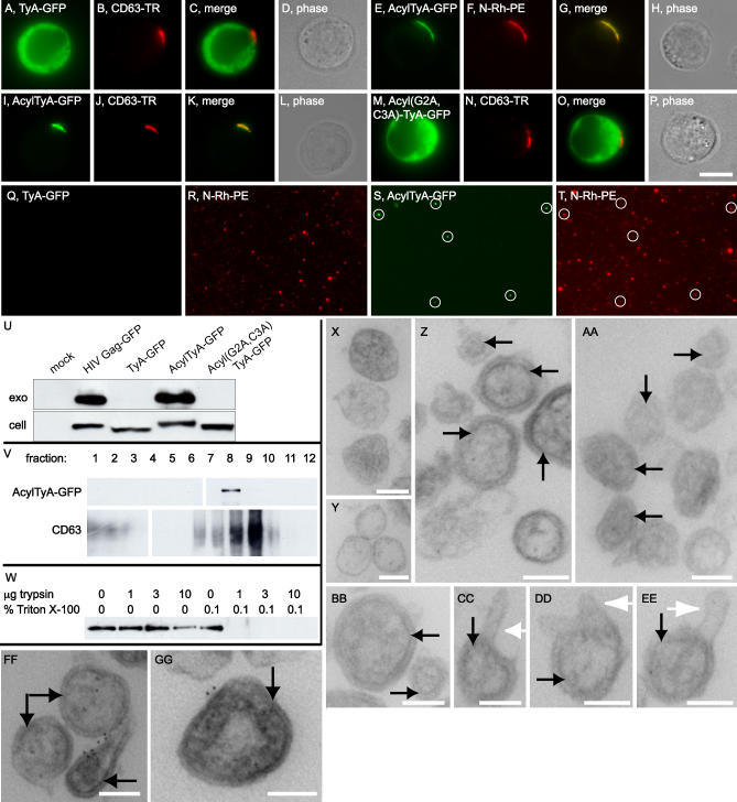
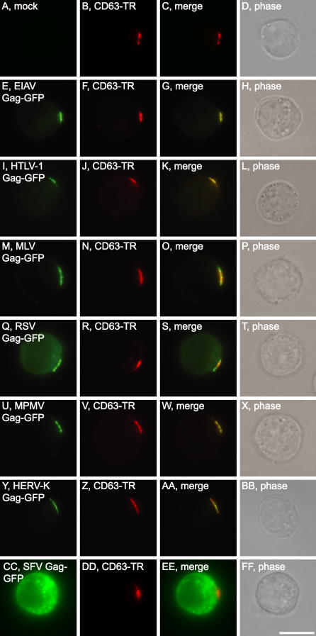
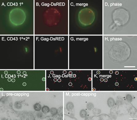
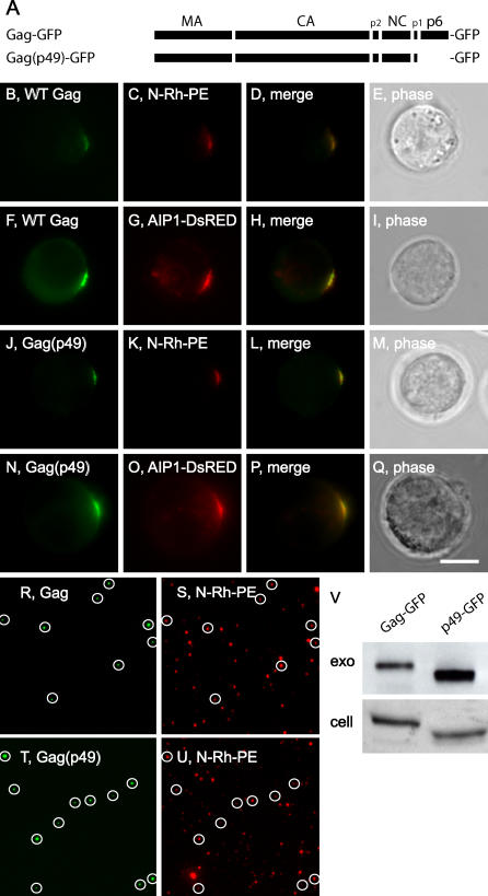
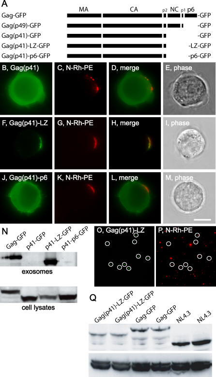
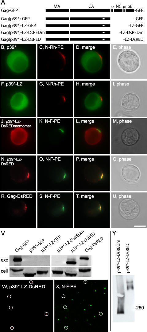
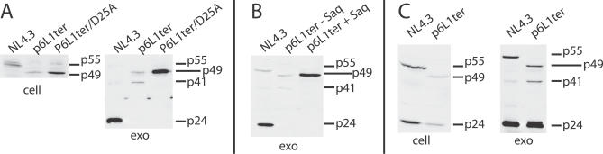
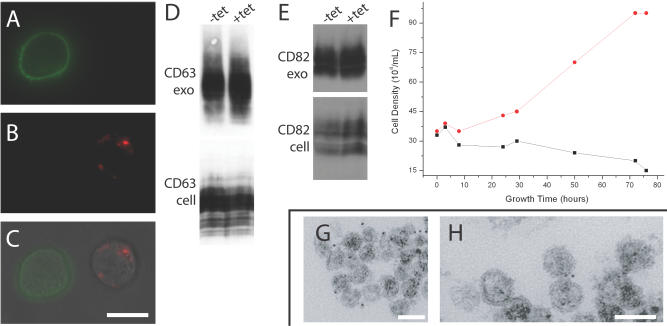
Exosomes are secreted organelles that have the same topology as the cell and bud outward (outward is defined as away from the cytoplasm) from endosome membranes or endosome-like domains of plasma membrane. Here we describe an exosomal protein-sorting pathway in Jurkat T cells that selects cargo proteins on the basis of both higher-order oligomerization (the oligomerization of oligomers) and plasma membrane association, acts on proteins seemingly without regard to their function, sequence, topology, or mechanism of membrane association, and appears to operate independently of class E vacuolar protein-sorting (VPS) function. We also show that higher-order oligomerization is sufficient to target plasma membrane proteins to HIV virus-like particles, that diverse Gag proteins possess exosomal-sorting information, and that higher-order oligomerization is a primary determinant of HIV Gag budding/exosomal sorting. In addition, we provide evidence that both the HIV late domain and class E VPS function promote HIV budding by unexpectedly complex, seemingly indirect mechanisms. These results support the hypothesis that HIV and other retroviruses are generated by a normal, nonviral pathway of exosome biogenesis.


Conflict of interest statement

Competing interests. The authors have declared that no competing interests exist.

Figures

Figure 1
Figure 1. Higher-Order Oligomerization Targets Proteins to ELDs
(A–X) N-Rh-PE–labeled Jurkat T cells were incubated with monoclonal antibodies specific for (A–H) CD43, (I–P) CD45, or (Q–X) CD59 and FITC-labeled polyclonal anti-mouse antibodies, on ice, and then either (A–D, I–L, and Q–T) fixed or (E–H, M–P, and U–X) incubated at 37 °C for 2 h and then fixed. (Y–LL) Higher-order oligomerization targets CD43 to exosomes. (Y–FF) N-Rh-PE–labeled Jurkat T cells were incubated with FITC-labeled monoclonal antibodies specific for CD43, then incubated with either (Y–BB) buffer alone or (CC–FF) unlabeled polyclonal rabbit anti-mouse IgG antibodies, all on ice. Cells were incubated for 24 h at 37 °C and then examined. (GG–KK) Fluorescence microscopy of exosomes from cells treated with (GG and HH) primary antibodies only or (II and JJ) primary and secondary antibodies. (KK) Numbers of FITC-positive exosomes secreted by cells exposed to (left bar) primary antibodies only or (right bar) primary and secondary antibodies. Bar indicates 10 μm. (LL) Samples from Jurkat cells incubated with either mouse anti-CD43 IgG only (1°) or with mouse anti-CD43 IgG and anti-mouse IgG (1° + 2°) were blotted with anti-mouse antibodies to determine the amount of CD43 secreted on exosomes, and anti-Lamp1 antibodies to determine the level of exosome secretion. Bar indicates 10 μm.
Figure 2
Figure 2. A Plasma Membrane Anchor Targets TyA to ELDs and Exosomes
(A–P) Fluorescence microscopy of (A–D) Jurkat T cells expressing TyA-GFP, fixed and stained with antibodies to detect surface CD63; (E–H) N-Rh-PE–labeled Jurkat T cells expressing AcylTyA-GFP; (I–L) Jurkat T cells expressing AcylTyA-GFP, fixed and stained with antibodies to detect surface CD63; (M–P) Jurkat T cells expressing Acyl(G2A,C3A)TyA-GFP, fixed and stained with antibodies to detect surface CD63. Bar indicates 10 μm. (Q–T) Fluorescence microscopy of exosomes secreted by N-Rh-PE–labeled Jurkat T cells expressing (Q and R) TyA-GFP or (S and T) AcylTyA-GFP. White circles mark exosomes that contain both N-Rh-PE and AcylTyA-GFP. (U) Anti-GFP immunoblot of exosomes (exo) and cell lysates (cell) prepared from Jurkat T cells (mock) and Jurkat T cells expressing HIV Gag-GFP, TyA-GFP, AcylTyA-GFP, or Acyl(G2A,C3A)TyA-GFP. (V) Exosomes secreted by Jurkat T cells expressing AcylTyA-GFP were purified by sucrose density centrifugation, fractions were collected from the bottom of the gradient, and equal amounts of each fraction were examined by immunoblot using antibodies specific for (upper panel) GFP and (lower panel) the exosomal marker CD63. Fractions 1–12 were of the densities 1.34, 1.33, 1.33, 1.32, 1.24, 1.20, 1.16, 1.13, 1.11, 1.10, and 1.09 g/ml, respectively. (W) Anti-GFP immunoblot of exosomes collected from Jurkat T cells expressing AcylTyA-GFP and incubated with different amounts of trypsin in the absence or presence of 0.1% Triton X-100. (X–GG) Immunoelectron microscopy of N-Rh-PE–labeled Jurkat T cells expressing (X and Y) Acyl(G2A,C3A)TyA or (Z–GG) AcylTyA. Black arrows denote electron-dense lamina under the membrane of exosomes secreted by cells expressing AcylTyA, white arrows denote exosome protrusions. (FF and GG) Six-nanometer immunogold is directed against rhodamine of N-Rh-PE. Bar indicates 100 nm.
Figure 3
Figure 3. Jurkat T Cells Selectively Sort Orthoretroviral Gag Proteins to ELDs
Fluorescence microscopy of Jurkat T cells (A–D) untransfected (mock) and (E–FF) transfected with plasmids designed to express GFP-tagged Gag proteins from (E–H) EIAV, (I–L) HTLV-1, (M–P) MLV, (Q–T) RSV, (U–X) MPMV, (Y–BB) HERV-K, and (CC–FF) SFV, each stained for surface CD63. Bar indicates 10 μm.
Figure 4
Figure 4. Jurkat T Cells Selectively Sort Orthoretroviral Gag Proteins to Exosomes
(A–P) Fluorescence microscopy of exosomes secreted by N-Rh-PE–labeled Jurkat T cells (A and B) untransfected (mock) or transfected with plasmids that express GFP-tagged Gag proteins from (C and D) EIAV, (E and F) HTLV-1, (G and H) MLV, (I and J) RSV, (K and L) MPMV, (M and N) HERV-K, or (O and P) SFV. White circles denote vesicles containing the Gag-GFP protein, almost all of which co-localize with the exosomal marker N-Rh-PE. (Q) Anti-GFP immunoblots of (upper panel) exosomes (exo) and (lower panel) cell lysates (cell) (using the same exosome:cell ratio for all samples) from mock-transfected (mock) Jurkat T cells and Jurkat T cells expressing GFP-tagged Gag proteins from HIV, EIAV, HTLV-1, MLV, RSV, MPMV, HERV-K, and SFV. Bar indicates 10 μm.
Figure 5
Figure 5. Higher-Order Oligomerization Targets CD43 to HIV VLPs
(A–H) Jurkat T cells expressing HIV Gag-DsRed were labeled with FITC-conjugated mouse anti-CD43 antibodies and either (A–D) buffer or (E–H) polyclonal rabbit anti-mouse IgG antibodies, grown for 24 h at 37 °C, and examined by fluorescence microscopy of. Bar indicates 10 μm. (I–K) Fluorescence microscopy of exosomes secreted by cells expressing HIV Gag-DsRED and incubated with FITC-primary and secondary antibodies to CD43. White circles mark exosomes that contain both CD43-antibody complexes and HIV Gag-DsRED. (L and M) Immunoelectron microscopy of N-Rh-PE–labeled Jurkat T cells expressing untagged, full-length HIV Gag, incubated with primary and secondary antibodies to CD43 and then either (L) fixed or (M) incubated at 37 °C for 2 h and then fixed. Samples were then incubated with immunogold to detect (6 nm) N-Rh-PE and (18 nm) CD43. Bar indicates 100 nm.
Figure 6
Figure 6. HIV Gag Is Sorted to ELDs and Exosomes Independently of Its p6 Domain
(A) Line diagram of HIV Gag showing the relative positions and lengths of its MA (matrix), CA (capsid), p2, NC (nucleocapsid), p1, and p6 domains, as well as the absence of p6 from HIV Gag(p49)-GFP. (B–Q) Fluorescence micrographs of Jurkat T cells expressing (B–I) HIV Gag-GFP and (J–Q) HIV Gag(p49)-GFP, that had either been (B–E and J–M) incubated previously with the exosomal lipid N-Rh-PE or (F–I and N–Q) co-transfected with a plasmid that expresses AIP1-DsRED. (R–U) Fluorescence micrographs of exosomes collected from N-Rh-PE–labeled Jurkat T cells expressing either (R and S) HIV Gag-GFP or (T and U) HIV Gag(p49)-GFP. White circles mark exosomes that contain both N-Rh-PE and Gag-GFP or Gag(p49)-GFP. (V) Anti-Gag immunoblot of exosomes (exo) and cell lysates (cell) from Jurkat T cells expressing either (left lanes) HIV Gag-GFP or (right lanes) HIV Gag(p49)-GFP. The same ratio of exosome lysate:cell lysate was used for both samples. Bar indicate 10 μm.
Figure 7
Figure 7. A Synthetic Leucine Zipper Can Suppress the Gag Budding Defect Caused by Loss of NC-p1-p6
(A) Line diagram of full-length HIV Gag and mutant Gag-GFP proteins. (B–M) Fluorescence micrographs of N-Rh-PE–labeled Jurkat T cells expressing (B–E) HIV Gag(p41)-GFP, (F–I) HIV Gag(p41)-LZ-GFP, or (J–M) HIV Gag(p41)-p6-GFP. (N) Anti-Gag immunoblot of exosome and cell lysates of Jurkat T cells expressing (left lanes) full-length HIV Gag-GFP, (center left lanes) HIV Gag(p41)-GFP, (center right lanes) HIV Gag(p41)-LZ-GFP, and (right lanes) HIV Gag(p41)-p6-GFP, all loaded at the same ratio of exosome:cell lysate. (O and P) Fluorescence micrographs of exosomes secreted by N-Rh-PE–labeled Jurkat T cells expressing HIV Gag(p41)-LZ-GFP. White circles mark exosomes that contain both N-Rh-PE and Gag(p41)-LZ-GFP. (Q) Budding of p6-deficient Gag proteins is not an overexpression artifact. Jurkat T cells were transfected twice separately with (left two lanes) pcDNA3/HIVGag(p41)-LZ-GFP, (middle two lanes) pcDNA3/HIVGag-GFP, or (right two lanes) the HIV provirus NL4.3ΔEnv::GFPkdel. Two days later, the cells were lysed, and equal amounts of each lysate were processed for immunoblot using antibodies specific for (upper panel) HIV Gag, and (lower panel) Hsp90 (loading control). Bar indicates 10 μm.
Figure 8
Figure 8. Higher-Order Oligomerization Targets HIV Gag to ELDs and Exosomes
(A) Line diagram of full-length HIV Gag and mutant Gag-GFP proteins. (B–I) Fluorescence micrographs of N-Rh-PE–labeled Jurkat T cells expressing (B–E) HIV Gag(p39*)-GFP or (F–I) HIV Gag(p39*)-LZ-GFP. (J–U) Fluorescence micrographs of N-F-PE–labeled Jurkat T cells expressing (J–M) HIV Gag(p39*)-LZ-DsREDmonomer, (N–Q) HIV Gag(p39*)-LZ-DsRED, or (R–U) HIV Gag-DsRED. (V) Anti-Gag immunoblot of exosome (exo) and cell lysates (cell) of Jurkat T cells expressing (lane 1) full-length HIV Gag-GFP, (lane 2) HIV Gag(p39*)-GFP, (lane 3) HIV Gag(p39*)-LZ-GFP, (lane 4) HIV Gag(p39*)-LZ-DsREDmonomer, (lane 5) HIV Gag(p39*)-LZ-DsRED, or (lane 6) HIV Gag-DsRED, all loaded at the same ratio of exosome:cell lysate. (W and X) Fluorescence micrographs of exosomes secreted by N-F-PE–labeled Jurkat T cells expressing HIV Gag(p39*)-LZ-DsRED. White circles mark exosomes that contain both N-F-PE and Gag(p39*)-LZ-DsRED. (Y) Anti-Gag immunoblot of native cell lysates generated from Jurkat T cells expressing (left lane) HIV Gag(p39*)-LZ-DsREDmonomer or (right lane) HIV Gag(p39*)-LZ-DsRED, separated by native gel electrophoresis. Bar indicates 10 μm.
Figure 9
Figure 9. Vesicular Secretion of a Synthetic Exosomal Cargo
(A–H) Fluorescence micrographs of N-F-PE–labeled Jurkat T cells expressing (A–D) Acyl-LZ-DsREDmonomer or (E–H) Acyl-LZ-DsRED. (I–L) Fluorescence micrographs of exosomes secreted by N-F-PE–labeled Jurkat T cells expressing (I and J) Acyl-LZ-DsREDmonomer or (K and L) Acyl-LZ-DsRED. White circles mark exosomes that contain both N-F-PE and Acyl-LZ-DsRED. Bar indicates 10 μm. (M) Anti-DsRED immunoblot of exosome and cell lysates of Jurkat T cells expressing (left lanes) Acyl-LZ-DsREDmonomer or (right lanes) Acyl-LZ-DsRED (equal ratios of exosome lysate:cell lysate).
Figure 10
Figure 10. HIV Virus Can Bud from Cells Independently of Its p6 Domain
(A) Anti-HIV Gag immunoblots of cell (cell) and exosome (exo) lysates (constant exosome:cell ratio) of 293T cells transfected with equal amounts of (left lanes) pNL4.3ΔEnv::GFPkdel, (middle lanes) pNL4.3ΔEnv::GFPkdel/p6L1ter, and (right lanes) pNL4.3ΔEnv::GFPkdel/p6L1ter/PRD25A. (B) Anti-HIV Gag immunoblots of exosomes secreted by 293T cells transfected with equal amounts of (left lane) pNL4.3ΔEnv::GFPkdel or (middle and right lanes) pNL4.3ΔEnv::GFPkdel/p6L1ter , incubated in the (middle lane) absence or (right lane) presence of 7.5-μm HIV protease inhibitor. (C) Anti-HIV Gag immunoblots of cell and exosome lysates from Jurkat T cells transfected with equal amounts of (left lanes) pNL4.3ΔEnv::GFPkdel and (right lanes) pNL4.3ΔEnv::GFPkdel/p6L1ter.
Figure 11
Figure 11. Impairing Class E VPS Function Does Not Block Exosome Budding
(A–C) Fluorescence micrographs of T-rex/DsRED-VPS4B/K180Q cells that had been (left cell) grown in the absence of tetracycline, labeled with the green plasma membrane dye PKH-67, and fixed, or (right cell) incubated overnight with tetracycline and fixed. Following fixation, the two cell populations were mixed and images were collected for (A) PKH-67 fluorescence, (B) DsRED-VPS4B/K180Q fluorescence, and (C) phase contrast, which was merged with the two fluorescent images. Bar indicates 10 μm. (D and E) Immunoblots of exosome (exo) and cell lysates generated from T-rex/DsRED-VPS4B/K180Q cells incubated with (+tet) or without (−tet) tetracycline overnight, blotted with antibodies specific for (D) CD63 and (E) CD82. (F) Growth curve (cell density [104 cells/ml] vs. time in hours) of T-rex/DsRED-VPS4B/K180Q cells in the (red circles) absence or (black squares) presence of tetracycline. (G and H) Immunoelectron micrographs of exosomes secreted by K562 cells expressing DsRED-VPS4B/K180Q, labeled with immunogold for the exosomal protein CD63. Bar indicates 100 nm.
Figure 12
Figure 12. Removal of p6 Allows HIV to Bud Independently of Class E VPS Function
Jurkat T cells were co-transfected the HIV proviruses (A) pNL4.3ΔEnv::GFPkdel or (B) pNL4.3ΔEnv::GFPkdel/p6L1ter/PRD25A, and plasmids designed to express (left lanes) no VPS4B protein (−), (center left lanes) DsRED-VPS4B (WT), (center right lanes) DsRED-VPS4B/K180Q (K180Q), or (right lanes) DsRED-VPS4B/E235Q (E235Q). Two days later, the cells and exosomes were collected, separated by SDS-PAGE (constant ratio of cells:exosomes in all experiments), and processed for immunoblot using antibodies specific for (upper and middle panels) HIV Gag and (lower panel) DsRED.

References

    1. Trams EG, Lauter CJ, Salem N, Jr, Heine U. Exfoliation of membrane ecto-enzymes in the form of micro-vesicles. Biochim Biophys Acta. 1981;645:63–70. - PubMed
    1. Thery C, Zitvogel L, Amigorena S. Exosomes: Composition, biogenesis and function. Nat Rev Immunol. 2002;2:569–579. - PubMed
    1. Booth AM, Fang Y, Fallon JK, Yang JM, Hildreth JE, et al. Exosomes and HIV Gag bud from endosome-like domains of the T cell plasma membrane. J Cell Biol. 2006;172:923–935. - PMC - PubMed
    1. Denzer K, Kleijmeer MJ, Heijnen HF, Stoorvogel W, Geuze HJ. Exosome: From internal vesicle of the multivesicular body to intercellular signaling device. J Cell Sci. 2000;113:3365–3374. - PubMed
    1. Saez F, Frenette G, Sullivan R. Epididymosomes and prostasomes: Their roles in posttesticular maturation of the sperm cells. J Androl. 2003;24:149–154. - PubMed

Publication types

MeSH terms

Substances

LinkOut - more resources