. 2007 Aug;149(2):378-86.
doi: 10.1111/j.1365-2249.2007.03427.x. Epub 2007 Jun 5.

Interleukin-13-induced type II polarization of inflammatory macrophages is mediated through suppression of nuclear factor-kappaB and preservation of IkappaBalpha in a T cell lymphoma

Affiliations

Interleukin-13-induced type II polarization of inflammatory macrophages is mediated through suppression of nuclear factor-kappaB and preservation of IkappaBalpha in a T cell lymphoma

P Deepak et al. Clin Exp Immunol. 2007 Aug.

Retraction in

  • Retraction statement.
    [No authors listed] [No authors listed] Clin Exp Immunol. 2009 Dec;158(3):334. doi: 10.1111/j.1365-2249.2009.04061.x. Clin Exp Immunol. 2009. PMID: 19906100 Free PMC article. No abstract available.

Abstract

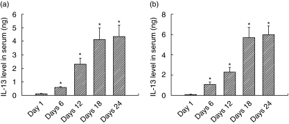
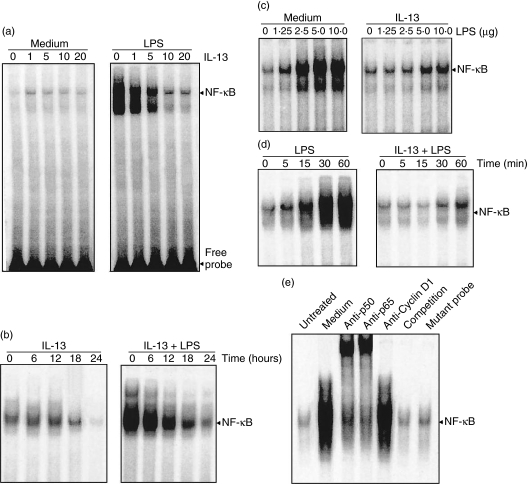
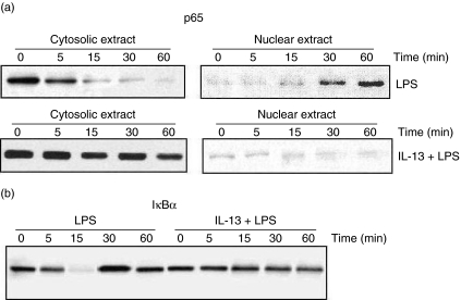
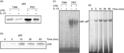
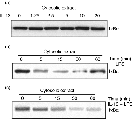
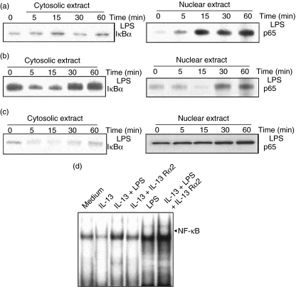
Spontaneously arising transplantable T cell lymphoma, designated as Dalton's lymphoma (DL), is characterized by a highly invasive and deleterious nature almost completely paralysing the host immune system. The level of interleukin (IL)-13 is elevated in serum and ascitic fluid of the DL-bearing host. IL-13 is a potent immunosuppressive cytokine and is an alternative activator of macrophages that suppresses the production of nitric oxide (NO) and expression of inducible nitric oxide synthase (iNOS), and proinflammatory cytokines. The expression of iNOS and proinflammatory cytokines are dependent largely upon the activation of nuclear factor-kappaB (NF-kappaB). Activation of NF-kappaB involves the degradation of cytoplasmic inhibitor IkappaBalpha, allowing the nuclear translocation of NF-kappaB and thereby transcription of the iNOS gene. Therefore, in this study we sought to determine whether the alternative activation or type II polarization of macrophages induced by IL-13 is mediated by the suppression of NF-kappaB and cytoplasmic preservation of IkappaBalpha. Western blot analysis and electrophoretic mobility shift assay (EMSA) indicate that tumour-associated macrophages (TAM) or polarized type II macrophages are due to preserved protein expression of IkappaBalpha, and therefore suppressed NF-kappaB nuclear translocation. These findings suggest that IL-13 may operate through the suppression of NF-kappaB activation and preservation of IkappaBalpha.


Figures

Fig. 1
Fig. 1
Interleukin (IL)-13 serum and ascitic fluid levels. Serum and ascitic fluid from a Dalton's lymphoma (DL)-bearing host were collected at the time-periods indicated and IL-13 concentration was measured. Data are representative of at least three experiments; four mice each time-period gives a mean IL-13 value ± standard error of the mean. (a) IL-13 serum level; (b) IL-13 ascitic fluid level. Data are taken as significant at P < 0·05.
Fig. 2
Fig. 2
Effect of interleukin (IL)-13 on lipopolysaccharide (LPS)-mediated nuclear factor kappa B (NF-κB) activation. (a) Inflammatory macrophages were preincubated with different concentrations of IL-13 (0–20 ng/ml) for 24 h at 37°C and stimulated with 10 ng/ml of LPS for 30 min. After that, nuclear extracts were prepared and analysed by electrophoretic mobility shift assay (EMSA), as described in Materials and methods. (b) Cells were preincubated with 10 ng/ml of IL-13 for 24 h at 37°C and stimulated with or without 10 ng/ml of LPS, and EMSA analysis was performed. (c) Cells were preincubated with 10 ng/ml of IL-13 for 24 h at 37°C and then stimulated with different concentrations of LPS (0–10 ng/ml) for 30 min at 37°C, and EMSA was performed. (d) Cells were preincubated with 10 ng/ml of IL-13 for 24 h, stimulated with 10 ng/ml of LPS for different time intervals (0–60 min) at 37°C, and the EMSA analysis was performed. (e) Nuclear extracts were prepared from unstimulated or LPS-stimulated cells, incubated for 20 min with the antibodies indicated and cold as well as mutated NF-κB probe, and then EMSA analysis was performed.
Fig. 3
Fig. 3
Effect of interleukin (IL)-13 on lipopolysaccharide (LPS)-mediated degradation of IκBα and p65 nuclear migration. Inflammatory macrophages preincubated with medium alone or medium containing 10 ng/ml of IL-13 for 24 h at 37°C, stimulated with 10 ng/ml of LPS for different time intervals (0–60 min), and Western blot analysis was performed for IκBα levels in cytosolic extract (a) and p65 in cytosolic and nuclear extracts (b).
Fig. 4
Fig. 4
Effect of interleukin (IL)-13 on lipopolysaccharide (LPS)-mediated degradation of p65 nuclear migration and nuclear factor kappa B activation (NF-κB) in tumour-associated macrophages (TAM). TAM were preincubated with medium alone for 24 h at 37°C, stimulated with LPS for different time-periods (0–60 min), and Western blot analysis and electrophoretic mobility shift assay (EMSA) were performed. (a) Western blot for p65 in non-stimulated TAM; (b) Western blot for p65 of LPS-stimulated TAM; (c) NF-κB activity of non-stimulated TAM; (d) NF-κB activity of LPS-stimulated TAM.
Fig. 5
Fig. 5
Effect of interleukin (IL)-13 on IκBα level in tumour-associated macrophages (TAM). TAM were preincubated with medium alone for 24 h at 37°C, and then treated either with or without 10 ng/ml of lipopolysaccharide (LPS) or with IL-13 + LPS for different time-periods (0–60 min) at 37°C, and Western blot analysis was performed. (a) IκBα level in TAM treated without LPS stimulation; (b) IκBα level in LPS-treated TAM; (c) IκBα level in TAM treated with IL-13 + LPS.
Fig. 6
Fig. 6
Effect of interleukin (IL)-13 neutralization on lipopolysaccharide (LPS)-dependent IκBα degradation and p65 nuclear translocation. Tumour-associated macrophages (TAM) were preincubated with medium alone or medium containing 10 ng/ml of IL-13 or IL-13 + 10 ng/ml of IL-13 Rα2 for 24 h at 37°C, stimulated with 10 ng/ml of LPS for different time-periods (0–60 min), and Western blot analysis was performed for IκBα in cytosolic extract and gradual increase in p65 level in nuclear extract of untreated TAM (a), increases in the level of IκBα in cytosolic extract and gradual decrease in p65 level in nuclear extract of IL-13-treated TAM (b), and the effect of IL-13 neutralization on IκBα level in cytosolic extract and p65 level in nuclear extract of TAM (c) were observed. Further, involvement of IL-13 in impairment of nuclear factor kappa B nuclear translocation were confirmed by electrophoretic mobility shift assay (EMSA) analysis of nuclear extracts of TAM preincubated with IL-13, LPS, IL-13 + LPS, IL-13 + IL-13 Rα2 or IL-13 + LPS + IL-13 Rα2 (d).

References

    1. Klein G. Comparative studies of mouse tumors with respect to their capacity for growth as ‘ascitic tumor’ and their average nucleic acid content. Exp Cell Res. 1951;2:518–24.
    1. Goldie H, Felix MD. Growth characteristics of free tumor cells transformed serially in the peritoneal fluid of mouse. Cancer Res. 1951;1:173–80. - PubMed
    1. Parajuli P, Singh SM, Kumar A, Sodhi A. Alterations in the tumoricidal function of murine tumor-associated macrophages during progressive growth of a tumor in vivo. Cancer J. 1997;10:222–7.
    1. Singh MP, Rai AK, Singh SM. Gender dimorphism in the progressive in vivo growth of a T-cell lymphoma: involvement of cytokines and gonadal hormones. J Reprod Immunol. 2005;65:17–32. - PubMed
    1. Minty A, Chalon P, Derocq J-M, et al. Interleukin-13 is a new human lymphokine regulating inflammatory and immune response. Nature. 1993;362:248–50. - PubMed

Publication types

MeSH terms