. 2007 Jun 5:7:10.
doi: 10.1186/1475-2867-7-10.

Selective cytotoxicity of pancratistatin-related natural Amaryllidaceae alkaloids: evaluation of the activity of two new compounds

Affiliations

Selective cytotoxicity of pancratistatin-related natural Amaryllidaceae alkaloids: evaluation of the activity of two new compounds

Carly Griffin et al. Cancer Cell Int. .

Abstract

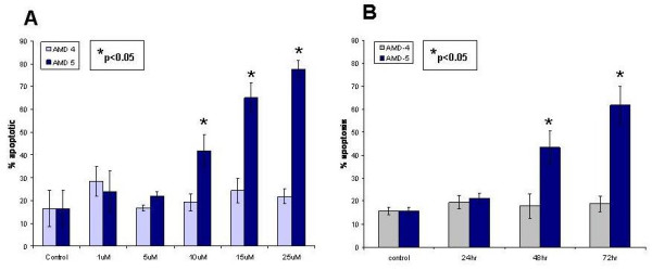
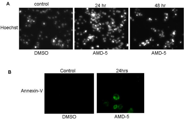
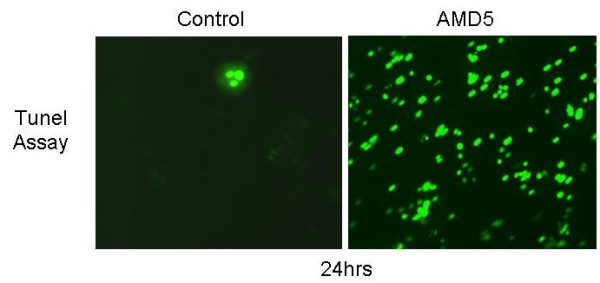
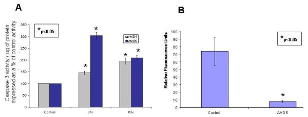
Background: Pancratistatin (PST), a compound extracted from an Amaryllidaceae (AMD) family plant, has been shown to specifically induce apoptosis in cancer cells with no/minimal toxic effect on normal cells. A systematic synthetic approach has indicated that the minimum cytotoxic pharmacophore comprises the trans-fused b/c-ring system containing the 2, 3, 4-triol unit in the C-ring. To further explore the structure-activity relationship of this group of compounds we have investigated the anti-cancer efficacy and specificity of two PST-related natural compounds, AMD4 and AMD5. Both of these compounds lack the polyhydroxylated lycorane element of PST instead having a methoxy-substituted crinane skeleton.

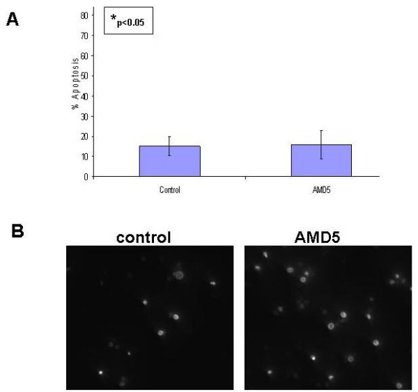
Results: Our results indicate that AMD5 has efficacy and selectivity similar to PST, albeit at a 10-fold increased concentration. Interestingly AMD4 lacks apoptotic activity.

Conclusion: Our results indicate that the phenanthridone skeleton in natural Amaryllidaceae alkaloids may be a significant common element for selectivity against cancer cells; furthermore, the configuration of the methoxy-side groups is responsible for higher binding affinity to the target protein/s thus making for a more efficient anti-cancer agent.


Figures

Figure 1
Figure 1
Chemical structure of native Pancratistatin (A) and of Amaryllidaceae alkaloids AMD4 and AMD5 (B). The phenanthridone skeleton is a conserved feature in alkaloids of the Amaryllidaceae family. The Amaryllidaceae compounds have a methoxy group that varies in orientation between AMD4 (α) and AMD5 (β).
Figure 2
Figure 2
A. Dose-response curve for Jurkat cells treated with various concentrations of either AMD4 or AMD5 for 48 hrs. Cells were stained with cell-permeable Hoechst 33342 dye; brightly stained and condensed nuclei were considered to be apoptotic while non-bright, smooth nuclei were considered healthy (non-apoptotic). The degree of apoptosis was calculated by the number of apoptotic cells counted over the total number of cells visible and displayed as a percentage. A minimum of 5 fields with at least 100 cells per field were counted and tabulated using Microsoft Excel software; values that are statistically significant to p < 0.05 are indicated with an asterisk. Micromolar is represented as uM in this figure only. B. Time Course: A measurement of the degree of apoptosis induced in Jurkat cells treated with 10 μM of either AMD4 or AMD5 over 72 hours. Following treatment, Jurkat cells were incubated with cell-permeable Hoechst 33342 dye; brightly stained condensed nuclei were considered to be apoptotic while non-bright, smooth nuclei were considered healthy. The degree of apoptosis was calculated by the number of apoptotic cells counted over the total number of cells visible displayed as a percentage. A minimum of 5 fields with at least 100 cells per field were counted and tabulated using Microsoft Excel software; values that are statistically significant to p < 0.05 are indicated with an asterisk.
Figure 3
Figure 3
A. Nuclear Morphology of Jurkat cells treated with AMD5 at 10 μM for either 24 hrs or 48 hrs. Jurkat cells were stained with cell-permeable Hoechst 33342 dye to observe apoptotic nuclear morphology. Apoptotic nuclei are brightly stained and condensed when compared to healthy nuclei. The control was treated with DMSO solvent. Magnification: 200×. B. Annexin-V Binding Assay: Jurkat cells were treated with 10 μM AMD5 for 24 hrs. Jurkat cells were incubated with Annexin-V 488 Alexa-Fluor conjugate to observe phosphatidyl-serine flipping from the inner to the outer leaf of the plasma membrane. This is a characteristic event of apoptosis that is made visible with use of a fluorescent microscope; cells undergoing apoptosis will have more Annexin-V substrate bound, and thus appear brighter than healthy cells. Magnification: 400×
Figure 4
Figure 4
TUNEL Assay: Jurkat cells were treated with AMD5 at 10 μM for 24 hrs. Following treatment, Jurkat cells were fixed and immuno-stained with anti-BdrU antibody to observe the extent of DNA damage as described in the Materials and Methods section. The level of staining indicates the degree of DNA damage induced by treatment, where more positively stained cells are in the final stages of apoptosis. Magnification: 200×
Figure 5
Figure 5
A. Caspase-3 Assay: Jurkat cells were treated with 10 μM AMD4 or AMD5 for the early time-points of 3 hrs and 6 hrs. Following treatment, Jurkat cells were incubated with DEVD-AFC peptide to detect caspase-3 activity; fluorescence was measured at excitation 400 nm and emission 505 nm. Activation of caspase-3 is a common event in cells undergoing apoptosis. The level of caspase-3 activity was calculated per microgram of protein and then displayed as a percent increase in fluorescence from caspase-3 activity in untreated cells (control). Values that are statistically significant to p < 0.05 are indicated with an asterisk. B. Mito-Casp Assay: Jurkat cells were treated with 10 μM AMD5 for 48 hrs. Following treatment, Jurkat cells were incubated with a mitochondrial membrane potential (MMP) sensitive dye, and fluorescence was measured at excitation 574 nm and emission at 595 nm. Loss of MMP indicates permeable mitochondria which allows for numerous cytotoxic materials to leak into the cytosol, ending in apoptosis. Loss of MMP is represented as a drop in relative fluorescence units per 10,000 cells. Values that are statistically significant to p < 0.05 are indicated with an asterisk.
Figure 6
Figure 6
A. Time Course: Degree of apoptosis in healthy peripheral mono-nucleated blood cells treated with 10 μM AMD5 for 48 hrs. Healthy human peripheral mono-nucleated blood cells were collected & treated for 48 hours with 10 μM AMD5. The cells were then stained with cell-permeable Hoechst 33342 dye to observe apoptotic nuclear morphology. Brightly stained and condensed nuclei were considered apoptotic, non-bright smooth nuclei were considered healthy. The degree of apoptosis was calculated by the number of apoptotic cells counted over the total number of cells visible displayed as a percentage. A minimum of 5 fields with at least 100 cells per field were counted and tabulated using Microsoft Excel software; values that are statistically significant to p < 0.05 are indicated with an asterisk. B. Nuclear Morphology: Healthy human peripheral mono-nucleated blood cells were collected & treated with 10 μM of AMD5 for 48 hrs. Cells were stained with cell permeable Hoechst 33342 dye to observe apoptotic nuclear morphology. Apoptotic nuclei are brightly stained and condensed when compared to healthy nuclei. The control was treated with DMSO solvent. Magnification: 200×.

Similar articles

Cited by

References

    1. Pettit GR, Pettit GR, III, Backhaus RA, Boyd MR, Meerow AW. Antineoplastic Agents 256. Cell Growth Inhibitory Isocarbostyrils from Hymenocallis. Journal of Natural Products. 1993;56:1682. doi: 10.1021/np50100a004. - DOI - PubMed
    1. Kekre N, Griffin C, McNulty J, Pandey S. Pancratistatin causes early activation of caspase-3 and the flipping of phosphatidyl serine followed by rapid apoptosis specifically in human lymphoma cells. Cancer Chemotherapy & Pharmacology. 2005;56:29–38. doi: 10.1007/s00280-004-0941-8. - DOI - PubMed
    1. McLachlan A, Kekre N, McNulty J, Pandey S. Pancratistatin: a natural anti-cancer compound that targets mitochondria specifically in cancer cells to induce apoptosis. Apoptosis. 2005;10:619–630. doi: 10.1007/s10495-005-1896-x. - DOI - PubMed
    1. McNulty J, Mao J, Gibe R, Mo R, Wolf S, Pettit GR, Herald DL, Boyd MR. Studies directed towards the refinement of the Pancratistatin cytotoxic pharmacophore. Bioorganic & Medicinal Chemistry Letters. 2001;11:169–172. doi: 10.1016/S0960-894X(00)00614-4. - DOI - PubMed
    1. McNulty J, Larichev V, Pandey S. A synthesis of 3-deoxydihydrolycoricidine: refinement of a structurally minimum Pancratistatin pharmacophore. Bioorganic & Medicinal Chemistry Letters. 2005;15:5315–5318. doi: 10.1016/j.bmcl.2005.08.024. - DOI - PubMed