

Ultrasound measurement of joint cartilage thickness in large and small joints in healthy children: a clinical pilot study assessing observer variability
- PMID: 17550627
- PMCID: PMC1869024
- DOI: 10.1186/1546-0096-5-3
Ultrasound measurement of joint cartilage thickness in large and small joints in healthy children: a clinical pilot study assessing observer variability
Abstract
Background: Loss of joint cartilage is a feature of destructive disease in JIA. The cartilage of most joints can be visualized with ultrasonography (US). Our present study focuses on discriminant validity of US in children. We studied reproducibility between and within a skilled and a non-skilled investigator of US assessment of cartilage thickness in small and large joints in healthy children.
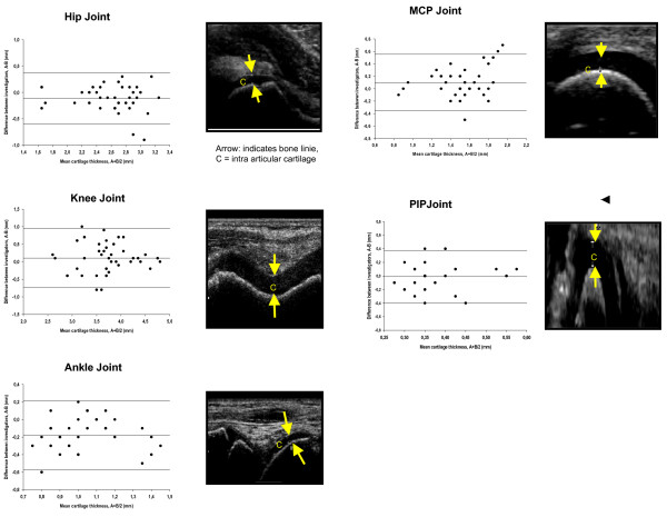
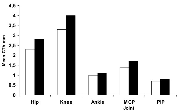
Methods and results: In 11 healthy children (5 girls/6 boys), aged 9.6 years (9.3-10 years), 110 joints were examined. Cartilage thickness of the right and left hip, knee, ankle, 2nd metacarpophalangeal (MCP), and 2nd proximal interphalangeal (PIP) joint independently. The joints were examined twice, two days apart by a skilled and a non-skilled investigator. Mean cartilage thickness in the five joints was: hip 2.59 +/- 0.41, knee 3.67 +/- 0.64, ankle 1.08 +/- 0.31, MCP 1.52 +/- 0.27 and PIP 0.73 +/- 0.15 mm. We found the same mean differences in CTh of 0.6 mm in the inter-observer part with regard of the PIP joint. Within investigators (intra-observer), the smallest mean difference of CTh was found in the MCP joint with -0.004 (skilled) and 0.013 mm (non-skilled).
Conclusion: We found the level of agreement between observers within a 95% Confidence Interval in assessment of cartilage thickness in hip-, knee-, ankle-, MCP-, and PIP joints in healthy children. Observer variability seems not to relate to joint size but to the positioning of the joints and the transducer. These factors seem to be of major importance for reproducible US measurements. The smallest difference in measurement of cartilage thickness between observers was found in the PIP joint, and within observers in the MCP joint and it seems that using EULAR standard US guidelines is feasible for a pediatric setting. The use of US in children is promising. Studies on larger groups of children are needed to confirm the validation and variability of US in children as well as determining the smallest detectable difference of US measures.
Figures
References
LinkOut - more resources
Full Text Sources
Miscellaneous
