

Eye-blink conditioning is associated with changes in synaptic ultrastructure in the rabbit interpositus nuclei
- PMID: 17551096
- PMCID: PMC1896088
- DOI: 10.1101/lm.348307
Eye-blink conditioning is associated with changes in synaptic ultrastructure in the rabbit interpositus nuclei
Abstract
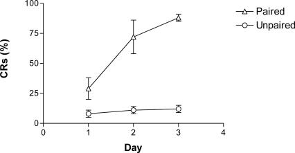
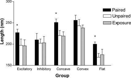
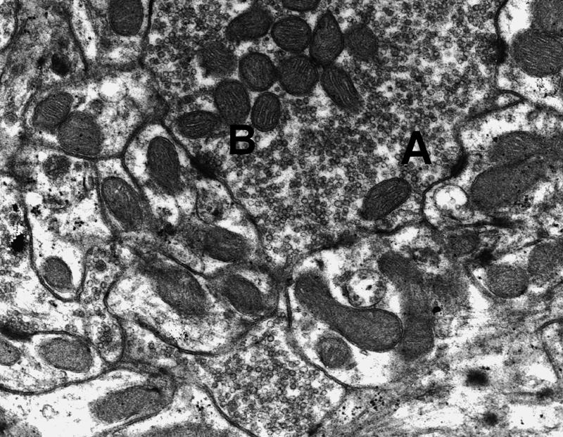
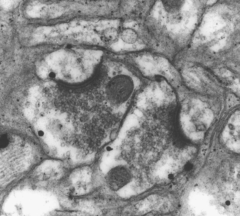
Eye-blink conditioning involves the pairing of a conditioned stimulus (usually a tone) to an unconditioned stimulus (air puff), and it is well established that an intact cerebellum and interpositus nucleus, in particular, are required for this form of classical conditioning. Changes in synaptic number or structure have long been proposed as a mechanism that may underlie learning and memory, but localizing these changes has been difficult. Thus, the current experiment took advantage of the large amount of research conducted on the neural circuitry that supports eye-blink conditioning by examining synaptic changes in the rabbit interpositus nucleus. Synaptic quantifications included total number of synapses per neuron, numbers of excitatory versus inhibitory synapses, synaptic curvature, synaptic perforations, and the maximum length of the synapses. No overall changes in synaptic number, shape, or perforations were observed. There was, however, a significant increase in the length of excitatory synapses in the conditioned animals. This increase in synaptic length was particularly evident in the concave-shaped synapses. These results, together with previous findings, begin to describe a sequence of synaptic change in the interpositus nuclei following eye-blink conditioning that would appear to begin with structural change and end with an increase in synaptic number.
Figures
Similar articles
-
Developmental changes in eye-blink conditioning and neuronal activity in the cerebellar interpositus nucleus.J Neurosci. 2000 Jan 15;20(2):813-9. doi: 10.1523/JNEUROSCI.20-02-00813.2000. J Neurosci. 2000. PMID: 10632611 Free PMC article.
-
Classical conditioning does not occur when direct stimulation of the red nucleus or cerebellar nuclei is the unconditioned stimulus.Brain Res. 1988 Feb 23;442(1):97-104. doi: 10.1016/0006-8993(88)91436-9. Brain Res. 1988. PMID: 3359261
-
The learning-related activity that develops in the pontine nuclei during classical eye-blink conditioning is dependent on the interpositus nucleus.Learn Mem. 1997 Mar-Apr;3(6):532-44. doi: 10.1101/lm.3.6.532. Learn Mem. 1997. PMID: 10456115
-
The role of the cerebellum in classical conditioning of discrete behavioral responses.Neuroscience. 2009 Sep 1;162(3):732-55. doi: 10.1016/j.neuroscience.2009.01.041. Epub 2009 Jan 27. Neuroscience. 2009. PMID: 19409234 Review.
-
Role of the nuclei in eyeblink conditioning.Ann N Y Acad Sci. 2002 Dec;978:93-105. doi: 10.1111/j.1749-6632.2002.tb07558.x. Ann N Y Acad Sci. 2002. PMID: 12582044 Review.
Cited by
-
Neurons in the barrel cortex turn into processing whisker and odor signals: a cellular mechanism for the storage and retrieval of associative signals.Front Cell Neurosci. 2015 Aug 21;9:320. doi: 10.3389/fncel.2015.00320. eCollection 2015. Front Cell Neurosci. 2015. PMID: 26347609 Free PMC article.
-
Coordinated Plasticity among Glutamatergic and GABAergic Neurons and Synapses in the Barrel Cortex Is Correlated to Learning Efficiency.Front Cell Neurosci. 2017 Jul 26;11:221. doi: 10.3389/fncel.2017.00221. eCollection 2017. Front Cell Neurosci. 2017. PMID: 28798668 Free PMC article.
-
Cerebellar Theta-Burst Stimulation Impairs Memory Consolidation in Eyeblink Classical Conditioning.Neural Plast. 2018 Oct 9;2018:6856475. doi: 10.1155/2018/6856475. eCollection 2018. Neural Plast. 2018. PMID: 30402087 Free PMC article.
-
Learning-related long-term potentiation of inhibitory synapses in the cerebellar cortex.Proc Natl Acad Sci U S A. 2008 Jan 15;105(2):769-74. doi: 10.1073/pnas.0706342105. Epub 2008 Jan 9. Proc Natl Acad Sci U S A. 2008. PMID: 18184813 Free PMC article.
-
No Medium-Term Spinocerebellar Input Plasticity in Deep Cerebellar Nuclear Neurons In Vivo?Cerebellum. 2017 Jun;16(3):638-647. doi: 10.1007/s12311-016-0839-0. Cerebellum. 2017. PMID: 28032320 Free PMC article.
References
-
- Bracha V., Irwin K.B., Webster M.L., Wunderlich D.A., Stachowiak M.K., Bloedel J.R. Microinjections of anisomycin into the intermediate cerebellum during learning affect the acquisition of classically conditioned responses in the rabbit. Brain Res. 1998;788:169–178. - PubMed
-
- Chen G., Steinmetz J.E. Microinfusion of protein kinase inhibitor H7 into the cerebellum impairs the acquisition but not the retention of classical eyeblink conditioning in rabbits. Brain Res. 2000a;856:193–201. - PubMed
-
- Chen G., Steinmetz J.E. Intra-cerebellar infusion of NMDA receptor antagonist AP5 disrupts classical eyeblink conditioning in rabbits. Brain Res. 2000b;887:144–156. - PubMed
Publication types
MeSH terms
Grants and funding
LinkOut - more resources
Full Text Sources