. 2007 Jun 5;14(6):390-9.
doi: 10.1101/lm.571007. Print 2007 Jun.

Metaplastic effect of apamin on LTP and paired-pulse facilitation

Affiliations

Metaplastic effect of apamin on LTP and paired-pulse facilitation

Laurence Ris et al. Learn Mem. .

Abstract

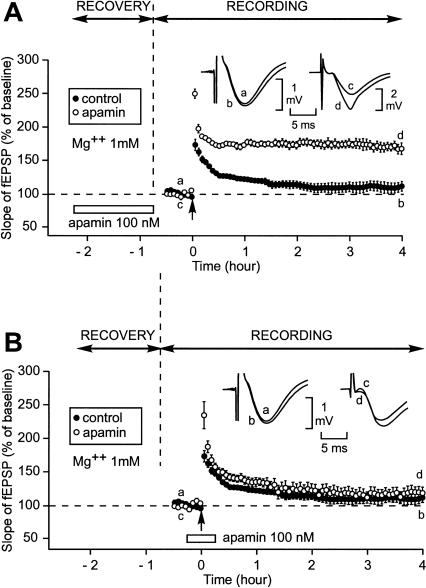
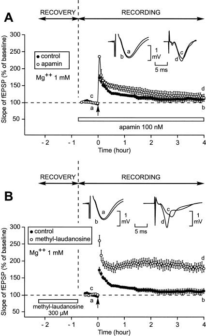
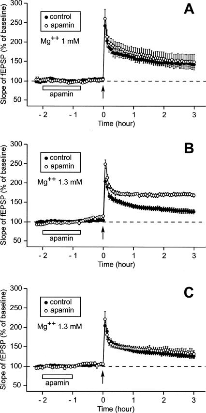
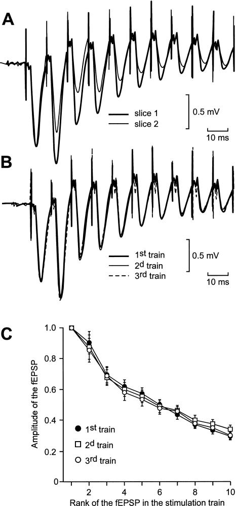
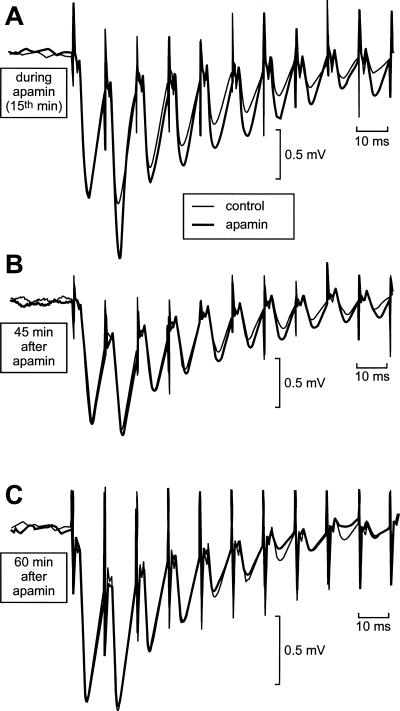
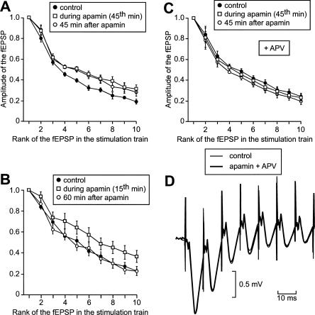
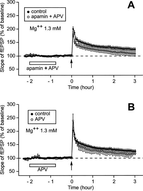
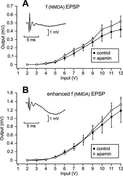
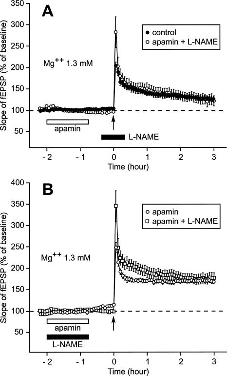
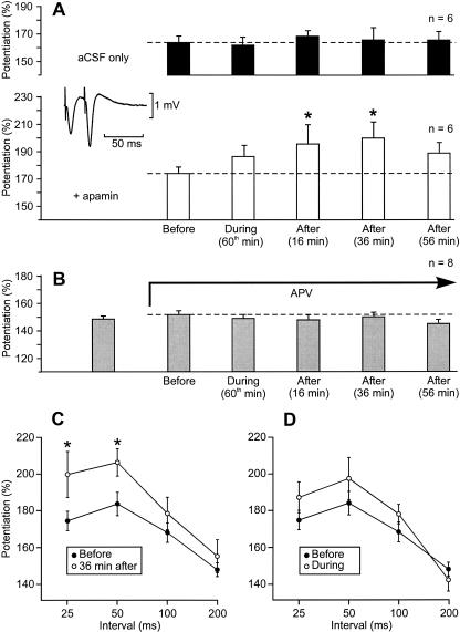
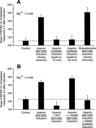
In area CA1 of hippocampal slices, a single 1-sec train of 100-Hz stimulation generally triggers a short-lasting long-term potentiation (S-LTP) of 1-2 h. Here, we found that when such a train was applied 45 min after application of the small conductance Ca(2+)-activated K(+ )(SK) channel blocker apamin, it induced a long-lasting LTP (L-LTP) of several hours, instead of an S-LTP. Apamin-induced SK channel blockage is known to resist washing. Nevertheless, the aforementioned effect is not a mere delayed effect; it is metaplastic. Indeed, when a single train was delivered to the Schaffer's collaterals during apamin application, it induced an S-LTP, like in the control situation. At the moment of this LTP induction (15th min of apamin application), the SK channel blockage was nevertheless complete. Indeed, at that time, under the influence of apamin, the amplitude of the series of field excitatory postsynaptic potentials (fEPSPs) triggered by a stimulation train was increased. We found that the metaplastic effect of apamin on LTP was crucially dependent on the NO-synthase pathway, whereas the efficacy of the NMDA receptors was not modified at the time of its occurrence. We also found that apamin produced an increase in paired-pulse facilitation not during, but after, the application of the drug. Finally, we found that the induction of each of these two metaplastic phenomena was mediated by NMDA receptors. A speculative unitary hypothesis to explain these phenomena is proposed.


Figures

Figure 1.
Figure 1.
Induction by a single train of high-frequency stimulation (100 Hz, 1 sec) (HFS) of a long-lasting LTP (L-LTP) instead of a short-lasting LTP (S-LTP) when apamin, an SK channel blocker, was applied during the recovery period but not when it was perfused around the time of LTP induction. (A) Slices were allowed to recover in interface and recordings were also carried out in interface. The figure shows the time course of the fEPSP slope 32 min before and 4 h after LTP induction by a single train (arrow) in the control situation (●) and when apamin was applied during the recovery period (○). (Inset) Sample fEPSP traces from individual experiments are shown; they were recorded just before (a,c) and 4 h after (b,d) the induction stimulation. Traces a and b were recorded in the control situation; c and d were recorded when apamin was applied during recovery. (B) Time course of fEPSP slope 32 min before and 4 h after delivery of 1-sec stimulation train in the control situation (●) and when apamin was applied 15 min before and 15 min after LTP induction (○). (Inset) Labeling similar to that used in A. (Left) A control experiment, (right) an experiment where apamin was applied around LTP induction.
Figure 2.
Figure 2.
Induction of an L-LTP instead of an S-LTP was not caused by apamin when this drug was applied during the whole recording period but was caused by (+)-methyl-laudanosine, another SK channel blocker, when this drug was applied during the recovery period. Contrarily to that induced by apamin, the SK channel blockage caused by (+)-methyl-laudanosine does not resist to a 10-min washout (Scuvée-Moreau et al. 2002; J. Scuvée-Moreau, J.-F. Liégeois, and V. Seutin, unpubl.). (A) Time course of fEPSP slope 32 min before and 4 h after delivery of 1-sec stimulation train in the control situation (●) and when apamin was applied during the whole recording period (○). (Inset) Examples of fEPSP recorded in one experiment just before (a,c) and 4 h after (b,d) induction in the control situation (left) and in presence of apamin (right). (B) Time course of fEPSP slope 32 min before and 4 h after delivery of 1-sec stimulation train in the control situation (●) and when (+)-methyl-laudanosine was applied during the recovery period (○). (Inset) Labeling similar to that used in A. (Left) A control experiment, (right) an experiment where (+)-methyl-laudanosine was applied during the recovery period.
Figure 3.
Figure 3.
Effect of prior application of apamin on the duration of LTP when the pre-LTP induction duration was as long as 2 h 30 min (after the recovery period). Each part of the figure compares the time course of the fEPSP slope 2 h 15 min before and 3 h after the delivery of a single train of HFS. (A) When Mg2+ concentration was 1 mM, an application of apamin lasting 1 h 15 min and ending 45 min before LTP induction did not modify the characteristics of LTP. In fact, the control LTP was improved as a result of the longer duration of the baseline. Compare this control LTP with that displayed in Fig. 1A. (B) When Mg2+ concentration was 1.3 mM, the duration of LTP was increased by prior application of apamin (with respect to the control LTP recorded at that Mg2+ concentration). (C) When Mg2+ concentration was 1.3 mM and when application of apamin was stopped 1 h before LTP induction (instead of 45 min in B), prior application of apamin no longer had any influence on LTP.
Figure 4.
Figure 4.
The profile of the global depolarization induced by an HFS train varied from one slice to another (A) but not when the stimulus was delivered thrice at 30-min intervals on the same slice (B,C). (A) Superimposition of the first 10 fEPSPs of two series of fEPSPs recorded in two distinct slices in response to the delivery of one train of HFS. The intensity was adjusted so that the amplitudes of the first fEPSP were the same in slice number 1 (thick trace) as in slice number 2 (thin trace). (B) Superimposition of the global depolarizations induced in a same slice by three HFS trains separated by 30 min. The intensity of stimulation was adjusted to obtain the same amplitude for the first fEPSP in each of the three global depolarizations. (C) In each slice from a group of 10, the amplitude of each of the first 10 fEPSPs elicited by three HFS trains at 30-min intervals was measured. The means ± SEM of the measurements were plotted in function of the rank of the corresponding fEPSP in the series.
Figure 5.
Figure 5.
Apamin causes an enhancement of the global depolarization induced by a train of HFS (100 Hz). After recovery and 15 min of baseline recording, apamin was applied for 1 h 15 min. (A,B,C) Each part of the figure compares the first 10 fEPSPs of two series of fEPSPs recorded on a same slice in response to the delivery of one train of HFS, one under the influence of apamin (thick traces), the other before any application of the drug (thin traces). (A) The global depolarization induced by the stimulation train was increased in presence of apamin (here at the 15th min after the start of the apamin perfusion). (B) The global depolarization induced by the stimulation train was still increased 45 min after the end of the application of apamin. (C) The global depolarization was no longer increased when elicited 60 min after the application of apamin had been stopped.
Figure 6.
Figure 6.
Apamin causes a long-lasting enhancement of the global depolarization elicited by a train of high-frequency stimulation, and this phenomenon is NMDA receptor-dependent. (A,B,C) The amplitude of each of the first 10 fEPSPs within the series of fEPSPs is plotted in function of its rank in the series. (A) Amplitudes of the fEPSPs induced by a stimulation train in a same group of six slices before apamin application, at the 45th min of the drug application, and 45 min after the end of apamin application. (B) Same display as in A, but the stimulation trains were applied on another set of eight slices before, 15 min after the start, and 60 min after the end of apamin perfusion. (C) Same display and same pattern of train delivery on another set of five slices, as in A, but when APV was present throughout the experiment. (D) Illustration of the lack of effect of apamin on the global depolarization induced by a high-frequency stimulation train when APV was coapplied with apamin. The first seven fEPSPs of two series of fEPSPs elicited before (thin trace) and 45 min after apamin application are superimposed.
Figure 7.
Figure 7.
Suppression of the metaplastic effect of apamin on LTP by APV. (A,B) Time course of fEPSP 2 h 15 min before and 3 h after one train of HFS. (A) When APV was coapplied with apamin, the LTP induced after prior application of apamin (○) was not different from the control LTP (●). (B) When APV was applied alone using the same timing as that used for apamin application, the LTP induced after prior application of APV (○) was not different from the control LTP (●).
Figure 8.
Figure 8.
Apamin did not produce any delayed change in the NMDA receptor-mediated fEPSP. (A) Amplitude of the NMDA receptor-mediated fEPSP in function of the stimulation intensity in the control situation (●) and 20 min after the end of apamin application (○). Apamin was applied for 1 h 15 min. The NMDA receptor-mediated component of the fEPSP was obtained in response to stimulation pulses delivered to the Schaffer collaterals of slices perfused for 20 min with aCSF containing CNQX, a blocker of AMPA receptors. (Inset) Sample trace of an NMDA receptor-mediated fEPSP recorded during an individual experiment. (B) Amplitude of the enhanced NMDA receptor-mediated fEPSP in function of the stimulation intensity in the control situation (●) and 40 min after the end of apamin application (○). After completion of the measurements summarized in A, the same slices were perfused with CNQX dissolved in Mg2+-free aCSF. This lifted the Mg2+ blockage in the NMDA receptors, so that all the NMDA receptors could be activated by glutamate. (Inset) Sample trace of an enhanced NMDA receptor-mediated fEPSP recorded during the same individual experiment as that shown in the inset of A.
Figure 9.
Figure 9.
NO-synthase signaling pathway is necessary for the expression of the metaplastic effect of apamin on LTP. (A) When L-NAME, an inhibitor of NO-synthase, was applied 20 min before and 20 min after LTP induction, it prevented prior application of apamin from transforming the S-LTP usually triggered by a single stimulation train into an L-LTP. In this case, the time course of the increase in the slope of the fEPSP triggered by one train of HFS delivered after prior application of apamin (○) was not different from that observed when no drug was applied (●). (B) When L-NAME was coapplied with apamin, it did not prevent apamin from having a metaplastic effect on LTP. In this case, the time course of the increase in the slope of the fEPSP triggered by a single stimulation train corresponded to the profile of an L-LTP whether apamin was applied 45 min before LTP induction with L-NAME (◽) or without (○).
Figure 10.
Figure 10.
Delayed influence of apamin on paired-pulse facilitation. (A) Influence of apamin (applied for 1 h 15 min) on the amplitude of paired-pulse facilitation tested with a 25-msec interval (empty columns). (*) A significant difference with respect to the measurement made before apamin application. The moment of testing is indicated under each column. When paired-pulse facilitation was measured in the absence of apamin application at the corresponding time points (black columns), the amplitude of the potentiation did not change over time. (aCSF) Artificial cerebrospinal fluid. (Inset) fEPSP elicited by paired-pulses separated by an interval of 25 msec. (B) The influence of apamin on paired-pulse facilitation was suppressed by APV. The testing pattern was the same as in A. (C) Amplitude of the paired-pulse facilitation in function of the interval separating the paired-pulses before (●) and 36 min after apamin application (○). (D) Amplitude of the paired-pulse facilitation in function of the interval separating the paired-pulses before (●) and at the 60th min of apamin application (○).
Figure 11.
Figure 11.
Summary bar graph gathering the results of experiments carried out to demonstrate that the effect of apamin on LTP was metaplastic (A) and to try to—at least partially—elucidate the mechanisms of this action (B). (A) This set of experiments was carried out using aCSF containing Mg2+ 1mM. The amplitude (±SEM) of the increase in the slope of fEPSP was measured 4 h after LTP induction. (B) In this second set of experiments, the concentration of Mg2+ was 1.3 mM and the measurements were made 3 h after LTP induction.

References

    1. Abel T., Nguyen P.V., Barad M., Deuel T., Kandel E.R., Bourtchouladze R. Genetic demonstration of a role for PKA in the late phase of LTP and in hippocampus-based long-term memory. Cell. 1997;88:615–626. - PubMed
    1. Abraham W.C., Bear M.F. Metaplasticity: The plasticity of synaptic plasticity. Trends Neurosci. 1996;19:126–130. - PubMed
    1. Abraham W.C., Tate W.P. Metaplasticity: A new vista across the field of synaptic plasticity. Prog. Neurobiol. 1997;52:303–323. - PubMed
    1. Arancio O., Kiebler M., Lee C., Lev-Ram V., Tsien R., Kandel E., Hawkins R. Nitric oxide acts directly in the presynaptic neuron to produce long-term potentiation in cultured hippocampal neurons. Cell. 1996;87:1025–1035. - PubMed
    1. Behnisch T., Reymann K.G. Inhibition of apamin-sensitive calcium dependent potassium channels facilitate the induction of long-term potentiation in the CA1 region of rat hippocampus in vitro. Neurosci. Lett. 1998;253:91–94. - PubMed

Publication types

MeSH terms

Substances

LinkOut - more resources