. 2007 Jun 6;2(6):e493.
doi: 10.1371/journal.pone.0000493.

Asymmetry of early endosome distribution in C. elegans embryos

Affiliations

Asymmetry of early endosome distribution in C. elegans embryos

Robert Andrews et al. PLoS One. .

Abstract

Background: Endocytosis is involved in the regulation of many cellular events, including signalling, cell migration, and cell polarity. To begin to investigate roles for endocytosis in early C. elegans development, we examined the distribution and dynamics of early endosomes (EEs) in embryos.

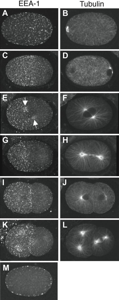
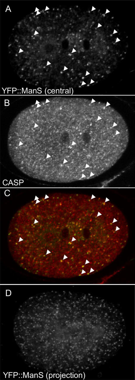
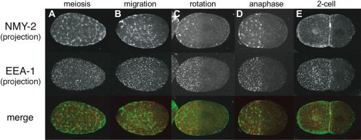
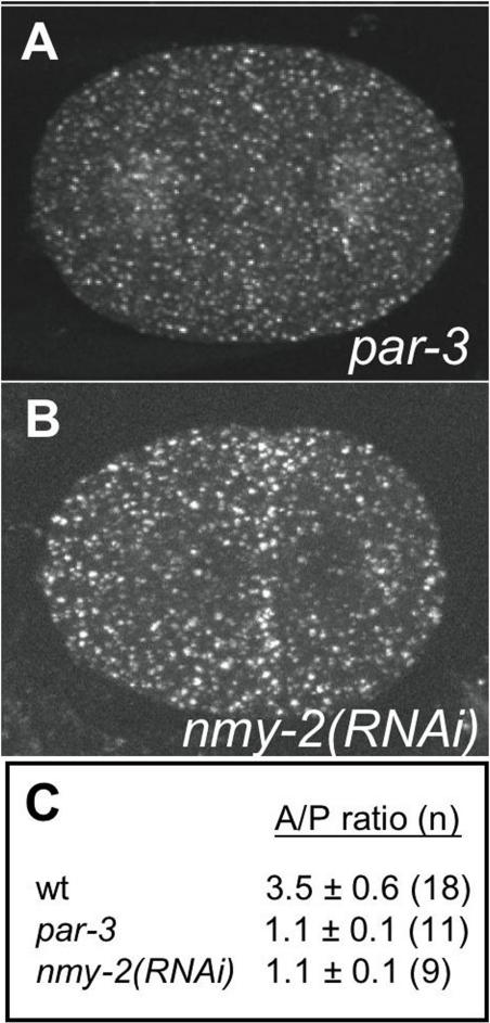
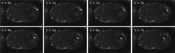
Methodology/principal findings: EEs are primarily found at the cell periphery with an initially uniform distribution after fertilization. Strikingly, we find that during the first cell cycle, EEA-1 positive EEs become enriched at the anterior cortex. In contrast, the Golgi compartment shows no asymmetry in distribution. Asymmetric enrichment of EEs depends on acto-myosin contractility and embryonic PAR polarity. In addition to their localization at the cortex, EEs are also found around the centrosome. These EEs move rapidly (1.3 microm/s) from the cortex directly to the centrosome, a speed comparable to that of the minus end directed motor dynein.

Conclusions/significance: We speculate that the asymmetry of early endosomes might play a role in cell asymmetries or fate decisions.


Conflict of interest statement

Competing Interests: The authors have declared that no competing interests exist.

Figures

Figure 1
Figure 1. Early endosomes become enriched in the anterior during the first cell cycle.
Shown are projections of EEs during the first two cell cycles visualised using anti-EEA-1 and a single focal plane of microtubules visualised using anti-tubulin. (A, B) meiosis. (C, D) polarity establishment; beginning of sperm aster growth. (E, F) prophase; arrows point to enrichment around the asters. (G, H) anaphase. (I, J) Early two cell stage. (K, L) Late two cell stage. (M) central focal plane of embryo in (A,B), showing that EEs are cortically associated. EEs are evenly distributed in meiosis (A), then become depleted from the posterior near the site of the sperm pronucleus (C). Anterior enrichment of EEs is maintained throughout the rest of the cell cycle and in the two cell stage. During meiotic divisions, there is a small clearing of EEs around the meiotic spindle (data not shown). Anterior, left.
Figure 2
Figure 2. The Golgi shows no asymmetry in distribution during the first cell cycle.
(A–C) Even distribution of Golgi structures in a wild-type telophase embryo. The Golgi is visualized using two markers: (A) YFP::ManS, a fusion of YFP and the Golgi resident mannosidase F58H1.1 and (B) anti-CASP/Y54F10AM.4b staining. CASP is an integral Golgi membrane protein . Arrowheads in (A), (B), and (C) (the merged image) show colocalization of puncta with the two markers. Central focal planes are shown. (D) even distribution of Golgi structures in a projection of 12 focal planes of YFP::ManS in a wild-type anaphase stage embryo.
Figure 3
Figure 3. Asymmetry of early endosomes is coincident with NMY-2 asymmetry GFP::NMY-2 embryos co-stained for EEs (anti-EEA-1) and NMY-2 (anti-GFP).
The area of enrichment of EEs coinncides with the NMY-2 domain at all stages.
Figure 4
Figure 4. NMY-2 and PAR-3 are required for EE asymmetry.
Projections of EEA-1 distributions in (A) par-3(it71) mutant, and (B) nmy-2(RNAi) embryo. Compare to similar staged wild-type images in Figure 1 (1G and 1I, respectively). Asymmetry is lost in par-3 mutant and nmy-2(RNAi) embryos. (C) Ratio of the number of EEA-1 positive puncta in an anterior region compared to a posterior region of the same size.
Figure 5
Figure 5. Centrosome directed movement of EEs.
EEs visualied live using GFP:EEA-1(FYVE*2). Shown are eight still images taken 1 second apart; arrow points to an EE that moves rapidly towards the centrosome.

References

    1. Emery G, Knoblich JA. Endosome dynamics during development. Curr Opin Cell Biol. 2006;18:407–415. - PubMed
    1. Goldstein B, Hird SN. Specification of the anteroposterior axis in Caenorhabditis elegans. Development. 1996;122:1467–1474. - PubMed
    1. Cowan CR, Hyman AA. Centrosomes direct cell polarity independently of microtubule assembly in C. elegans embryos. Nature. 2004;431:92–96. - PubMed
    1. O'Connell KF, Maxwell KN, White JG. The spd-2 gene is required for polarization of the anteroposterior axis and formation of the sperm asters in the Caenorhabditis elegans zygote. Dev Biol. 2000;222:55–70. - PubMed
    1. Sadler PL, Shakes DC. Anucleate Caenorhabditis elegans sperm can crawl, fertilize oocytes and direct anterior-posterior polarization of the 1-cell embryo. Development. 2000;127:355–366. - PubMed

Publication types

Substances