. 2007 Jun 6;2(6):e513.
doi: 10.1371/journal.pone.0000513.

The role of the substantia nigra pars compacta in regulating sleep patterns in rats

Affiliations

The role of the substantia nigra pars compacta in regulating sleep patterns in rats

Marcelo M S Lima et al. PLoS One. .

Abstract

Background: As of late, dopaminergic neurotransmission has been recognized to be involved in the generation of sleep disturbances. Increasing evidence shows that sleep disturbances in Parkinson's disease (PD) patients are mostly related to the disease itself, rather than being a secondary phenomenon. Evidence contained in the literature lends support to the hypothesis that the dopaminergic nigrostriatal pathway is closely involved in the regulation of sleep patterns.

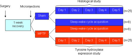
Methodology/principal findings: To test this hypothesis we examined the electrophysiological activity along the sleep-wake cycle of rats submitted to a surgically induced lesion of the SNpc by 1-methyl-4-phenyl-1,2,3,6-tetrahydropyridine (MPTP). We demonstrated that a 50% lesion of the substantia nigra pars compacta (SNpc) suffices to produce disruptions of several parameters in the sleep-wake pattern of rats. A robust and constant decrease in the latency to the onset of slow wave sleep (SWS) was detected throughout the five days of recording in both light [F((22.16)) = 72.46, p<0.0001] and dark [F((22.16)) = 75.0, p<0.0001] periods. Also found was a pronounced increase in the percentage of sleep efficiency during the first four days of recording [F((21.15)) = 21.48, p<0.0001], in comparison to the sham group. Additionally, the reduction in the SNpc dopaminergic neurons provoked an ablation in the percentage of rapid eye movement sleep (REM) during three days of the sleep-wake recording period with a strong correlation (r = 0.91; p<0.0001) between the number of dopaminergic neurons lost and the percentage decrease of REM sleep on the first day of recording. On day 4, the percentage of REM sleep during the light and dark periods was increased, [F((22.16)) = 2.46, p<0.0007], a phenomenon consistent with REM rebound.

Conclusions/significance: We propose that dopaminergic neurons present in the SNpc possess a fundamental function in the regulation of sleep processes, particularly in promoting REM sleep.


Conflict of interest statement

Competing Interests: The authors have declared that no competing interests exist.

Figures

Figure 1
Figure 1. Schematic representation of the experimental design.
After the intranigral microinjections of saline or MPTP, the rats were distributed for sleep-wake cycle recording (n = 8/group), midbrain TH immunohistochemical examination (n = 5) and nigral TH protein expression study (n = 5). Histological and western blotting experiments used animals that had their brains collected along the 5 days of recording (n = 5/day/group) at the same hour they were lesioned, according to the time-points schedule.
Figure 2
Figure 2. Dopaminergic neurons present in the SNpc are reduced by half after MPTP intranigral microinjection.
A representative immunohistochemistry labeling of TH-ir neurons is shown in animals at the end of 5-day sleep-wake cycle recordings; (a) sham group (the inset square shows the specific region depicted in the panel below), (b) MPTP group (the inset square shows the specific region depicted in the panel below), (c) sham group in higher magnification (d) MPTP group in higher magnification, (e) bilateral quantification of the loss of TH-ir neurons in the SNpc, (f) the loss of TH-ir neurons in the SNpc correlated closely with the decrease of REM sleep on the first day of recording, subsequent to MPTP microinjection. The values are expressed as mean±S.E.M. **p<0.01, ANOVA followed by the Newman–Keuls test.
Figure 3
Figure 3. Dual effects upon the latencies to SWS and REM sleep after selective SNpc lesion.
(a) Latency to SWS showed to be decreased at all time-points after MPTP exposure, in both light and dark periods. (b) Latency to REM sleep was increased in the light and dark periods after MPTP only on the first day of recording. The values are expressed as mean±S.E.M. *p<0.05, ***p<0.0001 compared to baseline, ANOVA followed by the Tukey test.
Figure 4
Figure 4. Nigral disruption promoted an increase in the sleep efficiency during the first four days of recording.
SNpc dopaminergic neuronal loss promoted a sustained increase in the percentage of sleep efficiency on the first 3 days of the sleep-wake recording, in both light and dark periods. On the fourth day, the increase in this parameter occurred only in the dark period. The values are expressed as mean±S.E.M. **p<0.05 compared to baseline, #p<0.05 compared to the respective sham group, ANOVA followed by the Tukey test.
Figure 5
Figure 5. Slight increase in the percentage of SWS after SNpc lesion.
The percentage of SWS was increased on the second and fourth days, only in the dark periods, indicating sleepiness effect in the activity period of the rodent. The values are expressed as mean±S.E.M. *p<0.05 compared to those of baseline, #p<0.05 compared to the respective sham group. ANOVA followed by the Tukey test.
Figure 6
Figure 6. SNpc neurons are fundamental in the generation of REM sleep.
The percentage of REM sleep, of the MPTP group, was reduced on the first three days of recording, in both light and dark periods. In contrast, the percentage of REM sleep significantly rose on the fourth day, probably as a compensatory rebound mechanism. The values are expressed as mean±S.E.M. *p<0.05 compared to those of baseline, #p<0.05 compared to the respective sham group, ψp<0.0007 compared to the MPTP group day 1. ANOVA followed by the Tukey test.
Figure 7
Figure 7. Western blotting analysis of the Nigral TH protein expression along the sleep-wake cycle recording.
(a) Serial dilution of nigral TH protein was loaded across the lanes and visualized with enhanced chemiluminescence. A standard curve was generated from densitometric values obtained from computer analysis of digitized film from immunoblot. A linear relationship between optical density and the amount of protein was observed with a strong correlation. (b) TH protein expression was significantly decreased in the MPTP group in comparison to the sham on the first day after the microinjection. Two days after MPTP, TH presented an expression of 98% in comparison to the sham group, on the respective day. A similar situation was observed for the subsequent time-points analyzed. The values are expressed as mean±S.E.M. *p<0.05 compared to those of baseline. ANOVA followed by the Newman-Keuls test.

References

    1. Adler CH. Nonmotor complications in Parkinson's disease. Mov Disord. 2005;20:S23–S29. - PubMed
    1. Gagnon JF, Bedard MA, Fantini ML, Petit D, Panisset M, et al. REM sleep behavior disorder and REM sleep without atonia in Parkinson's disease. Neurology. 2002;59:585–589. - PubMed
    1. Hobson D, Lang A, Wayne MW, Razmy A, Rivest J, et al. Excessive daytime sleepiness and sudden-onset sleep in Parkinson Disease. A survey by the Canadian Movement Disorder Group. JAMA. 2002;287:455–463. - PubMed
    1. Sandyk R. Treatment with weak electromagnetic fields restores dream recall in a parkinsonian patient. Int J Neurosci. 1997;90:75–86. - PubMed
    1. Hobson JA, Pace-Schott EF, Stickgold R, Kahn D. To dream or not to dream? Relevant data from new neuroimaging and electrophysiological studies. Curr Opin Neurobiol. 1998;8:239–244. - PubMed

Publication types

Substances